Antimicrobial Performance of Thermoplastic Elastomers Containing Zinc Pyrithione and
Silver Nanoparticles
Michele Pittola*, Daiane Tomacheskia,b, Douglas Naue Simõesa,b, Vanda Ferreira Ribeiroa,b, Ruth
Marlene Campomanes Santanab
Received: January 18, 2017; Revised: May 11, 2017; Accepted: June 20, 2017
The purpose of the present study was to evaluate the antimicrobial potential of styrene-ethylene/ butylene-styrene based thermoplastic elastomers (TPE) incorporated with zinc pyrithione (ZnPT) and silver nanoparticles (AgNano). Japan Industrial Standard was applied to evaluate the antimicrobial potential of incorporated TPE compounds against Staphylococcus aureus (S. aureus) and Escherichia coli (E. coli). Antifungal action was evaluated against Aspergillus niger, Candida albicans and
Cladosporium cladosporioides. Samples prepared with ZnPT eliminated 99.9% of the E. coli and 99.7 % of the S. aureus population, and presented an inhibition zone in the fungal assay. Samples prepared with AgNano eliminated 99.7% of the E. coli and 95.5 % of the S. aureus population. There was no inhibition zone in samples containing AgNano; however, these samples did not present fungal growth on their surfaces. TPE samples containing ZnPT showed biocidal activity against the microorganisms tested and can be used to develop antimicrobial products.
Keywords: Antimicrobial polymer, silver nanoparticles, thermoplastic elastomers, zinc pyrithione
* e-mail: [email protected]
1. Introduction
Styrene-ethylene/butylene-styrene (SEBS) based thermoplastic elastomers (TPE) are employed in a wide range of products with elastic properties, such as remote control keyboards, cell phone covers, tooth brush cables, toys and others. The World Health Organization has disclosed that the most common route for the transmission of diseases is by contact with surfaces contaminated with infectious droplets from coughs, sneezes or speech produced by contaminated persons; that can remain on surfaces for days1, and hands of health-care workers2.
With that in mind, assuming that a bioilm can build up
within a few hours3 and considering that these devices are
not usually cleaned properly, the SEBS-based TPE materials can become a place for microbial growth, leading to material degradation and ultimately the spread of infections. In this sense, the production of SEBS-based TPE materials with antimicrobial properties is important in order to maintain microbial cells at low counts4.
An efective biocide should be able to migrate to the
polymer surface, where it can act inhibiting susceptible
surface-colonizing cells, and hence delay bioilm accumulation5.
Antimicrobial additives can be categorized as organic and inorganic. Among organic additives, 5-chloro-2-(2,4-dichlorophenoxy) phenol (Triclosan®), isothiazolone and
zinc pyrithione (ZnPT) have shown eicacy and fast results
toward a large range of microorganisms, such as Candida albicans, Escherichia coli, Pseudomonas aeruginosa,
Salmonella choleraesuis and Staphylococcus aureus 6. The
industrial use of ZnPT is more common as anti-dandruf in
shampoo and soap, and in body wash due to its low water solubility, lack of color and odor when applied in cosmetics7-9.
As antifungal and antibacterial, this substance has been used in paints10, textiles11 and polymeric matrices as polyurethane
foam6 and polyvinyl chloride (PVC)12. The biocide action
of ZnPT relies on the inhibition of membrane transport and
elux pumps of microorganisms taunting the accumulation
of toxic substances inside the cells13,14.
The inorganic additive used in the present study was a nanoform of silver (AgNano). One of the mechanisms of silver ion release from polymeric matrix occurs through contact of metallic silver with dissolved oxygen in moisture15,16. Once
released, AgNano will act on the microbial cells through several modes of action17, such as: (a) nanoparticles can bind
to proteins of vital enzymes presents in the mycelial18 and
bacterial19 cell wall, (b) also can damage the cellular structures
and biomolecules20, (c) cause toxicity by the generation
of reactive oxygen species21, besides (d) afecting in the
molecular and cellular routes of bacteria. Recently, silver nanoparticles have been pointed out as the most innovative
and eicient antibacterial form16, since they provide better
dispersion in the polymeric matrix22. However, it is reported
that in incorporated polymers, ZnPT leaches from the bulk toward to polymer surface in a faster way than silver. These
diferences in release rates could make silver additive less readily efective, but with bigger long-term efects than ZnPT23.
aSofter Brasil Compostos Termoplásticos, Campo Bom, RS, Brazil
Besides the antimicrobial eicacy, in order to be widely
applied in consumer goods, antimicrobial TPEs needs to maintain their mechanical properties and color qualities. Toward to this end, this article evaluated the antimicrobial potential of TPE compounds incorporated with zinc pyrithione (ZnPT) and nanosilver (AgNano) in inhibiting the growth of microbial strains. This study describes: (i) the mechanical and color characteristics of metal-incorporated TPE compounds, (ii) the bactericidal performance of incorporated TPE compounds against Gram-positive and Gram-negative bacteria, (iii) the fungicide performance of incorporated TPE compounds against the yeast Candida albicans and ilamentous fungi Aspergillus niger and Cladosporium cladosporioides.
2. Material and Methods
2.1 Additives
Two additives were tested, a zinc pyrithione in a plastic compound (polypropylene) in a formulation with high concentration of additives to improve handling (masterbatch) (FBP 435, ZnPT) and colloidal suspension of silver nanoparticles (AgNano 6011, AgNano), both supplied by Ipel (Itibanyl Special Products Ltd., São Paulo, Brazil). The additives were included at a proportion of 1.5% by weight (as recommended by the supplier) in a TPE formulation compounded by styrene-ethylene/butylene-styrene copolymer (SEBS, 32% styrene, ethylene/butylene 32/68, linear, Mw 214.8 g mol-1, M
w/Mn = 1.3), polypropylene homopolymer
(PP, melt low index 1.5 g 10 min-1 at 230°C, 2.16 Kg), white
mineral oil (64% parainic and 36% naphtenic), at the ratio
of 30/20/50, respectively. Antioxidant Pentaerythritol Tetrakis (3-(3,5-di-tert-butyl-4-hydroxyphenyl)propionate) (0.1%) was added to avoid thermal degradation during processing.
2.2 Preparation of the compounds
The samples were prepared using a co-rotating double screw extruder (L/D 40 and 16 mm screw diameter (AX
Plásticos)), with a temperature proile ranging from 170 °C
to 190°C, speed of 300 rpm, feed rate of 1.5 kg h-1 andmelt
discharge temperature of 200°C. The extrusion parameters were kept constant throughout the tests. Test samples in 2 mm thick plate form were prepared using injection molding machine (Haitian, PL860) at 190°C and an injection pressure of 17 bars. After molding, the test specimens were conditioned at 23 ± 2°C and 50 ± 5% relative humidity for 24 h before testing. A Standard sample with the same composition of the compounds without the inclusion of additives was also prepared.
2.3 Characterization of the compounds
2.3.1 Mechanical properties
The tensile at break, modulus at 100%, and elongation at break properties of the compounds were analyzed according to ASTM D 412 method. Dumbbell samples types C were tested in universal testing machine EMIC DL 2000 with a 0.1 kN load cell at room temperature. The cross-head speed and gauge length of the apparatus were 500 mm min-1 and
25 mm, respectively. The determination of the hardness Shore A of the compounds were performed according to ASTM D 2240, using 6 mm square samples and a Durometer Bareiss HPE-A, with a reading time of 3 seconds. For all the measurements mentioned above the result was the
mean ± standard deviation of ive test samples. The density
measurement was performed in accordance to ASTM D 792, method A, namely hydrostatic method, with 2 mm square samples. For density the result was the mean ± standard deviation of three test samples.
2.3.2 Color analysis of treated samples
Color fastness was determined by exposing the samples to an Osram Ultra-Vitalux 300W UV lamp for 96 hours, color
fastness delta values (∆E) were obtained using a portable
colorimeter Delta Color Colorium 2 model. CIELAB color space was used to determine parameters L*, a*, and b*. L* value ranges from 0 (black) to 100 (white); a* value ranges from -80 (green) to 100 (red); and b* value ranges from -80
(blue) to 70 (yellow). ∆E was calculated as a global parameter
of color alteration according to equation 1.
*
*
*
*
( )
E
L
2a
2b
21
D
=
Q
D
V
+
Q
D
V
+
Q
D
V
2.3.3 Analysis of fourier transform infrared spectroscopy
Fourier transformed infrared spectroscopy attenuated
total relection (FTIR-ATR) of surface samples was recorded
on a PerkinElmer spectroscope (Frontier). Each spectrum was recorded with a total of 10 scans at a resolution of 4 cm-1 at room temperature. Spectrum software was used for
spectra analysis.
2.4 Antimicrobial studies
2.4.1 Microbial strains and culture conditions
and three fungal species Aspergillus niger ATCC 6275 (A. niger), Candida albicans ATCC 10231 (C. albicans) and
Cladosporium cladosporioides IMI 178517 (C. cladosporioides) were obtained from the Andre Tosello Fundation culture collection. The stock cultures were freeze-dried and stored at 20°C. Before use, bacterial cultures were resuscitated and grown in Brain Heart Infusion Broth (Oxoid) and in Potato Dextrose Agar (Oxoid) for fungus overnight at 37°C. Both media were prepared following the manufacturer’s protocol. All media were sterilized by autoclaving at 121°C for 15 min.
2.4.2 Antibacterial test
Japan Industrial Standard (JIS) Z 280124 was applied
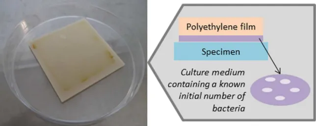
to evaluate antibacterial eiciency of samples against S. aureus and E. coli strains. Prior the assay, the TPE specimen (square - 50 mm x 50 mm) were disinfected with ethanol and then exposed to ultraviolet (UV) light with the wavelength between 300 and 400 nm for 2 h. The distance between the UV light and the specimen was kept at 10 cm. After that, the samples were placed in a sterile Petri dish followed by an inoculation of 7.6 x 10⁶ CFU cm-2 of E. coli and 3.5 x 10⁶ CFU
cm-2 of S. aureus suspension on the specimen surface, and
covered with polyethylene ilm (as shown in Figure 1). All
of them were incubated for 24 h at 35 ± 1°C. The reduction in bacterial population (percentage, %), was calculated from
the diference between the number of colony forming units
(CFU) per square centimeter at zero hour (initial) and after 24 hours of incubation, equation (2):
%
( )
Ef
Pi
Pi
Pf
2
=
-Q V
Where Ef is the reduction in bacterial population (percentage, %), Pi and Pf are, respectively, initial and
inal bacterial population (colony forming units per square
centimeter, CFU cm-2). The result is the mean ± standard
deviation of three test samples.
Antibacterial efectiveness - R, was validated in accordance with JIS Z 2801, with the equation (3):
( )
R
=
Ut
-
At
3
Where Ut is the average of logarithm numbers of viable bacteria after inoculation on control (additive free) sample after 24 h and At is the average logarithm numbers of viable bacteria after inoculation in antibacterial samples after 24
h. To be considered efective, R must be ≥ 2.0.
2.4.3 Fungal growth test
The Brazilian Association of Technical Standards (ABNT) NBR 1527525 was used to evaluate the compounds’
antifungal abilities toward the fungi A. niger, C. albicans
and C. cladosporioides. Three test samples(square - 25 mm x 25 mm) for each additive concentration were sterilized with ultraviolet (UV) light and then placed in a sterile Petri dish with agar and 100 µL of 1x10⁵-1x10⁶ spores mL-1 of
fungus suspension were inoculated on the specimen surface. All of them were incubated for seven days at 30 ± 2 °C. The presence of an inhibition zone (after 48 h incubation) and hyphal growth (after seven days incubation) were evaluated with a stereoscopic microscope. The results were expressed in millimeters of diameter of inhibition zone and the percentage of the specimen area covered by the fungus.
2.5 Statistical analysis
Statistical analysis of variance (ANOVA) and t-test was applied in tensile strength at break, modulus at 100%, elongation at break, hardness, density and antibacterial results using MYSTAT, student version 12 (Systat Software,
Inc., CA, USA). The level of signiicance was set at 0.05.
3. Results and Discussion
In the materials industry, the control of standard characteristics such as mechanical properties is important to ensure the product quality. Therefore, the incorporation of an additive in polymeric materials should allow for an improvement in performance without prejudice to the original characteristics.
Table 1 shows the variations in mechanical properties after the incorporation of 1.5% (w/w) of ZnPT and AgNano.
There were no signiicant changes in tensile and elongation at
break values of metal-incorporated samples when compared to the Standard compounds (tensile - p=0.47; elongation at break p=0.09). However, the modulus, density and hardness values in ZnPT incorporated compounds presented
a signiicant diference (p<0.05) when compared with
Standard and AgNano compounds, which could be due to the polypropylene fraction in masterbatch, present in ZnPT additive, that have higher modulus, density and hardness than the SEBS (Table 1).
In order to verify if the addition of ZnPT and AgNano
may cause any color modiication or molecular organization diference in TPE incorporated materials, a color fastness
and a FTIR-ATR analysis were performed.
As seen in Table 2, with the incorporation of ZnPT a loss of transparency (L*) was noted in TPE samples, which may be related to the high amount of polypropylene present in the masterbatch. After 96 hours of UV light, the sample incorporated with ZnPT turned completely yellow. This color
modiication is visually perceptible in Figure 2 and proven
by improving the yellowness value (b*), that leapt from 1.9
to 22. This modiication in color of ZnPT-loaded material
can be related to its extreme instability12 accelerated by UV
Figure 1. Representative scheme of antibacterial assay composed by TPE specimen, bacterial suspension, and the polyethylene ilm.
Table 1. Mechanical properties of Standard sample and sample incorporated with 1.5 % of ZnPT and AgNano.
Compounds Tensile strength at break, MPa Modulus at 100%, MPa
Elongation at
break, % Density, g/cm
3 Hardness, Shore A
Standard 10.3 ± 1.0 2.5 ± 0.0 771 ± 39 0.889 ± 0.001 64 ± 2
ZnPT 10.6 ± 0.5 2.7 ± 0.1(a) 747 ± 25 0.893 ± 0.001(a) 70 ± 2(a)
AgNano 10.9 ± 0.5 2.5 ± 0.0 793 ± 23 0.890 ± 0.001 65 ± 2
(a) Statistically diferent from the Standard (p < 0.05)
Figure 2. Color variation comparing Standard TPE samples and TPE incorporated with 1.5% of ZnPT and 1.5% AgNano, before and after 96 hours of UV light exposure.
Yellowness of the sample incorporated with AgNano was accentuated after 96 hours of UV light, the yellowness value (b*) changed from 11.1 to 20.1. The decrease in transparency and increase in yellowness (b*) has been already reported by Choi16, and Martinez-Abad27 and is associated to silver
oxidation, as shown in equation (4).
( )
O
H O
Ag
Ag
H O
4
4
4
6
4
( .) ( .)
aq s
aq
2 3
2
"
+
+
+
+ +
Q V
In SEBS compounds, color variation is normal and could be ascribed to degradation of styrene and conjugated bond sequences in the polymer backbone, hence becoming similar to stilbene (which is yellow)28,29. In the Standard sample, the
global color (∆E) change after 96 h under UV light was of
2.4, results up to 2.0 are visually imperceptible.
At infrared assay, there were no diference between the
spectra from Standard and AgNano (Figure 3a and Figure
Table 2. Color analysis of Standard samples and TPE incorporated with 1.5% of ZnPT and 1.5% of AgNano before and after 96 h of UV light exposure.
Samples L* a* b* ∆E
Standard 72.23 0.44 4.03
-Standard (after 96 h) 72.88 0.75 6.33 2.4(a)
ZnPT 72.05 -2.74 1.86 3.17(b)
ZnPT (after 96 h) 69.66 1.66 22.04 20.8(c)
AgNano 67.91 0.15 11.14 10.56(b)
AgNano (after 96 h) 68.33 2.98 20.14 9.4(d)
Note: L 0 (black) to 100 (white); ∆a* -80 (green) to 100 (red); ∆b* -80 (blue) to 70 (yellow); ∆E global parameter of color alteration.
Figure 3. FTIR-ATR in the region from 1700 -1 to 600 cm-1 of
samples (a) Standard, (b) 1.5% AgNano and (c) 1.5% ZnPT.
3b). Diferences were only noticed in the ZnPT spectra, this
fact may be due to the easy leaching of zinc pyrithione to the surface becoming detectable. In Figure 3c, an increment can be observed in the band intensity between 1300 cm-1 and 800
cm-1,which could be assigned to an increase in isotactic PP
amount from the masterbach30. An increase in band intensity
at 763 cm-1 was attributed to aromatic =C-H out-of-plane
deformation vibrations from the pirithione structure. The weak band at 1536 cm-1 depicts that the coordination of
oxygen atoms to Zn is in chelated form, if they were in ionic form, a peak at 1576 cm-1 would be observed31.
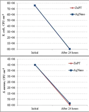
The reduction in E. coli population did not difer signiicantly between ZnPT and AgNano incorporated TPE
samples (E. coli - t = 2.60; df = 2.01; p = 0.12). The S. aureus counts were signiicantly diferent between ZnPT and AgNano
incorporated compounds (S. aureus - t =4.43; df =2.01; p =
0.05), with better efectiveness of ZnPT (Figure 4).
Figure 4. Variation in the number of colony forming units (CFU) of: (a) Escherichia coli and (b) Staphylococcus aureus population
between the initial and inal time in ZnPT and AgNano TPE
incorporated samples.
A good inhibitory efect towards Gram-negative and
Gram-positive bacteria was obtained in both additives tested, even after undergoing typical polymer industrial processing.
Moreover, a better bactericide efect of ZnPT compared to
AgNano was observed. Dagostin and coworkers6 also found
a better antimicrobial result of ZnPT incorporated into polyurethane foam matrices than Triclosan® and isothiazolone. Previous studies report that pyrithione can penetrate bacterial cytosol besides acting in cell membrane32. Once inside the
cell, the chelating mechanism of ZnPT promotes a disorder in the cell envelope, owing to the leakage of intracellular components as well as the inhibition of nutrient uptake7.
In the present investigation, metal uptake by bacteria was not explored. However, Gram-negative bacteria were most susceptible to the action of both additives while S. aureus was more susceptible to ZnPT. The diferential efectiveness
observed between the additives tested herein may be related to the way in which these metals operate. Nanosilver is positively charged and bacterial cell wall is negative, which leads to an electrical disturbance; pyrithione is a chelating agent that can sequestrate metal ions which are important in bacterial cell conformation7. This property also causes an electrical
disorder in bacteria membranes, mainly Gram-negative ones, due to the action on lipopolysaccharide groups, present in the outer membrane of these microorganisms33. It was
shown that zinc pyrithione leads to decrease in intracellular adenosine triphosphate amount in Gram-negative bacteria species Escherichia coli and Pseudomonas aeruginosa 7.
Also, the treatment with ZnPT induces the production of reactive oxygen species (ROS)34,35, which are related to the
antibacterial action 36. Thus, the ROS from zinc ions, such
as hydrogen peroxide, would be associated to the toxic
efects of ZnPT37.
According to Standard JIS Z 280124, in order to accept
the material as being antibacterial, the efectivenessvalue must be equal to or higher than 2. In the present study, the antimicrobial incorporated compounds did not reach the R value of 2.0 in the measurement against the S. aureus
population.However, against the E. coli population, the ZnPT
compounds presented better antibacterial efectiveness (R =
3.1)thanAgNano (R = 2.0) compounds Table 3.
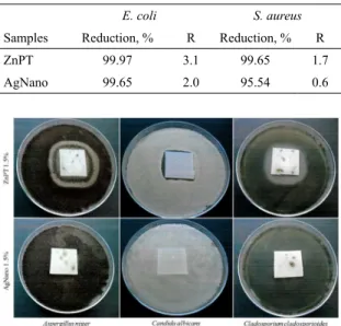
Results of antifungal activity are presented inFigure 5. The ZnPT antifungal activity against A. niger (Figure 5a),
C. albicans (Figure 5b) and C. cladosporioides (Figure 5c) are indicated by the appearance of inhibition zones of 7 mm, 2 mm and 6 mm, respectively. There was no inhibition zone in samples containing AgNano; however, these samples did not present fungal growth on their surfaces (Figure 5).
Inhibition zone in sample with ZnPT and other organic antifungal agents have been ascribed to the fact that this component leaches from the polymer to the ambient 5,38,39.
According to Coulthwaite et al.5 a zone of inhibition means
that free molecules of the biocide are on the surface of the
polymer and are released in suicient quantities into the
Table 3. Percentage of bacterial population reduction after 24 h of
incubation and (R) antibacterial efectiveness values.
E. coli S. aureus
Samples Reduction, % R Reduction, % R
ZnPT 99.97 3.1 99.65 1.7
AgNano 99.65 2.0 95.54 0.6
Figure 5. Antifungal activity of ZnPT and AgNano incorporated samples: (a) Aspergillus niger, (b) Candida albicans, (c) Cladosporium cladosporioides.
are present. The characteristics of the polymeric matrix
related to its water difusion ability 40,41, and the structure
of the polymer system42 can inluence biocidal action, since
it may facilitate the release of the antifungal agent. For example, Silaparson et al.43 show that it is more diicult
for Triclosan® to be difused through amorphous and rigid
thermoplastics (such as polystyrene and polyvinyl chloride) than through soft and crystalline thermoplastics (such as polypropylene and polyethylene).
The antifungal capability of ZnPT is widely known44.
Chandler and Segel13 have demonstrated that inhibition of
fungi growth by pyrithione is the result of the reduction in membrane transport systems. In addition, the source of
ZnPT antifungal eicacy is related to the balance between
the acquisition, storage and usage of metals by the fungus45.
In that case, the metal starvation34 or high intracellular
availability8,45 will afect the activity of iron-sulfur proteins
involved in diverse metabolic functions required for microbial growth46. Ion exchange is a slow process and this can enlarge
the biocidal efect of metal-based antimicrobials47. Silver
nanoparticles have been indicated to prevent the growth of
C. albicans through modiications in cell dynamics18,42,48,.
On the basis of the results, we infer that better biocide
efect of ZnPT than the AgNano can be related to the diferent
mechanisms that these substances use to reach the polymer
surface. Although few studies report the speciic mechanisms
of ZnPT in polymers, it is known that organic additives as ZnPT, n-octylisothiazolin-one (OIT) and Triclosan® are known as migratory and easily leached from polymer matrices, forming an inhibition zone in fungal assays23.
Even with no inhibition zone, the sample incorporated with AgNano presented no mycelia growth on its surface. For silver nanoparticles is reported that to achieve biocide properties in polymer, silver ions must migrate to the polymer surface to get in contact with fungi and bacteria cells, for this, water must permeate the polymer chain to oxidize the silver nanoparticles (as shown in equation 4) 16,40,49,50.
4. Conclusions
Overall data obtained in our study showed a good antimicrobial activity of ZnPT-incorporated TPE materials against all the fungus and bacterial species tested. However, the precise mechanisms require further investigation. Samples prepared with AgNano also presented bactericidal
action, however with no fungistatic efect. No relevant
modiication in mechanical properties was observed, showing
that there was a small interaction between the additive and
polymer. Owing to the color modiication in TPE materials
containing both additives, the industrial utilization of these incorporated materials will be restricted to applications in which pigmentation would not represent a market problem.
5. Acknowledgements
The authors would like to thank FINEP for the inancial
support (03.13.0280.00) and Softer Brasil Compostos Termoplásticos LTDA for infrastructure support. A special thanks to the additive supplier Ipel Itibanyl Produtos Especiais Ltda.
6. References
1. Leung WK, Lau APS, Yeung KL. Bactericidal and sporicidal performance of a polymer-encapsulated chlorine dioxide-coated surface. Journal of Applied Microbiology. 2009;106(5):1463-1472.
2. Prüss A, Giroult E, Rushbrook P, eds. Safe management of wastes from health-care activities. Geneva: World Health Organization; 1999.
3. Carpentier B, Cerf O. Bioilms and their consequences, with
particular reference to hygiene in the food industry. Journal of Applied Bacteriology. 1993;75(6):499-511.
4. Page K, Wilson M, Parkin IP. Antimicrobial surfaces and their potential in reducing the role of the inanimate environment in the incidence of hospital-acquired infections. Journal of Materials Chemistry. 2009;19(23):3819-3831.
5. Coulthwaite L, Bayley K, Liauw C, Craig G, Verran J. The
efect of free and encapsulated OIT on the biodeterioration of
plasticised PVC during burial in soil for 20 months. International Biodeterioration & Biodegradation. 2005;56(2):86-93.
6. Dagostin VS, Golçalves DL, Pacheco CB, Almeida WB, Thomé IP, Pich CT, et al. Bactericidal polyurethane foam mattresses:
7. Dinning AJ, Al-Adham ISI, Eastwood IM, Austin P, Collier PJ. Pyrithione biocides as inhibitors of bacterial ATP synthesis.
Journal of Applied Microbiology. 1998;85(1):141-146.
8. Reeder NL, Kaplan J, Xu J, Youngquist RC, Wallace J, Hu P, et
al. Zinc pyrithione inhibits yeast growth through copper inlux
and inactivation of iron-sulfur proteins. Antimicrobial Agents and Chemotherapy. 2011;55(12):5753-5760.
9. Tozer SA, Kelly S, O’Mahony C, Daly EJ, Nash JF. Aggregate
exposure modelling of zinc pyrithione in rinse-of personal
cleansing products using a person-orientated approach with
market share reinement. Food and Chemical Toxicology. 2015;83:103-110.
10. Bressy C, Hugues C, Margaillan A. Characterization of chemically active antifouling paints using electrochemical impedance spectrometry and erosion tests. Progress in Organic Coatings. 2009;64(1):89-97.
11. Windler L, Height M, Nowack B. Comparative evaluation of antimicrobials for textile applications. Environment International. 2013;53:62-73.
12. Burley JW, Cliford PD. Extending the use of zinc-containing
biocides in PVC. Journal of Vinyl & Additive Technology. 2004;10(2):95-98.
13. Chandler CJ, Segel IH. Mechanism of the antimicrobial action
of pyrithione: efects on membrane transport, ATP levels, and
protein synthesis. Antimicrobial Agents and Chemotherapy. 1978;14(1):60-68.
14. Guthery E, Seal LA, Anderson EL. Zinc pyrithione in alcohol-based products for skin antisepsis: Persistence of antimicrobial
efects. American Journal of Infection Control. 2005;33(1):15-22.
15. D’Arcy N. Antimicrobials in plastics: a global review. Plastics Additives and Compounding. 2001;3(12):12-15.
16. Choi O, Deng KK, Kim NJ, Ross L Jr, Surampalli RY, Hu Z.
The inhibitory efects of silver nanoparticles, silver ions, and
silver chloride colloids on microbial growth. Water Research. 2008;42(12):3066-3074.
17. Dakal TC, Kumar A, Majumdar RS, Yadav V. Mechanistic Basis of Antimicrobial Actions of Silver Nanoparticles. Frontiers in Microbiology. 2016;7:1831.
18. Kim KJ, Sung WS, Suh BK, Moon SK, Choi JS, Kim JG, et al. Antifungal activity and mode of action of silver nano-particles on Candida albicans. Biometals. 2009;22(2):235-242.
19. Morones JR, Elechiguerra JL, Camacho A, Holt K, Kouri JB,
Ramírez JT, et al. The bactericidal efect of silver nanoparticles. Nanotechnology. 2005;16(10):2346-2353.
20. Ansari MA, Khan HM, Khan AA, Ahmad MK, Mahdi AA, Pal R, et al. Interaction of silver nanoparticles with Escherichia coli and their cell envelope biomolecules. Journal of Basic Microbiology. 2014;54(9):905-915.
21. Ninganagouda S, Rathod V, Singh D, Hiremath J, Singh AK, Mathew J, et al. Growth Kinetics and Mechanistic Action of Reactive Oxygen Species Released by Silver Nanoparticles from Aspergillus niger on Escherichia coli. BioMed Research International. 2014;2014:753419.
22. Ellison J, Wykof G, Paul A, Mohseni R, Vasiliev A. Eicient
dispersion of coated silver nanoparticles in the polymer matrix.
Colloids and Surfaces A: Physicochemical and Engineering Aspects. 2014;447:67-70.
23. Nichols D. Biocide in plastics. Shawbury: Rapra Review Reports 15(12); 2004. 116 p.
24. Japanese Industrial Standard (JIS). JIS Z 2801- Antimicrobial
products - test for antimicrobial activity and eicacy. Tokio: Japanese Industrial Standard; 2010.
25. Brazilian Association of Technical Standards (ABNT). NBR 15275 - Biological assays - Insole, synthetic laminated and sole - Determination of the resistance to the microbial attack. Rio de Janeiro: ABNT; 2014.
26. Kappock PS, Flaherty P, inventors; Arch Chemicals, Inc., assignee. Discoloration prevention in pyrithione-containing coating compositions. EP1348743 B1. 1997 Feb 21.
27. Martínez-Abad A, Ocio MJ, Lagaron JM. Morphology, physical properties, silver release, and antimicrobial capacity of ionic
silver-incorporated poly(l-lactide) ilms of interest in
food-coating applications. Journal of Applied Polymer Science. 2014;131(21):41001.
28. Allen NS, Edge M, Wilkinson A, Liauw CM, Mourelatou D, Barrio J, et al. Degradation and stabilisation of styrene-ethylene-butadiene-styrene (SEBS) block copolymer. Polymer Degradation and Stability. 2001;71(1):113-122.
29. Yousif E, Haddad R. Photodegradation and photostabilization of polymers, especially polystyrene: review. SpringerPlus. 2013;2:398.
30. Luongo JP. Infrared study of polypropylene. Journal of Applied Polymer Science. 1960;3(9):302-309.
31. Gönen M, Balköse D, Inal F, Ülkü S. Zinc Stearate Production by Precipitation and Fusion Processes. Industrial & Engineering Chemistry Research. 2005;44(6):1627-1633.
32. Dinning AJ, Al-Adham ISI, Austin P, Collier PJ. A novel assay for the distribution of pyrithione biocides in bacterial cells.
Letters in Applied Microbiology. 1998;27(1):1-4.
33. Clifton LA, Skoda MWA, Brun APL, Ciesielski F, Kuzmenko I,
Holt SA, et al. Efect of divalent cation removal on the structure
of Gram-negative bacterial outer membrane models. Langmuir. 2015;31(1):404-412.
34. Yasokawa D, Murata S, Iwahashi Y, Kitagawa E, Kishi K, Okumura Y, et al. DNA microarray analysis suggests that zinc pyrithione causes iron starvation to the yeast Saccharomyces cerevisiae.
Journal of Bioscience and Bioengineering. 2010;109(5):479-486.
35. Oyama TM, Saito M, Yonezawa T, Okano Y, Oyama Y. Nanomolar concentrations of zinc pyrithione increase cell susceptibility to oxidative stress induced by hydrogen peroxide in rat thymocytes. Chemosphere. 2012;87(11):1316-1322.
37. Linley E, Denyer SP, McDonnell G, Simons C, Maillard JY. Use of hydrogen peroxide as a biocide: new consideration of its mechanisms of biocidal action. Journal of Antimicrobial Chemotherapy. 2012;67(7):1589-1596.
38. Sørensen G, Nielsen AL, Pedersen MM, Poulsen S, Nissen H, Poulsen M, et al. Controlled release of biocide from silica microparticles in wood paint. Progress in Organic Coatings. 2010;68(4):299-306.
39. Jämsä S, Mahlberg R, Holopainen U, Ropponen J, Savolainen A,
Ritschkof AC. Slow release of a biocidal agent from polymeric
microcapsules for preventing biodeterioration. Progress in Organic Coatings. 2013;76(1):269-276.
40. Kumar R, Münstedt H. Silver ion release from antimicrobial polyamide/silver composites. Biomaterials. 2005;26(14):2081-2088.
41. Damm C, Münstedt H, Rösch A. Long-term antimicrobial polyamide 6/silver-nanocomposites. Journal of Materials Science. 2007;42(15):6067-6073.
42. Wady AF, Machado AL, Zucolotto V, Zamperini CA, Berni E, Vergani CE. Evaluation of Candida albicans adhesion and
bioilm formation on a denture base acrylic resin containing
silver nanoparticles. Journal of Applied Microbiology. 2012;112(6):1163-1172.
43. Silapasorn K, Sombatsompop K, Kositchaiyong A, Wimolmala
E, Markpin T, Sombatsompop N. Efect of chemical structure
of thermoplastics on antibacterial activity and physical
difusion of triclosan doped in vinyl thermoplastics and their
composites with CaCO3. Journal of Applied Polymer Science. 2011;121(1):253-261.
44. Reeder NL, Xu J, Youngquist RS, Schwartz JR, Rust RC, Saunders CW. The antifungal mechanism of action of zinc pyrithione. British Journal of Dermatology. 2011;165(Suppl 2):9-12.
45. Li L, Miao R, Bertram S, Jia X, Ward DM, Kaplan J. A role for iron-sulfur clusters in the regulation of transcription factor YAP5-dependent high iron transcriptional responses in yeast.
Journal of Biological Chemistry. 2012;287(42):35709-35721.
46. Li J, Kogan M, Knight SA, Pain D, Dancis A. Yeast mitochondrial protein, Nfs1p, coordinately regulates iron-sulfur cluster proteins, cellular iron uptake, and iron distribution. Journal of Biological Chemistry. 1999;274(46):33025-33034.
47. Kwakye-Awuah B, Williams C, Kenward MA, Radecka I.
Antimicrobial action and eiciency of silver-loaded zeolite
X. Journal of Applied Microbiology. 2007;104(5):1516-1524.
48. Panáček A, Kolář M, Večeřová R, Prucek R, Soukupová J,
Kryštof V, et al. Antifungal activity of silver nanoparticles against Candida spp. Biomaterials. 2009;30(31):6333-6340.
49. Radheshkumar C, Münstedt H. Antimicrobial polymers from polypropylene/silver composites - Ag+ release measured by
anode stripping voltammetry. Reactive and Functional Polymers. 2006;66(7):780-788.
50. Triebel C, Vasylyev S, Damm C, Stara H, Özpinar C, Hausmann S, et al. Polyurethane/silver-nanocomposites with enhanced silver ion release using multifunctional invertible polyesters.