Leandro Augusto HILGERT(a)
Soraya Coelho LEAL(a)
Gabriela Mesquita Lopes FREIRE(b)
Jan MULDER(c)
Jo E. FRENCKEN(c)
(a) Universidade de Brasília, Faculty of Health Sciences, Department of Dentistry, Brasília, DF, Brazil.
(b) Private Practice, Brasília, DF, Brazil.
(c) Radboud University Medical Center, College of Dental Sciences, Department of Oral Function and Prosthetic Dentistry, Nijmegen, The Netherlands.
3-year survival rates of retained
composite resin and ART sealants using
two assessment criteria
Abstract: The aim was to test the null-hypothesis that there is no difference in the cumulative survival rate of retained composite resin (CR) sealants and a high-viscosity glass-ionomer Atraumatic Restorative Treatment (ART) sealant in irst permanent molars calculated according to the traditional and the modiied retention assessment criteria over a period of 3 years. This cluster-randomized controlled clinical trial consisted of 123 schoolchildren, 6–7-years-old. At baseline, high-caries risk pits and issures of fully erupted irst permanent molars were treated with CR and ART sealants. Evaluations were performed after 0.5, 1, 2 and 3 years. Retention was scored for free-smooth surface and for each of three sections into which the occlusal surface had been divided. The modiied criterion differed from the traditional in that it determined an occlusal sealant to be a failure when at least one section contained no visible sealant material. Data were analysed according to the PHREG model with frailty correction, Wald-test, ANOVA and t-test, using the Jackknife procedure. The cumulative survival rates for retained CR and ART sealants in free-smooth and occlusal surfaces for both criteria were not statistically signiicantly different over the 3 years. A higher percentage of retained CR sealants on occlusal surfaces was observed at longer evaluations. Cumulative survival rates were statistically signiicantly lower for the modiied criterion in comparison to the traditional. The modiied retention assessment criterion should be used in future sealant-retention studies.
Keywords: Pit and Fissure Sealants; Dental Caries; Dental Atraumatic Restorative Treatment; Pediatric Dentistry.
Introduction
Dental carious lesions predominantly occur in pits and issures of occlusal surfaces in recently erupted molars.1,2 Placing sealants on occlusal
surfaces of permanent molars is an effective method for preventing and controlling carious lesion development in pits and issures.3 Currently,
the main materials used for sealing pits and fissures are resin- and glass-ionomer-based.4,5 Retention of sealants is considered a surrogate
endpoint for determining the caries-preventive effectiveness of sealants since it is believed that loss of retention would determine sealant failure. However, obtaining a real clinical endpoint by comparing sealed and
Declaration of Interests: The authors certify that they have no commercial or associative interest that represents a conflict of interest in connection with the manuscript.
Corresponding Author:
Leandro Augusto Hilgert E-mail: [email protected]
https://doi.org/10.1590/1807-3107BOR-2017.vol31.0035
Submitted: April 14, 2016
non-sealed surfaces is dificult for ethical reasons, since the effectiveness of sealants for high-caries risk populations has already been established.5,6
Resin-based sealants usually present higher retention rates than glass-ionomer (GIC) based sealants.6 Despite differences in their retention,
systematic reviews have reported no evidence of caries-preventive superiority of either material.3,4,7,8
In the past, low- and medium-viscosity glass-ionomers were used as sealant material but these have been replaced by high-viscosity glass-ionomers, as the latter have increased mechanical properties, which have enhanced sealant retention.9 Retention of
high-viscosity glass-ionomers may further be increased through the development of new formulas in the glass-ionomer technology, such as has been reported regarding its powder composition.10
In calculating survival of sealants’ retention, the traditional criterion considers a sealant a failure if all material has disappeared from the total sealed tooth surface. However, this criterion is questioned since absence of material in just a section of the occlusal surface re-exposes that section to the oral environment, which increases the chance that a new carious lesion is initiated or that an existing one progresses. This has led Chen et al.11 to suggest a new way of determining
sealant retention. The so called: ‘modiied’ retention criterion fails a sealant for its retention on the occlusal surface when only one section is re-exposed. Only a few studies have investigated the effect of the modiied retention criterion on carious lesion development and, consequently, the need for resealing re-exposed pits and issures.11,12
In 2009, a cluster-randomized clinical trial was undertaken to compare the effectiveness of three treatment protocols among primary school going children.13 Investigating the caries-preventive effect
on permanent first molars of a supervised tooth brushing (STB) programme, a composite resin (CR) sealant and a newly formulated high-viscosity glass-ionomer sealant applied with inger pressure (Atraumatic Restorative Treatment or ART sealant) were part of the trial.14 The study provided the
opportunity to test the following null-hypothesis: there is no difference in the cumulative survival rate of retained CR and newly formulated high-viscosity
glass-ionomer cement ART sealants in permanent molars calculated according to the traditional and the modiied retention assessment criteria over a period of 3 years. Furthermore, secondary analyses tested whether or not the level of sealant retention is related to the prevalence of cavitated dentine carious lesions over a period of 3 years.
Methodology
Sampling procedure
The reporting of the study is based on the CONSORT Statement.15
This equivalence cluster-randomized controlled clinical trial used a parallel group design. It was carried out in four public primary schools of Paranoá, a deprived suburban area of Brasilia, Brazil. The sample of this study was obtained from an oral health epidemiological survey among 6- and 7-year-old children attending all the schools in this area.16
The inclusion criteria were: a. good general health; b. at least two cavitated dentine carious lesions in vital pain-free primary molars assessed according to the ICDAS II index;17 c. erupted irst permanent molars
with pits and issures fully visible and accessible; d. high-caries risk surfaces, determined by ICDAS II codes 2 and 3 or a combination of ICDAS II code 1 and medium or deep issures assessed according to the Symons criteria;18 and e. having a signed
consent form.
The study covered two groups to treat high-caries risk surfaces of irst permanent molars. These were: CR sealants and ART sealants. The sampling unit was the school (two schools per cluster). Two of the schools were equipped with a dental unit and were allocated to the CR sealants group. The other two schools (ART group) were allocated by coin toss. A CONSORT lowchart that depicts the study design is presented in Figure 1.
Implementation
Sealants were placed by three trained and calibrated paedodontists, aided by trained dental assistants, between May and July 2009 at the school premises.
Children received an oral hygiene kit (toothbrush, luoridated dentifrice, plaque-disclosing dentifrice and dental loss) and instructions on how to use its content at the start of the study. These instructions
Inclusion criteria: - good general health;
- ≥2 cavitated dentine lesions in deciduous molars;
- erupted first permanent molars; - high-caries risk surfaces: ICDAS 1 with medium or deep fissures, or ICDAS 2 or ICDAS 3.
Epidemiological survey (Nchild= 613)
Included (Nchild=123)
Allocation (cluster-randomization)
ART sealant Composite
Resin sealant Evaluation
Nchild = 78 NSoc = 169 NSsm = 92
Nchild = 45 NSoc = 69 NSsm = 47 Baseline
Nchild = 123 NSoc = 238 NSsm = 139
Cumulative Drop-out (Nchild = 2)
Nchild = 76 NSoc = 168 NSsm = 86
Nchild = 45 NSoc = 69 NSsm = 47 6 months
Nchild = 121 NSoc = 237 NSsm = 133
Cumulative Drop-out (Nchild = 12)
Nchild = 69 NSoc = 152 NSsm = 82
Nchild = 42 NSoc = 65 NSsm = 45 1 year
Nchild = 111 NSoc = 217 NSsm = 127
Cumulative Drop-out (Nchild = 21)
Nchild = 64 NSoc = 143 NSsm = 77
Nchild = 38 NSoc = 60 NSsm = 41 2 years
Nchild = 102 NSoc = 203 NSsm = 118
Cumulative Drop-out (Nchild = 37)
Nchild = 54 NSoc = 120 NSsm = 71
Nchild = 32 NSoc = 51 NSsm = 32 3 years
Nchild = 86 NSoc = 171 NSsm = 103
were repeated during the evaluation sessions. Children were encouraged to brush twice daily.
Composite resin sealant group (CR)
Children were positioned in a dental chair. Isolation was obtained with cotton wool rolls and a suction device. Under good visibility from the operation lamp, the surface to be treated was cleaned with a rotating brush, acid-etched for 30 s with a 37% phosphoric acid gel (Acigel, SSWhite, Rio de Janeiro, Brazil), rinsed and dried, using a 3-way syringe. The sealant material, Fluoroshield (Dentsply, Petrópolis, Brazil), was placed in a dappen glass, transported to pits and issures with a ball-ended probe (Dulex, Rio de Janeiro, Brazil) and light-cured for 40 s (Ultralux, Dabi Atlante, Ribeirão Preto, Brazil). Occlusion was checked with carbon paper and adjusted where necessary with rotary instruments.
High-viscosity glass-ionomer ART sealant group (ART)
Pits and issures were cleaned with a toothbrush and toothpaste before the children lay on a portable bed. Isolation was obtained using cotton wool rolls. The occlusal surface was further cleaned with a dental probe and cotton wool pellets under artiicial light provided by a portable headlamp. The surface was
conditioned with polyacrylic acid for 10–15 s, washed with wet cotton wool pellets and dried with dry cotton wool pellets. Ketac Molar Easymix (3M ESPE, Seefeld, Germany) was hand-mixed according to the manufacturer’s instructions, applied on the surface with an ART applier instrument (Henry Schein, Chicago, USA) and pressed into the pits and issures with a petroleum jelly-coated inger for 15 s.19 Excess material was removed with the
ART carving instrument after the occlusion was checked with carbon paper. The sealant was coated with petroleum jelly and the children were told not to eat for 1 hour.
Evaluation
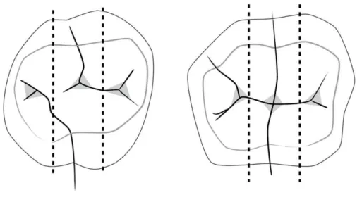
For evaluating sealant retention and carious lesion development, the occlusal surfaces of the first permanent molars were divided into three sections (mesial-central-distal) (Figure 2) but the free-smooth surfaces were not. Each of these sections and free-smooth surfaces were assessed for the presence of carious lesions using ICDAS II. Retention was scored according to the codes: 0 (a sealant is present, good seal in the main pits and issures initially sealed); 1 (partial loss of sealant that exposes the main pits and issures); or 6 (no sealant is visible, main pits and issures are completely exposed). The same
two independent evaluators (dentists) performed the evaluations at the school premises after 6 months, 1, 2 and 3 years.
The evaluators were trained and calibrated before each evaluation session by an experienced dental epidemiologist (JF). Battery-illuminated dental mirrors (Kudos®, Hong Kong, China), CPITN probe (Golgran®, São Caetano do Sul, Brazil) and compressed air aided the evaluation. A total of 67 surfaces were re-examined for reproducibility testing. The kappa coeficient value for the inter-evaluator consistency test in assessing retention over the four evaluation times was 0.77 while the percentage of agreement was 86.6%.
Statistical analyses
The sample for this investigation was obtained from a main study that investigated the effectiveness of protocols for treating dentine carious lesions in primary teeth.13 Focusing on the carious-lesion
preventive effect of CR (79%) and ART (94%) sealants after 5 years,20 a power of 80%, a dropout rate of 30%
and a correction for dependency of measurements of 20% gave a sample size of 117 sealants per group. The statistical analyses were performed by a biostatistician using SAS version 9.2 software (Cary, NC, USA). The dependent variables were survival rate of retained sealants calculated according to the traditional and the modiied retention criteria:
a. Traditional criterion: a failure is determined by the total loss of sealant material on the entire surface (code 6). For the occlusal surface, all three sections should present a code 6 for the surface to be considered a failure.
b. Modiied criterion: a failure is determined when at least one section of an occlusal surface presents no visible sealant material (code 6). Treatment group (CR, ART), age, gender, type of jaw, operator and baseline caries experience (D2MFT, D3MFT and d3mft) were the independent
variables. D2 represents ICDAS II codes 1-6 and
D3/d3 represents ICDAS II codes 4-6. ANOVA and
chi-square tests were used in testing for differences between the independent variables at baseline and the treatment groups, and for the non-response analysis. The Proportional Hazard Rate Regression
model (PHREG)21 with frailty correction22 was used to
estimate cumulative survival retention rates. The Wald test (chi-square) was used to test for differences in survival rates and for estimating effects of the independent variables. The Jackknife method23 was
applied in calculating standard errors for comparison of survival rates between treatment groups per interval using a t-test. Statistical signiicance was set at α = 0.05.
Results
Disposition of subjects
A total of 123 children (62 boys, 61 girls) with a mean age of 6.8 years were enrolled in the study. At baseline, 238 occlusal surfaces and 139 free-smooth surfaces in irst permanent molars of these children met the inclusion criteria. Mean age, mean d3mft,
D2MFT and D3MFT counts of the participating
children according to treatment group are presented in Table 1. A statistically signiicant difference at baseline between the treatment groups was found for age. Children in the ART sealant group were approximately two months older than children of the CR sealant group. From the 377 sealed surfaces at baseline, 274 surfaces (72.7%) were examined after 3 years and almost 30% of the sample was lost to follow-up during that period (Figure 1). Non-response analyses revealed no effect for treatment group (p = 1.00), age (p = 0.88), gender (p = 0.85), baseline d3mft (p = 0.78), baseline D2MFT (p = 0.16) and baseline
D3MFT (p = 0.38) counts.
Table 1. Mean and standard deviations (SD) of age, D2MFT, D3MFT and d3mft counts of participating children at baseline according to treatment group.
Variable
CR sealant ART sealant
(Nchild = 78) (Nchild = 45)
Mean SD Mean SD
Age 6.72 0.33 6.88 0.33
D2MFT 3.33 1.04 3.00 1.19
D3MFT 0.26 0.55 0.33 0.56
d3mft 5.86 3.08 5.80 2.33
Cumulative retention of sealants
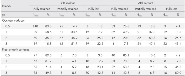
Frequency distributions of fully and partially retained and fully lost sealants for occlusal and free-smooth surfaces over the 3-year follow-up period by treatment group are presented in Table 2. The prevalence of fully retained sealants in occlusal surfaces after 3 years was low: 15.8% for CR and 7.8% for ART sealants. The percentages of fully retained sealants in free-smooth surfaces after 3 years were 49.3 for CR and 43.8 for ART sealants.
The cumulative survival rates of the two types of sealants retained in occlusal and free-smooth surfaces over 3 years using the traditional retention criterion are presented in Table 3. For occlusal surfaces, there
was no statistically signiicant difference between survival rates of retained CR and ART sealants over the total 3-year follow-up period (p = 0.05). However, a statistically signiicant difference was observed at the 1y, 2y and 3y evaluation intervals, which showed a higher retention survival rate for CR sealants. For the cumulative survival rates of retained sealants on free-smooth surfaces, no statistically signiicant difference was observed between the treatment groups over the 3-year period (p = 0.34) nor at any evaluation interval.
The inluence of independent variables on the survival model for retained occlusal sealants using the traditional retention criterion showed the
Table 2. Frequency distributions of fully and partially retained and fully lost sealants for occlusal and free-smooth surfaces according to treatment group and interval.
Interval CR sealant ART sealant
Fully retained Partially retained Fully lost Fully retained Partially retained Fully lost
yrs NSoc % NSoc % NSoc % NSoc % NSoc % NSoc %
Occlusal surfaces
0.5 140 83.3 25 14.9 3 1.8 53 76.8 13 18.8 3 4.4
1 89 58.6 51 33.6 12 7.9 32 49.2 21 32.3 12 18.5
2 50 35.0 67 46.9 26 25.2 12 20.0 32 53.3 16 26.7
3 19 15.8 62 51.7 39 32.5 4 7.8 24 47.1 23 45.1
Free-smooth surfaces
0.5 77 89.5 6 7.0 3 3.5 40 85.1 5 10.6 2 4.2
1 67 81.7 5 6.1 10 12.2 33 73.3 4 8.9 8 17.8
2 55 71.4 4 5.2 18 23.4 22 53.6 4 9.8 15 36.6
3 35 49.3 6 8.5 30 42.3 14 43.8 2 6.3 16 50.0
NSoc: number of occlusal surfaces; NSsm: number of free-smooth surfaces; CR: composite resin; ART: atraumatic restorative treatment.
Table 3. Cumulative survival rates (%) and standard errors (SE) of fully and partially retained sealants calculated according to the traditional criterion in occlusal and free-smooth surfaces over a period of 3 years.
Variable Occlusal surfaces Free-smooth surfaces
Interval CR sealant ART sealant CR sealant ART sealant
yrs % SE % SE % SE % SE
0.5y 98.2 1.0 95.7 2.4 96.5 1.9 95.7 3.1
1y 91.6a 2.4 82.2b 5.5 88.0 3.6 82.4 5.7
2y 80.6c 4.0 67.7d 6.5 77.5 4.9 64.9 7.8
3y 66.3e 4.9 50.8f 8.0 56.3 6.7 49.5 9.2
following indings: no effects of gender (p = 0.61), baseline d3mft (p = 0.47), baseline D2MFT (p = 0.07),
baseline D3MFT (p = 0.41) counts, type of jaw
(p = 0.97), operator (p = 0.14) or age (p = 0.76) were found. For free-smooth surfaces, no effects of baseline d3mft (p = 0.60), baseline D2MFT
(p = 0.83), baseline D3MFT (p = 0.28) counts, type
of jaw (p = 0.09) or operator (p = 0.48) were found. A statistically signiicant effect of gender (p = 0.02) and age (p = 0.04) was observed for these surfaces. Boys presented higher retention rates than girls and they were slightly younger than the girls. In an adjusted retention survival model, including the variables gender, age and treatment group, no effects of treatment group (p = 0.77), age (p = 0.07) and gender (p = 0.05) were found.
The cumulative survival rates of both types of sealants retained in occlusal surfaces over the 3-year period according to the modiied retention criterion are presented in Table 4. For occlusal surfaces, no statistically significant difference between cumulative survival rates of retained CR and ART sealants over the total 3-year follow-up period (p = 0.05) was observed. A statistically signiicant difference was observed at the 2y and 3y evaluation intervals, showing a higher survival rate for CR than for ART sealants. Analyses of the inluence of independent variables on the survival model for occlusal sealant retention using the modiied retention criterion showed no effects of gender (p = 0.70), baseline d3mft (p = 0.07) and
baseline D2MFT counts (p = 0.09) or age (p = 0.87).
Statistically significant effects were found for
operator (p = 0.01), type of jaw (p = 0.04) and baseline D3MFT counts (p = 0.04). One of the operators
performed a higher number of CR sealants than the other two. For CR sealants, the retention rate in irst molars of the lower jaw was higher than in those of the upper jaw (p < 0.01). Type of jaw had no effect on the retention rate of ART sealants (p = 0.28). The adjusted retention survival model, including operator, type of jaw, baseline D3MFT
count and treatment group, showed no effect of treatment group (p = 0.22) and baseline D3MFT
count (p = 0.15). Operator (p = 0.03) and type of jaw (p = 0.03) still presented a signiicant effect.
For both the CR and ART sealants, the survival model presented lower retention rates with the use of the modiied retention criterion than with the traditional retention criterion over the 3-year period (p < 0.01).
Loss of sealant retention and cavitated dentine carious lesion development in occlusal surfaces
Table 5 presents the cumulative survival rate of cavitated dentine carious lesion-free occlusal surfaces by evaluation interval,14 the frequency
distribution of the location of the cavitated dentine carious lesions and the number of surfaces having a retention failure according to the traditional and the modiied retention criterion at the time that those carious lesions were detected.
Of the 18 cavitated dentine carious lesions that were detected (12 in the CR sealant group and 6 in the ART sealant group), 15 (83.3%) were located on the distal section of the occlusal surface. Eight cavitated dentine carious lesions occurred in lower first permanent molars and 10 such lesions in comparable teeth in the upper jaw. According to the traditional retention criterion, 8 of the 18 cavitated dentine carious lesions in occlusal surfaces (44.4%) were found on surfaces that had been determined as ‘failed’ for retention at the time that the carious lesion had been detected, while 14 of the 18 cavitated dentine carious lesions in occlusal surfaces (77.8%) occurred on ‘failed’ retention surfaces when the modiied retention criterion was applied.
Table 4. Cumulative survival rates (%) and standard errors (SE) of fully and partially retained sealants calculated according to the modified criterion in occlusal surfaces over a period of 3 years.
Occlusal surface
Interval CR sealant ART sealant
yrs % SE % SE
0.5 91.1 2.3 85.5 5.8
1 70.1 4.5 60.0 7.5
2 47.6a 5.3 29.2b 6.1
3 29.8c 5.4 15.5d 5.0
Discussion
Methodology
Sampling of the population of this study was based on the selection and randomization process of the main study, which compared treatment protocols in primary molars. From the included high-caries risk children, only irst permanent molars that had erupted and also presented a high-caries risk proile at surface-level were sealed. Cluster-randomization by school was deined by the fact that two of the four public primary schools had a dental room with a fully equipped dental unit and these schools were allocated to the CR sealant group. However, as no dentist had been employed at these schools for many years, children from these schools had no expected advantage in terms of oral health care or knowledge over the children of the other two schools. Although socio-economic status was not assessed, there is no
reason to believe that any difference among schools and treatment groups occurred in this aspect, since all children came from the same area of social and economical deprivation.
The sample size of the two sealant groups differed. This is not unusual in clinical trials. The main reason for the difference is most likely that the school, rather than the number of children or the number of tooth surfaces that required a sealant, was chosen as the unit of randomization. The schools that had a dental unit and that, consequently, were used for placing CR sealants contained many more children than the schools in which the ART sealant was placed. But it is very unlikely that the unequal sample size caused a bias in the comparison of results of the two sealant groups. It is argued that, whilst an equal sample size provides the highest level of power, a deviation from it only reduces the power slightly, provided the sample size is not very low. For comparing survival results Table 5. Cumulative survival of cavitated dentine carious lesion-free occlusal surfaces [%(SE)], frequency distribution of cavitated dentine carious lesion location, percentage of occlusal sections with partial or fully lost sealants and retention failures at lesion detection according to the traditional and modified retention criteria.
Variables
Interval
Cumulative survival of DCav-free occlusal
surfaces NDCavnew
New lesion location % of occlusal sections with partial and
fully lost sealants
Number of occlusal surfaces considered a ‘retention failure’ when a cavitated dentine carious lesion was assessed, according to
the retention assessment criteria
yrs % SE Tooth Occlusal
section Traditional Modified
CR sealant
0.5 99.4 0.1 1 46 od 10.3 0 1
1 98.1 0.6 2 16 od 26.1 0 2
36 od
2 95.4 2.0 4
16 od, od
43.8 1 3
26 od
46 od
3 91.4 2.9 5
16 oc, od
63.6 3 3
26 od
36 oc
46 od
ART sealant
0.5 97.1 2.0 2 26 od 15.5 1 1
36 od
1 97.1 2.0 0 - - 32.8 -
-2 93.9 2.0 2 36 oc, od 56.1 2 2
3 90.2 5.0 2 16 od 73.8 1 2
26 od
between groups, the standard error, a correction for the dependency of data within a child and the number of sealants in the group with the lowest size, are important factors. In the present study, the Jackknife standard error was calculated for compensation of the dependency of data and the lowest group size was 69 and 47 sealants in occlusal and free-smooth surfaces, respectively, falling short of the requested sample size but being large enough for a comparison between groups. Although lower than anticipated, these numbers are suficiently high to allow for a controlled comparison between the two sealant groups.
At baseline, all caries experience counts (d3mft,
D2MFT and D3MFT) were similar between treatment
groups. A slight, but signiicant difference of age was found. Children that received ART sealants were, on average, almost two months older than children of the CR sealant group. In the retention survival models, age was a significant factor for free-smooth surfaces only. These results support the assumption that there is no reason to presume bias in the composition of the treatment groups at start. The quality of the results is further increased by the observation that there was no difference in independent variables observed between the study and the non-response groups.
Using initial signs of carious lesions and a issure depth classiication (caries risk assessment at surface-level) as criteria for sealing pits and issures allowed for a more realistic assessment of the effectiveness of a sealant compared to a situation in which the caries risk is determined at child-level only.24,25 The latter approach often includes pits
and issures that are shallow and/or deep but free of a carious lesion. Without the inclusion of an assessment of the caries situation at surface level, a true comparison between sealants of different materials is not possible. However, sealing occlusal surfaces of molar teeth without a carious lesion assessment at surface level is not uncommon in sealant studies.26,27,28
Resin-based sealants are preferably performed under rubber dam isolation. In the present study, rubber dam isolation was not used, as had been reported for many other sealant studies.20,27,29,30,31,32
T h is dev iat ion f rom t he goi ng protocol for resin-based procedures is justiied since it was
shown that rubber dam does not improve the retention of resin-based sealants.33
It was possible to blind treatment for the children, as only one kind of treatment was performed at each of the schools. Operators were not blinded since the sealing protocols were different. Owing to the different clinical aspects of the CR and ART sealants, it was not possible to blind the evaluators. The statistician was blinded by not knowing the meaning of the treatment group codes. The loss-to-follow-up rates were high despite the many efforts made to trace children for examination. Considering the nature of this clinical trial, we we consider its internal validity to be substantial but its external validity to be low.34
Outcomes
Cumulative survival of retained sealants
The null-hypothesis failed to be rejected. No differences were found in the survival rates of retained CR and the newly formulated high-viscosity glass-ionomer ART sealants in both occlusal and free-smooth surfaces in permanent molars over the total period of 3 years according to both the traditional and the modiied retention criteria. This outcome is different from that usually reported in retention studies between resin-based and glass-ionomer-based sealant materials in occlusal surfaces according to the traditional retention criterion. A reason for this situation might be the use of a potentially mechanically stronger high-viscosity glass-ionomer, the different application procedure that pushes the glass-ionomer into the pits and issures with a inger (ART) and the fact that the generally accepted difference in retention rates between the two sealant materials has been derived from comparing results from studies that were not comparable and that had applied different retention assessment criteria.6 However,
age: 7.8 years);20 higher retention survival rate for
CR sealants (81%) than for ART sealants (56%) after 4 years (mean age: 8 years);12 and higher retention
survival rate for CR sealants (73%) than for ART sealants (50%) after 2 years (mean age: 7.8 years).29
The children in the present study were one year younger than the children in the studies referred to. At that age, a difference of one year can make a difference, perhaps not so much in the retention survival rate of the high-viscosity glass-ionomer, but more likely in that of the resin-composite sealant, as the latter requires a moist-free environment, which, in general, is more dificult to obtain in on average 6.8 year olds (present study) than in on average 7.8–8 year olds.12,29 Whether age, therefore, is a reason for
the difference in outcomes in the comparison between the CR and ART sealants of the present study and the Liu et al.29 and Zhang et al.12 studies, both of which
used Clinpro and Ketac Molar Easymix as sealant materials, is dificult to say. The pattern of retention survival rates between the two types of sealants in the Beiruti et al.20 study is strange, with a sudden
steep drop in retention survival rates between years 2 and 3, and, therefore, is left out of the comparison.
The fact that the difference between the two types of sealants over the total 3-year period in the present study was of borderline signiicance and that a signiicant difference in the cumulative retention survival rate between the two types of sealants was found at the evaluation intervals of 1, 2 and 3 years, being higher for CR sealants, should not go undiscussed. That information and the 1-year younger age of the children in the present study makes it fair to conclude that, even in true comparison studies, the cumulative retention survival rate of CR sealants is probably higher than that of high-viscosity glass-ionomer ART sealants. It appears that the newly formulated high-viscosity glass-ionomer, used in the present study, did not increase the retention survival rate suficiently, at least, to equal that of composite resin.
The present study did not show a significant difference between the cumulative retention survival rate of CR and ART sealants in free-smooth surfaces over the total 3-year period and at any of the three time intervals. This inding is different from those reported by Zhang et al.12 In that study, the cumulative retention
survival rate in free-smooth surfaces after 4 years was signiicantly higher in CR (81%) than in high-viscosity glass-ionomer ART sealants (57%). As very few true comparison studies have been carried out using CR and ART sealants in free-smooth surfaces, it is not possible to speculate about the difference in the study outcomes.
Sealant retention assessment criteria
In the present study, the pattern of results obtained after using the modiied retention criterion for calculating cumulative survival rates of the two types of sealants was similar to that of the traditional criterion. The main difference between the two retention assessment criteria concerned the signiicantly lower cumulative survival rates of retained sealants obtained when the modiied retention criterion was applied. This inding was expected considering the change in the deinition of ‘failed’ retained sealant, and it is in line with results reported after 2 years.11 As the pattern of results
between the two retention assessment criteria is similar and as the modiied retention criterion fails a sealant earlier, would it not be better to assess sealant retention through applying the modiied retention criterion instead of the traditional one, which has been used for decades? Using the former would allow the dental professional to intervene earlier by either resealing or applying other caries-preventive measures, but only if needed, if the child and/or tooth surfaces are still at a high-caries risk.
Relationship between loss of sealant retention and cavitated dentine carious lesion
Sealant retention has been used for decades as a surrogate endpoint for determining its caries-preventive effectiveness. To what extent the retention of a sealant is a prerequisite for its preventive effect has not been reported frequently.5,35,36 In the present study, 84%
of the occlusal surfaces sealed with CR and 92% of those sealed according to ART were either partially or completely re-exposed after 3 years while in only approximately 9% of the sealed occlusal surfaces did a cavitated dentine carious lesion develop during the 3-year study period.14 This indicates that loss of
in re-exposed occlusal surfaces. Furthermore, the manner in which retention survival of sealants was calculated appears to be related to the prevalence of cavitated dentine carious lesions. Using the modiied retention criterion, a higher percentage of cavitated dentine carious lesions (78%) occurred in an occlusal surface that was assessed a failure compared to the traditional criterion (44%). Despite the fact that the percentage of cavitated dentine carious lesions was low, the inding suggests that the modiied retention criterion is more suitable for indicating re-exposed occlusal surfaces at risk for cavitated dentine carious lesion development.
What appears to be clear from the present study is that solely using loss of sealant retention as a reason to reseal is an over-treatment with a questionable cost-effectiveness ratio. Therefore, sealant retention can only be considered a surrogate endpoint and, perhaps, should not be considered an endpoint at all, as was advocated recently.5 Another remarkable
observation was that, although only occlusal surfaces in irst permanent molars at high-caries risk were sealed, the level of caries risk in these molars, and perhaps in the mouth, probably became substantially lower over the study years. Whether this is due to the placement of the sealants (despite the low retention over the inal evaluated intervals) or to the improved oral health habits of the children over the years is dificult to say. Notwithstanding, this observation
fosters the understanding that sealants are truly important but should be considered an interim treatment only for children with molar teeth that have a high risk for carious lesion development.
Conclusions
Cumulative survival rates of retained CR and ART sealants for both occlusal and free-smooth surfaces were not signiicantly different from each other over the total follow-up period of 3 years. The modiied retention criterion presented significantly lower retention rates than the traditional criterion. Despite low retention rates, survival rates of cavitated dentine carious lesion-free occlusal surfaces were high after 3 years. Using retention survival rates as a surrogate endpoint to determine sealant effectiveness is questioned.
Acknowledgements
The authors thank all dentists and dental assistants that provided treatments and examined children; the local Educational Department; directors, teachers, and students of the schools; 3MESPE for providing the high-viscosity glass-ionomer; FAPDF (193.000.381/2008) and Radboud University Nijmegen (R00001285) for providing inancial support; ABCD-DF for logistic support. The authors declare no potential conlicts of interest.
1. Carvalho JC, Ekstrand KR, Thylstrup A. Dental plaque and caries on occlusal surfaces of first permanent molars in relation to stage of eruption. J Dent Res. 1989;68(5):773-9. https://doi.org/10.1177/00220345890680050401
2. Vehkalahti MM, Solavaara L, Rytömaa I. An eight-year follow-up of the occlusal surfaces of first permanent molars. J Dent Res. 1991;70(7):1064-7. https://doi.org/10.1177/002203 45910700071001
3. Ahovuo-Saloranta A, Forss H, Walsh T, Hiiri A, Nordblad A, Mäkelä M et al. Sealants for preventing dental decay in the permanent teeth. Cochrane Database Syst Rev. 2013;3(3):CD001830-0. https://doi.org/10.1002/14651858. CD001830.pub4
4. Beiruti N, Frencken JE, Hof MA, Palenstein Helderman WH. Caries-preventive effect of resin-based and glass ionomer sealants over time: a systematic review. Community Dent Oral Epidemiol. 2006;34(6):403-9. https://doi.org/10.1111/ j.1600-0528.2006.00321.x
5. Mickenautsch S, Yengopal V. Validity of sealant retention as surrogate for caries prevention: a systematic review. PLoS One. 2013;8(10):e77103. https://doi.org/10.1371/ journal.pone.0077103
6. Kühnisch J, Mansmann U, Heinrich-Weltzien R, Hickel R. Longevity of materials for pit and fissure sealing: results from a meta-analysis. Dent Mater. 2012;28(3):298-303. https://doi.org/10.1016/j.dental.2011.11.002
7. Yengopal V, Mickenautsch S, Bezerra AC, Leal SC. Caries-preventive effect of glass ionomer and resin-based fissure sealants on permanent teeth: a meta analysis. J Oral Sci. 2009;51(3):373-82. https://doi.org/10.2334/josnusd.51.373 8. Mickenautsch S, Yengopal V. Caries-preventive effect of
glass ionomer and resin-based fissure sealants on permanent teeth: an update of systematic review evidence. BMC Res Notes. 2011;4(1):22. https://doi.org/10.1186/1756-0500-4-22 9. Hof MA, Frencken JE, Palenstein Helderman WH,
Holmgren CJ. The atraumatic restorative treatment (ART) approach for managing dental caries: a meta-analysis. Int Dent J. 2006;56(6):345-51. https://doi.org/10.1111/j.1875-595X.2006.tb00339.x
10. Peez R, Frank S. The physical-mechanical performance of the new Ketac Molar Easymix compared to commercially available glass ionomer restoratives. J Dent. 2006;34(8):582-7. https://doi.org/10.1016/j.jdent.2004.12.009
11. Chen X, Du M, Fan M, Mulder J, Huysmans MC, Frencken JE. Effectiveness of two new types of sealants: retention after 2 years. Clin Oral Investig. 2012;16(5):1443-50. https://doi.org/10.1007/s00784-011-0633-9
12. Zhang W, Chen X, Fan M, Mulder J, Frencken JE. Retention rate of four different sealant materials after 4 years. Oral Health Prev Dent. Forthcoming 2017.
13. Mijan M, Amorim RG, Leal SC, Mulder J, Oliveira L, Creugers NHJ et al. The 3.5-year survival rates of primary molars treated according to three treatment protocols: a controlled clinical trial. Clin Oral Investig. 2014;18(4):1061-9. https://doi.org/10.1007/s00784-013-1077-1
14. Hilgert LA, Leal SC, Mulder J, Creugers NHJ, Frencken JE. Caries-preventive effect of supervised toothbrushing and sealants. J Dent Res. 2015;94(9):1218-24. https://doi. org/10.1177/0022034515592857
15. Moher D, Hopewell S, Schulz KF, Montori V, Gøtzsche P, Devereaux PJ et al. CONSORT 2010 explanation and elaboration: updated guidelines for reporting parallel group randomised trials. BMJ. 2010;340:c869. https://doi. org/10.1136/bmj.c869
16. Amorim RG, Figueiredo MJ, Leal SC, Mulder J, Frencken JE. Caries experience in a child population in a deprived area of Brazil, using ICDAS II. Clin Oral Investig. 2012;16(2):513-20. https://doi.org/10.1007/s00784-011-0528-9 17. International Caries Detection and Assessment
System – ICDAS. Coordinating Committee. Criteria manual: International Caries Detection and Assessment System (ICDAS II). 2009 [cited 2016 Mar 20] Available from: http://www.icdas.org/uploads/ICDAS% 20Criteria%20 Document%20corrected%202013.pdf
18. Symons AL, Chu CY, Meyers IA. The effect of fissure morphology and pretreatment of the enamel surface on penetration and adhesion of fissure sealants. J Oral Rehabil. 1996;23(12):791-8. https://doi.org/10.1046/j.1365-2842.1996. d01-202.x
19. Frencken JE, Amerogen E, Phantumvanit P, Songpaisan Y, Pilot T. Manual for the atraumatic restorative treatment approach to control dental caries. 3rd ed. Groningen: Collaborating Centre for Oral Health Services Research; 1997. 20. Beiruti N, Frencken JE, Hof MA, Taifour D, van Palenstein
Helderman WH. Caries-preventive effect of a one-time application of composite resin and glass ionomer sealants after 5 years. Caries Res. 2006;40(1):52-9. https://doi. org/10.1159/000088907
21. Cox DR. Regression models and life-tables. J R Stat Soc Series B Stat Methodol. 1972;34(2):187-220.
22. Hougaard P. Frailty models for survival data. Lifetime Data Anal. 1995;1(3):255-73. https://doi.org/10.1007/ BF00985760
23. Efron B. The jackknife, the bootstrap, and other
resampling plans. Philadelphia: Society for Industrial and Applied Mathematics; 1982.
24. Heller KE, Reed SG, Bruner FW, Eklund SA, Burt BA. Longitudinal evaluation of sealing molars with and without incipient dental caries in a public health program. J Public Health Dent. 1995;55(3):148-53. https://doi. org/10.1111/j.1752-7325.1995.tb02358.x
25. Splieth CH, Ekstrand KR, Alkilzy M, Clarkson J, Meyer-Lueckel H, Martignon S, et al. Sealants in dentistry: outcomes of the ORCA Saturday Afternoon Symposium 2007. Caries Res. 2010;44(1):3-13. https://doi.org/10.1159/000271591 26. Cagetti MG, Carta G, Cocco F, Sale S, Congiu G, Mura A et
al. Effect of fluoridated sealants on adjacent tooth surfaces: a 30-mo randomized clinical trial. J Dent Res. 2014;93(7 Suppl):59-65S. https://doi.org/10.1177/0022034514535808 27. Muller-Bolla M, Lupi-Pégurier L, Bardakjian H, Velly AM.
Effectiveness of school-based dental sealant programs among children from low-income backgrounds in France: a pragmatic randomized clinical trial. Community Dent Oral Epidemiol. 2013;41(3):232-41. https://doi.org/10.1111/cdoe.12011
28. Oliveira DC, Cunha RF. Comparison of the caries-preventive effect of a glass ionomer sealant and fluoride varnish on newly erupted first permanent molars of children with and without dental caries experience. Acta Odontol Scand. 2013;71(3-4):972-7. https://doi.org/10.3109/00016357.2012.741695 29. Liu BY, Xiao Y, Chu CH, Lo ECM. Glass ionomer ART
sealant and fluoride-releasing resin sealant in fissure caries prevention: results from a randomized clinical trial. BMC Oral Health. 2014;14(1):54. https://doi. org/10.1186/1472-6831-14-54
30. Zhang W, Chen X, Fan M-W, Mulder J, Huysmans M-CCDNJM, Frencken JE. Do light cured ART conventional high-viscosity glass-ionomer sealants perform better than resin-composite sealants: a 4-year randomized clinical trial. Dent Mater. 2014;30(5):487-92. https://doi.org/10.1016/j.dental.2014.01.016
32. Ulusu T, Odabaş ME, Tüzüner T, Baygin O, Sillelioğlu H, Deveci C et al. The success rates of a glass ionomer cement and a resin-based fissure sealant placed by fifth-year undergraduate dental students. Eur Arch Paediatr Dent. 2012;13(2):94-7. https://doi.org/10.1007/ BF03262852
33. Lygidakis NA, Oulis KI, Christodoulidis A. Evaluation of fissure sealants retention following four different isolation and surface preparation techniques: four years clinical trial. J Clin Pediatr Dent. 1994;19(1):23-5.
34. Schulz KF, Grimes DA. Blinding in randomised trials: hiding who got what. Lancet. 2002;359(9307):696-700. https://doi.org/10.1016/S0140-6736(02)07816-9 35. Rock WP, Anderson RJ. A review of published fissure