Enamel remineralization: controlling
the caries disease or treating early
caries lesions?
§Abstract: The emphasis currently given to new technologies for enamel remineralization suggests that the changes in the understanding of the dental caries disease, which occurred in the last century, were either not yet adopted or were forgotten. Just like in the past, when the disease was “treated” by restoring cavities, there is presently a misunderstand-ing on the concept of incipient lesion remineralization. The aim of this paper was to review some concepts about caries, the natural phenom-enon of enamel remineralization and the effect of luoride (F) on it, and also to discuss the clinical relevance of remineralizing products recently launched in the marketplace aiming to “treat early caries lesions”.
Descriptors: Dental caries; Dental enamel; Tooth remineralization; Fluorides.
Jaime Aparecido Cury(a) Livia Maria Andaló Tenuta(b)
(a) Professor; (b)Assistant Professor of
Biochemistry and Cariology, Piracicaba Dental School, State University of Campinas (UNICAMP), Piracicaba, SP, Brazil.
Corresponding author: Jaime Aparecido Cury
Faculdade de Odontologia de Piracicaba Caixa Postal 52
CEP: 13414-903 Piracicaba - SP - Brazil E-mail: [email protected]
Received for publication on May 02, 2009 Accepted for publication on May 12, 2009
§ Paper presented at the “Oral Health
Dental caries: the disease and
its signals
For many years in the past, the dental profession had dificulty to distinguish dental caries as a dis-ease from its signals: the caries lesions. Therefore when a cavity was illed, not only the professional believed that he was performing the best treatment, but the patient also believed that he/she was being treated for the disease.
A cavity, however, is only the terminal stage of a progressive mineral loss provoked in enamel by caries as a silent disease, dependent on the presence of bioilm and sugar exposure. Bioilm (plaque) and sugar are respectively necessary and determinant factors for the manifestation of the disease,1 i.e.,
bacteria need to accumulate on dental surfaces and be frequently exposed to dietary sugars for dental caries to develop. The disease progresses silently un-til the changes provoked in enamel by bioilm-sugar are clinically visible, i.e., a white spot lesion is pres-ent. If the disease is not controlled, dental destruc-tion will not be prevented (Graph 1).2
When white spots were recognized as caries le-sions that could develop into cavities, a clinical dis-cussion on strategies to repair or revert the changes provoked by the disease in enamel through a non-invasive treatment started in Dentistry. However, if the disease signs are the only factors taken into con-sideration, the strategies used to treat patients with white spot lesions or early signs of the disease may repeat the same mistakes made in the past when ill-ing was considered the solution for caries.3,4
Furthermore, there is clear evidence showing that the main effect found when a white spot lesion be-comes clinically less evident (and does not progress to a cavity) is the result of mechanical abrasion of the enamel surface, and not only of repair (“remin-eralization”) of the mineral loss suffered by enamel.2
Understanding that the “treatment of a white spot” was not only the result of true reprecipitation of minerals (remineralization) was very important be-cause it emphasized the relevance of plaque control not only to arrest the lesion but also to restore the physiological equilibrium between tooth minerals and oral luids (and allow natural remineralization from saliva) and, as a result, control the disease.5
The consequence of disease control would be seen clinically by a change in the appearance of white spots, from chalky and rough (active) to bright and smooth (inactive). Eventually, the white spot could even disappear.
Although arrestment of caries lesions is not merely a “remineralization” of white spots, this should not be used to cast doubt on the natural phe-nomenon of enamel remineralization which occurs in the oral cavity, and its enhancement by luoride.6
Enamel remineralization
At physiological conditions, the oral luids (sa-liva, bioilm luid) have calcium (Ca) and phosphate (Pi) in supersaturated concentrations with respect to the mineral composition of enamel and, as a result, these ions are continually deposited on the enamel surface or are redeposited in enamel areas where they were lost. This can be considered a natural de-fense phenomenon promoted by saliva to preserve the mineral structure of enamel in the mouth.6
Therefore, remineralization would be best deined as the redeposition of minerals lost by enamel, and this term has been used as a synonymous of enamel repair or rehardening.
Mineral loss (demineralization) or gain (reminer-alization) by enamel is a dynamic physicochemical process occurring when oral bacteria form a bioilm
Clinically not visible White spot lesions
Cavities Fillings
‘Secondary caries’ Fillings replacement
Dental loss Implants
D
e
ntal
des
tru
ctio
n
Life time
on the enamel surface and this bioilm is exposed to fermentable dietary carbohydrates, sucrose being the most cariogenic of them.7 Thus, every time sugar
penetrates into a cariogenic bioilm and is converted to acids by bacterial metabolism, the bioilm luid becomes undersaturated with respect to the enamel mineral, and demineralization occurs.8 A critically
low pH for tooth dissolution is maintained for a cer-tain time, but it returns to physiological values when exposure to sugar ceases. Therefore, when the pH is raised and the supersaturating conditions are re-stored, a certain amount of the mineral lost can be recovered by enamel. This process has been named remineralization. Redeposition of the mineral lost by enamel can occur by Ca and Pi found in the bioilm luid or by direct action of salivary Ca and Pi soon after the bioilm is removed by toothbrushing. The amount of Ca and Pi gained, however, is lower than that lost, and the net result is a small mineral loss.
If the factors responsible for the disease – bioilm accumulation and frequent sugar exposure – are not controlled, enamel mineral loss cannot be stopped (Graph 1).2 Repeated events of mineral dissolution
will eventually surpass the capacity of oral luids to repair mineral loss, and the disease will show its irst clinical signs: white spot lesions. Differences in the progression rate of caries lesions in different individuals or populations can also be the result of other factors which modulate the caries process, since caries is a “multifactorial” or “complex
dis-ease”, and the risk of developing new lesions is nev-er znev-ero.5 In some individuals, lesions will progress
slowly, and the disease might not be clinically de-tected throughout their lifetime. In others, they will progress rapidly, and the manifestation of the dis-ease will become clinically evident (white spots) be-fore cavitation (Graph 2). Additionally, the progres-sion from non-cavitated to cavitated leprogres-sions could be arrested, and the strategies used should consider if the lesions are “active or not”.9 This classiication
is aimed to correctly diagnose caries as a disease, as the basis for treatment decision.9
Fluoride effect on the dynamics of
the caries process
Nowadays there is a consensus that the predomi-nant effect of luoride (F) is not systemic, by pre-eruptively changing the enamel structure, but main-ly localmain-ly, by interfering with the caries process.10
Hence, F must be present in the right place (bioilm luid or saliva), and at the right time (when bioilm is exposed to sugar or right after bioilm removal) to interfere with de- and remineralization events. For this effect, even below ppm values of F available are effective.
Thus, as described previously, enamel is dissolved by the lowering of pH in dental plaque due to acid production every time sugar is ingested (Figure 1). However, if F is present in the bioilm luid, and the pH is not lower than 4.5, hydroxyapatite (HA) is dissolved at the same time that luorapatite (FA) is formed.6 The net result is a decrease in enamel
dissolution, since a certain amount of Ca and Pi, which was lost as HA, is recovered by enamel as FA. This mineral gain as FA during the pH drop (Fig-ure 1) has not been considered as remineralization but rather as a decrease in demineralization because the mineral redeposited is different from that lost. Furthermore, FA is deposited on the surface layer of enamel while HA is dissolved from the subsurface.
This indirect effect of F reducing enamel demin-eralization when the pH drops is complemented by its natural effect on remineralization when the pH rises (Figure 2) enhancing the redeposition of Ca and Pi present in the bioilm luid on demineralized enamel. If the demineralized enamel is cleaned by
Mineral
lo
ss
Life time
C
B
A
Clinically not visible White spot lesions
Detected by instruments
brushing, saliva is able to remineralize it,11 but in the
presence of F this effect is enhanced.12 As a result,
small amounts of Ca and Pi lost by enamel during the pH drop can be more eficiently recovered if F is still present in the oral environment (bioilm luid or saliva) after the cariogenic challenge. This effect should be considered natural, not induced, because it occurs irrespective of patient compliance or den-tist intervention if, for example, a F-dentifrice is be-ing used and F is made available to the oral cavity.
By acting on the dynamics of the caries process,10
F is very effective in slowing down (retarding) the progress of caries lesions. However, since it does not have a direct effect on the etiological factors respon-sible for the disease (bioilm and sugar), it will not avoid it, and invariably the disease will leave scars on teeth, clinically visible or not (Graph 3).
Nonetheless, the relative contribution of F – re-ducing demineralization or enhancing remineral-ization – to the inal effect on caries is not known because it is impossible to isolate these effects con-sidering the dynamics of the process.13 Indeed, some
F
-Saliva Biofilm
Acid
Sucrose
Demineralization
+
F
pH<5.5; pH>4.5
Glucose Fructose Ca++ Ca++ Ca++ Ca++ HF H+ H+
H2PO4
PO43
F- F
-F -F -Sucrose 40 Tooth minutes
Ca10(PO4)6(OH)2 HA
FA
Ca10(PO4)6F2 UndersaturatedFA
SupersaturatedFA
Undersaturated HA
10 4.5 5.0 5.5 6.0 6.5 7.0 20 30 pH
Figure 1 - Enamel demineralization in the presence of F in dental biofilm. Sugars (sucrose, glucose, fructose) are converted to acids in the biofilm. When the pH decreases to below 5.5, undersaturation with respect to hydroxyapatite (HA) is reached in the biofilm fluid, resulting in mineral dissolution. However, if the pH is higher than 4.5 and F is present, the biofilm fluid is supersaturated with respect to fluorapatite (FA) and there is reprecipitation of minerals in the enamel. As a consequence, the net demineralization is reduced.10
Saliva Biofilm Tooth
Acid Acid
Sucrose
Remineralization
+
F
pH>5.5
Ca10(PO4)6(OH)2 HA
FA Ca10(PO4)6F2
Salt
Ca++
Ca++
Ca++
Ca++
PO43
PO43
PO43
PO43
F -F -F -F -F -F -F -F -Sucrose minutes Supersaturated HA and FA
Supersaturated FA 10 4.5 5.0 5.5 6.0 6.5 7.0
20 30 40
pH
authors believe that the most important F effect is a reduction of enamel demineralization14 while
oth-ers consider that F speeds up the remineralization process, and this is the main mechanism of caries control.15
The effect of F on the reduction of enamel demin-eralization or enhancement of remindemin-eralization can be clearly shown in vitro using pH-cycling models which simulate clinical situations of predominance of demineralization (De > Re) or of remineralization (Re > De).16 Graph 4 shows the effect of luoride on
sound enamel when De > Re (patient under caries activity). The eficacy of F in decreasing enamel sur-face hardness loss and in reducing lesion depth are shown, as is the gain of F by enamel, which is a con-sequence of mineral change if F is present during the caries process.
Fluoride effect on caries lesions
Since F enhances enamel remineralization, its clinical use to repair early caries lesions was advo-cated (“luoride therapy”). However, the effect of F in the dynamics of the caries process and its success in controlling caries should not be confused with its arrestment or reversal effect on caries lesions. Furthermore, it should be emphasized that shallow demineralized enamel areas remineralize faster than deep ones.
Arrestment of white spot caries lesions and the changes observed in a demineralized enamel
sur-face and subsursur-face were well documented 25 years ago.17 Change in enamel lesions from a whitish to
a shiny appearance was explained in terms of wear and polishing of the dull, partly dissolved surface of the active lesions, rather than redeposition of min-eral lost.2 However, the porosity of the deeper parts
of the lesions was reduced, suggesting a partial rem-ineralization of the lesion body.
Therefore the presence of a surface layer does not prevent subtle alterations at the crystals level inside the caries lesions after removing the bioilm accu-mulated on the enamel surface.2 Re-establishing the
physiological conditions between enamel and oral luids will lead to the redeposition of minerals in the demineralized areas. However, while subsurface le-sions remineralize in vitro within weeks, years are required for a complete remineralization in vivo.18
The effect of F on enamel remineralization is eas-ily shown in vitro.16 Graph 5 shows the effect of F on
enamel with a “caries lesion” simulating a clinical situation under low caries challenge (Re > De). The enamel surface was rehardened, the lesion depth was reduced and there was an increase in F concen-tration in the remineralized enamel. However, the lesion was not totally repaired.
Therefore, if the bioilm accumulated on the enamel surface presenting non-cavitated active car-ies lesions is controlled by brushing with a F-con-taining dentifrice, it is not surprising that the lesions can not only be arrested,19-21 but also partially
re-paired (Graph 6). Even cavitated lesions can be ar-rested.2
In summary, the effect of F in enhancing enamel remineralization is well known from several in vi-tro and in situ studies,22 but considering the best
knowledge about dental caries, any “remineralizing therapy” should follow two fundamental principles:
Dental bioilm, the necessary factor responsible 1.
for caries lesions, should be controlled by tooth-brushing.
Fluoride should be used either to arrest existing 2.
lesions or to reduce the progression of new ones. It should be emphasized that the source of min-erals to rebuild demineralized enamel are salivary Ca and Pi since saliva is supersaturated with respect to tooth mineral, favoring mineral precipitation.
Mineral
lo
ss
Life time
C B A
White spot lesions
Clinically not visible Cavities
Therefore, the normal endogenous concentrations of Ca and Pi found in saliva are high enough to in-duce remineralization, but the latter can be signii-cantly enhanced by exogenous F, supplied by differ-ent ways of oral delivery.23
News technologies for
enamel remineralization
Enamel remineralization has been studied for about 100 years, and it has been suggested that “the non-invasive treatment of early caries lesions by rem-ineralization has the potential to be the major ad-vance in the clinical management of the disease”.24
This subject was recently reviewed,24 as well
as the effect of the three remineralizing products launched in the marketplace after 2000, all based on calcium phosphate remineralization systems. One technology involves casein phosphopeptide stabi-lized amorphous calcium phosphate (CPP-ACP; Re-caldentTM), the second is an unstabilized amorphous
calcium phosphate (ACP, EnamelonTM) and the third
is a bioactive glass containing calcium sodium phos-phosilicate (NovaMinTM). Since all systems rely on
calcium and phosphate compounds, their effect is mainly based on an enhancement of the natural ca-pacity of saliva to remineralize mineral loss.
The clinically-based evidence to support these
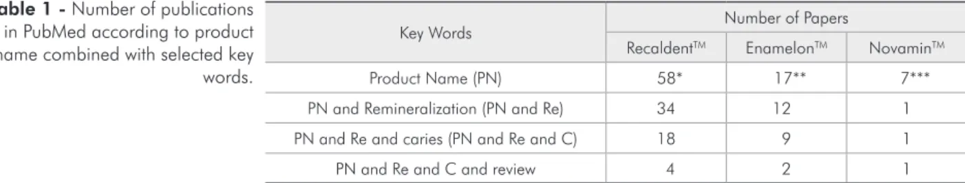
products is currently not abundant. A literature search of the PubMed database conducted in April, 2009 using the key words Recaldent or Enamelon or Novamin alone or combined with remineralization AND caries AND review resulted in the number of papers listed in Table 1.
Recaldent is the most studied system. The avail-able reviews on it state either that it could signii-cantly slow the progression of coronal caries and promote the regression of lesions,24 or that there
is insuficient clinical trial evidence, in quantity and also quality, to make any recommendation on
R
elativ
e
valu
es
pH cycling (DE>RE)
SML (%) Lesion depth (µm) F (µg F/cm2)
0 30.0 40.0 80.0
70.0 90.0 100.0
60.0
50.0
20.0
10.0
Placebo 225 ppm F 71.2
47.1 62.9
32.5
4 9.1
Graph 4 - Enamel surface microhardness loss (SML, %), le-sion depth (µm) and total fluoride in enamel (µgF/cm²) after a demineralizing (De > Re) pH-cycling regimen, according to the treatments (mean ± SD; n = 10). The differences were statistically significant (p < 0.05).16
R
elativ
e
valu
es
pH cycling (RE>DE)
SMR (%) Lesion depth (µm) F (µg F/cm2)
0 30.0 40.0 80.0
70.0 90.0 100.0
60.0
50.0
20.0
10.0
Placebo 225 ppm F
7.9 48.7
28.2 28.4
7.6 80.3
Graph 5 - Enamel surface microhardness recovery (SMR, %), lesion depth (µm) and total fluoride in enamel (µgF/cm²) after a remineralizing (Re > De) pH-cycling regimen, ac-cording to the treatments (mean ± SD; n = 10). The differ-ences were statistically significant (p < 0.05).16
D
ental
des
ctru
ctio
n
Time
C D E
B
A Clinically not visible
Cavities
White spot lesions
the clinical use of this product.25 As for Enamelon,
except for a descriptive publication made by the manufacturers of this commercial product,26 the
only other review, published by Reynolds24 (2008),
concluded that there is evidence that Enamelon has an anticariogenic effect on root caries. Except for the paper by Reynolds24 (2008), no other paper
was found on NovaminTM AND remineralization in
PubMed, but a great number of abstracts have been presented in the last IADR meetings (54 in 2008 and 24 in 2009).
As Reynolds24 (2008) concluded, “calcium
phos-phate-based remineralization technologies show promise as adjunctive treatments to luoride therapy in the non-invasive management of early caries le-sions”, since they do not have general recommenda-tion, but some people could beneit from them. Oth-erwise, they would not be a solution to the problem of controlling caries disease.
Conclusions
Inicipient caries lesions regress or clinically dis-1.
appear due to mechanical removal of affected enamel and true reprecipitation of minerals (remineralization), both of which are dependent on the control of caries as a disease.
Remineralization occurs naturally by the action 2.
of saliva.
Fluoride slows down caries progression by inter-3.
fering with the dynamics of the process, reduc-ing enamel demineralization and enhancreduc-ing its remineralization.
Remineralization is activated by luoride. 4.
Any strategy to reduce the progression of caries 5.
lesions should be based on the control of caries as a bioilm-dependent disease.
The repair of early caries lesions may be speed-6.
ed up by an exogenous Ca and Pi supply, but the clinical signiicance of the remineralization should be better evaluated.
The calcium phosphate-based remineralization 7.
technology could help some patients but it does not have general recommendation.
Key Words Number of Papers
RecaldentTM EnamelonTM NovaminTM
Product Name (PN) 58* 17** 7***
PN and Remineralization (PN and Re) 34 12 1
PN and Re and caries (PN and Re and C) 18 9 1
PN and Re and C and review 4 2 1
1st publication in: *2001; **1998 and ***1952 (anti-allergic drug).
Table 1 - Number of publications in PubMed according to product name combined with selected key words.
References
1. Fejerskov O, Thylstrup A. Different concepts of dental caries and their implications. In: Textbook of clinical cariology. 2nd ed. Munksgaard, Copenhagen; 1994. chapter 9.
2. Fejerskov O, Nyvad B, Kidd E. A. M. Pathology of dental caries. In: Fejerskov O, Kidd E (ed). Dental caries – The dis-ease and its clinical management. 2nd ed. Oxford: Blackwell
Munksgaard; 2008. chap. 3.
3. Ardu S, Castioni NV, Benbachir N, Krejci I. Minimally inva-sive treatment of white spot enamel lesions. Quintessence Int. 2007;38(8):633-6.
4. Diefenderfer KE, Stahl J. Caries remineralization therapy: implications for dental readiness. Mil Med. 2008;173(1 Sup-pl):48-50.
5. Fejerskov O. Changing paradigms in concepts on dental caries: consequences for oral health care. Caries Res. 2004;38(3):182-91. Review.
6. ten Cate JM, Larsen MJ, Pearce EIF, Fejerskov O. Chemical interactions between the tooth and oral fluids. In: Fejerskov O, Kidd E (ed). Dental caries – The disease and its clinical management. 2nd ed. Oxford: Blackwell Munksgaard; 2008.
chap. 12.
7. Dawes C. What is the critical pH and why does a tooth dis-solve in acid? J Can Dent Assoc. 2003;69:722-4.
composi-tion after APF applicacomposi-tion and F dentifrice use. J Dent Res. 2004;83:71-5.
9. Nyvad B. Diagnosis versus detection of caries. Caries Res. 2004;38(3):192-8. Review.
10. Cury JA, Tenuta LM. How to maintain a cariostatic fluo-ride concentration in the oral environment. Adv Dent Res. 2008;20(1):13-6.
11. Edgar WM, Higham SM. Role of saliva in caries models. Adv Dent Res. 1995;9:235-8.
12. Dijkman A, Huizinga E, Ruben J, Arends J. Remineraliza-tion of human enamel in situ after 3 months: the effect of not brushing versus the effect of an F dentifrice and an F-free dentifrice. Caries Res. 1990;24:263-6.
13. Arends J, ten Bosh JJ. In vivo de- and remineralization of den-tal enamel. In: Leach SA. Factors relating to demineralization and remineralization of the teeth. Oxford: IRL Press Limited; 1985. p. 1-11.
14. ten Cate JM. Current concepts on the theories of the mechanism of action of fluoride. Acta Odontol Scand. 1999;57(6):325-9. Review.
15. Featherstone JD. Dental caries: a dynamic disease process. Aust Dent J. 2008;53(3):286-91.
16. Moi GP, Tenuta LM, Cury JA. Anticaries potential of a fluor-ide mouthrinse evaluated in vitro by validated protocols. Braz Dent J. 2008;19(2):91-6.
17. Holmen L, Thylstrup A, Artun J. Surface changes during the arrest of active enamel carious lesions in vivo. A scanning elec-tron microscope study. Acta Odontol Scand. 1987;45(6):383-90.
18. Arends J, Gelhard TBFM. In vivo remineralization of human enamel. In: Leach SA, Edgard WM. Demineralization and remineralization of the teeth. Oxford: IRL Press Limited; 1983. p. 1-16.
19. Machiulskiene V, Richards A, Nyvad B, Baelum V. Prospec-tive study of the effect of post-brushing rinsing behaviour on dental caries. Caries Res. 2002;36(5):301-7.
20. Baelum V, Machiulskiene V, Nyvad B, Richards A, Vaeth M. Application of survival analysis to carious lesion transi-tions in intervention trials. Community Dent Oral Epidemiol. 2003;31(4):252-60.
21. Lima TJ, Ribeiro CCC, Tenuta LMA, Cury JA. Low-fluoride dentifrice and caries lesions control in children with different caries experience: a randomized clinical trial. Caries Res. 2008;42(1):46-50.
22. Featherstone JDB. Clinical aspects of de/remineralization of teeth. Adv Dent Res. 1995;49(3):175-340.
23. Ellwood R, Fejerskov O, Cury JA, Clarkson B. Fluoride in caries control. In: Fejerskov O, Kidd E (eds). Dental caries: The disease and its clinical management. 2nd ed. Oxford: Blackwell
Munksgaard; 2008. p. 287-323.
24. Reynolds EC. Calcium phosphate-based remineralization sys-tems: scientific evidence? Aust Dent J. 2008;53(3):268-73. Review.
25. Azarpazhooh A, Limeback H. Clinical efficacy of casein de-rivatives: a systematic review of the literature. J Am Dent Assoc. 2008;139(7):915-24; quiz 994-5. Review.
26. Winston AE, Bhaskar SN. Caries prevention in the 21st