Operative procedural errors must be well analyzed in order to avoid influence negatively the root canal treatment (RCT) prognosis. The successful RCT prevents tooth loss and avoids pain and apical periodontitis. This review aimed to categorize common operative procedure errors and clinical factors associated with RCT. Based on this, will be approached common errors of procedures within the clinical operative sequence: endodontic treatment planning, pulp and periapical disease diagnosis, anaesthesia, access cavity preparation, isolation with rubber dam, root canal preparation, root canal filling and retreatment, restoration of endodontically treated teeth, postoperative pain, follow up of endodontically treated teeth. The professional must remind that in each phase of RCT an operative error may have adverse implication on prognosis, and these errors characterize risk factors to failure. The knowledge of probable operative procedural errors and its consequences are essentials to avoid future problems to the tooth health.
Common Operative Procedural Errors
and Clinical Factors Associated
w i t h R o o t C a n a l T r e a t m e n t
Carlos Estrela1, Jesus Djalma Pécora2, Cyntia R.A. Estrela3, Orlando A. Guedes3, Brunno S.F. Silva4, Carlos José Soares5, Manoel Damião Sousa-Neto2
1Department of Stomatologic Sciences, Dental School, UFG - Universidade Federal de Goiás, Goiânia, GO, Brazil
2Department of Restorative Dentistry, School of Dentistry of Ribeirão Preto, USP - Universidade de São Paulo, Ribeirão Preto, SP, Brazil 3Department of Oral Sciences, Dental School, UNIC - Universidade de Cuiabá, Cuiabá, MT, Brazil 4Department of Oral Diagnosis, Dental School, UniEVANGÉLICA, Anápolis, GO, Brazil
5Department of Restorative Dentistry and Dental Material, Dental School, UFU - Universidade Federal de Uberlândia, Uberlândia, MG, Brazil Correspondence: Prof. Dr. Carlos Estrela, Praça Universitária s/n, Setor Universitário, 74.605-220 Goiânia, GO, Brasil. Tel: +55-62-3209-6254. E-mail: [email protected]
Key Words: success, failure, outcome, root canal treatment, operative error.
Introduction
Contemporary endodontics has experienced scientific and technological innovations substantiated with imaging exams (1-5), instrument design and kinematics, root canal preparation (RCP) and filling techniques (6-11). These implements have been incorporated daily to clinical protocols. The new technologies and therapeutic methods require some time for a precise analysis of the risks and benefits to be integrated into clinical practice (12).
Parallel to scientific and technological advances, accidents and complications during root canal treatment (RCT) may occur any time. The professional should be aware to avoid these unpleasant events, since operative procedural errors characterize iatrogenic risk factors that may result in RCT failure (12-15).
An independent analysis of these errors must be made during the planning of operative procedures. Studies involving the prevalence of apical periodontitis in endodontically treated teeth reported that the quality of root canal filling and coronal restoration influence on the success or failure (16-25). The outcomes of RCT are an indicator to sustain or to adjust the therapeutic protocol (12).
Several factors and the ongoing clinical conditions are important to determine the challenges and difficulties in the moment of RCT. For example, presence of infection or inflammation; primary or secondary infection; apical periodontitis; symptomatology; sinus tract; endodontic and
periodontal lesion; missed root canal, apical transportation, coronal and root perforation, endodontic instrument fracture; traumatic dental injury; root resorption; radicular fracture; incomplete access to pulp chambers and root canal orifices; limits of root canal filling, overfilling; quality of coronal restoration, etc. (25-43).
In addition to all these factors, the patient’s systemic conditions must be included. Systemic and periodontal diseases have to be prudently verified during the endodontic planning, since they may be risk factors of RCT failures. Pulp and periapical diseases diagnosis previous to RCT are an important predictive referential to prognosis. The health of the professional (stress, work environment) is a human aspect many times neglected and which may be a risk factor to operative errors. The prediction of RCT success constitutes a factual challenge, due to all the biologic (local and systemic) and technical factors that involve operative procedures. RCT success includes knowledge and domain of root canal anatomy, control of microorganisms, technical-scientific mastering of therapeutic protocols (psychomotor skill) and a positive host-immune response (12).
C. Estrela et al.
preparation, root canal filling and retreatment, restoration of endodontically treated teeth, postoperative pain and follow up of endodontically treated teeth. This review aimed to categorize common operative procedure errors and clinical factors associated with RCT.
Endodontic Treatment Planning
The absence of clinical planning may induce the professional to operative procedural errors (12). In this perspective, it may be illusory to verify the configuration of the pulp cavity illustrated by periapical radiography. The periapical radiographs show 2-dimensional representations of 3-dimensional structures. The tooth morphological features may not be revealed by radiographic exams (1-5,44,45). Periapical radiolucencies and root resorptions may not be visible radiographically, although they may exist clinically (5,44-47).
The root canal anatomy is very complex and requires meticulous study before any therapeutic intervention (48,49). Innovative alternatives to study internal root canal anatomy, such as cone beam computed tomography (CBCT) and micro-computed tomography have been well discussed (1-5,48-54). CBCT images may reveal aspects unable to identify by periapical radiography and may favor a more predictable planning and treatment (1-5,46,47). Map-reading strategy in CBCT images reduces problems associated to overlapping of anatomical structures (43). The navigation dynamics favors a precise identification of roots position, frequency of root canals, presence of the isthmus and apical foramina (53,54). The root apices of the maxillary posterior teeth and the maxillary sinus floor may present intimate relationship that favors development of inflammatory, infectious and/or traumatic alterations in the maxillary sinus (MS) or vice-versa (55-59). Operative procedural errors during RCT, such as over-instrumentation, over-irrigation, overfilling and aggressive surgical procedures constitute risk factors by introduction of foreign bodies into the MS (58).
Full access to all root canal walls has a clinical impact on sanitization process and root canal filling and consequently on the successful treatment (60). Negligence, lack of planning and unfamiliarity with the internal anatomy contributes significantly to the failure of RCT. Each patient must be judiciously analyzed for anatomic, ethnic and genetic features.
Choice of the clinical protocol to be employed considering certain diagnostic hypotheses means that priorities need to be established, weighing the trade-off between the risks of treatment against the benefit of the eventual outcome. A decision made should avoid annoyances, including judicial processes. All care is small when it involves the health of individual.
Systemic and local factors related to the patient must be well analyzed. The doubtful prognoses may constitute an impediment for the precise implementation of the RCT. Among them, local factors include anatomic-pathologic aspects, modifications of the internal anatomy, excessive dilacerations, dental development anomalies, calcification of the pulp chamber, endodontic accidents, the loss of working length, ledge formation, root perforation, endodontic instrument fracture, presence of extensive posts, sealer or glass ionomer in root canal retreatments (12).
It is important to consider that not all the periapical radiolucencies are associated with infected root canals (61-65). Periapical lesions of non-endodontic origin may lead to misdiagnosis and consequently therapeutic errors (61-65). The case selection must be well considered and planned. In addition, the planning of the time required for the operative procedures, the probable number of interviews, the instruments and materials necessary to RCT are singular, but relevant to the planning.
A protocol for clinical attendance is necessary and basic, with ability to prevent cross-infection. It is essential to admit that all patients must be considered as infective agents and in the same way as the dental team, infectant vectors of cross-infection. Cross-infection can be prevented by the adoption of different behavior patterns and using protocol barriers (66).
Previous planning indicates a path to go through to attain a certain objective. An important strategy includes training in order to simulate future difficulties, with prediction of decisions to be made in case something happens out of the established protocol (67).
Pulp and Periapical Disease Diagnosis
Decision making achieves higher success rate in RCT when it is established from a correct diagnosis (12,25,26,60). The clinical examination consists in the analysis of visual physical features of the tooth and surrounding tissues, palpation, percussion and pulpal vitality tests and radiographic aspects. During anamnesis, the clinical characteristics of the pain (type - provoked or spontaneous; duration - short or long; frequency - continuous or intermittent and site - local or diffuse) constitute important referential to clinical diagnosis (26).Operative Errors and Clinical Factors in endodontics diagnosis of the pulp pain (a diagnostic hypothesis) (26).
Thus, it only leaves to the analysis of signs and the patient’s clinical symptoms, without correlation with possible histopathological events (60,68).
Pulp or periapical inflammatory diseases are usually identified by the consequences of aggressions to the tissues. The previous clinical conditions found in the tooth need to be identified during the diagnosis. It includes dental caries, pain, inflammation, primary or secondary infection, symptomatic/asymptomatic AP, periapical abscess with/ without sinus tract, open/closed cavity, history of traumatic dental injury (12,26,60). The clinical diagnosis of pulp and periapical diseases involve: hyperreactive pulpalgia, symptomatic pulpitis, asymptomatic pulpitis, pulp necrosis, symptomatic apical periodontitis of traumatic or infectious origin, asymptomatic apical periodontitis, periapical abscess without sinus tract, periapical abscess with sinus tract (26). Most occurrences of pulp pain have been associated with symptomatic pulpitis and the periapical pain with symptomatic apical periodontitis of infectious origin (26). The general clinical condition of the patient (systemic health) and local characteristics (tooth condition) favors prediction of a possible outcome of RCT. Prognosis of a RCT also depends on the level of scientific knowledge and ability of the endodontist.
Endodontics challenges begin when diseases from non-endodontic origin suggest a typical case of periapical lesion (61-65). Mandibular or maxillary areas surrounding the root apexes presenting radiolucent or radiopaque images may be a sign of non-endodontic disease. Thus, the differential diagnosis of diseases from endodontic or non-endodontic origin should be cautiously made to avoid misdiagnosis (61-65).
Anesthesia
Successful RCT involves an appropriate control of pain. Tooth pain represents the main reason why patients seek dental care. An understandable anxiety for both, patients and endodontists is the pain experienced before, during or after RCT (69). Many patients considered pain and dentistry synonymous (70). Pain is a complex phenomenon and dental pain, a multifactorial or multidimensional experience that involves sensory and emotional responses, conceptual and motivational aspects (71,72).
All patients consciously report having or not pain experience during or after RCT. The control of patient’s pain and anxiety favors a smooth progress of clinical procedures. The professional must recognize the pathologies that produce intensive pain (symptomatic pulpitis, symptomatic apical periodontitis and periapical abscess without fistula). The knowledge of anatomic structures, the anesthesia techniques, the mechanism of action, the effect
and complications of the anesthetics constitute essential abilities of the professional. The worst memory for a patient is the experience of pain during or after RCT (67).
Isolation of Tooth with Rubber Dam
The control of microorganisms in the infected root canals is an aim for the endodontic therapy (26-36,73-77). To guarantee complete sanitization during the operative procedure the isolation of tooth with rubber dam is recommended, which also minimizes cross infection, prevent accidents like aspiration and deglutition of endodontic instruments and the cytotoxic effect of irritant substances on vital tissues (sodium hypochlorite and calcium hydroxide) (66,67).Sodium hypochlorite is one of the best-known irrigants used by dentists all over the world (73,75,77-80). This irrigant solution may cause damage in contact with vital tissues (75,80-83). The accidents involving sodium hypochlorite may have some complications: severe pain; ballooning or immediate edema in soft tissue; extension of edema to a large area of the face, like the cheeks; periorbital ecchymosis on skin or mucosa as a result of profuse interstitial bleeding; profuse intraoral bleeding directly from root canal; palate mucosa necrosis; tissue necrosis; neurological complications; paresthesia; allergy; dermatological problems and life-threatening airway obstruction (80-83).
Thus, tooth isolation with rubber dam should be performed properly, in order to avoid serious problems to the professional and the patient’s health. Failure to use rubber dam has been shown to influence the choice of root canal irrigant. It has a negative impact on treatment outcome and places the patient at risk of swallowing or aspirating materials and instruments (84). An important care during tooth isolation with rubber dam is the selection and good adaptation of the clamp. Coronary fracture in tooth with little coronary structure due to the clamp’s force constitutes an extremely unpleasant accident and must be avoided. Different manners to tooth isolation were incorporated to the endodontics in order to obtain absolute isolation of the tooth from the oral cavity. Whenever needed, the tooth must be reconstructed before its isolation with rubber dam (67). Additional strategies to complement the rubber dam isolation may be required for young patients or during trans-surgery procedures, like the light curing gingival barrier or cyanoacrylate adhesive to block the possible blood or fluids penetration through the rubber dam (85). Thus, ergonomics and time economy are also included among the advantages.
Access Cavity Preparation
C. Estrela et al.
opportunity to point out some of the factors that could complicate access cavity preparation of the root canals. The presence of nodules in the pulp chamber, calcifications, incomplete dentinogenisis (class 2), resorptions, dislocated tooth (tooth out of the dental arc inclination) or a restoration covering the crown may hamper the access cavity preparation and induce accidents. Before starting the access cavity, it is prudent to verify in the imaging exam the size and shape of the pulp chamber, tooth inclination in the arch. Meticulous analysis of the root canal anatomy should be performed while RCT planning (12,14,15, 54).
Operative procedures earlier to the access cavity preparation involve the removal of all carious tissue, restoration of defects and weakened dentin structure, which could change the coronal references. A coronal reconstruction before the access cavity is required in some situations of large coronary destruction. The perfect access cavity can be prepared by meeting the following objectives: direct access to the root canal, complete elimination of the roof of pulp chamber, respect for the pulp chamber floor, ledge formation on the proximal walls of the pulp chamber and proper selection of drills. An incomplete access cavity reduces the emptying quality and may alter the form of root canal preparation. An exaggerated access cavity favors perforations, compromise the biomechanics performance of root treated teeth and exposes the tooth to coronary/root fracture (67).
The analysis of the coronary chamber in the contemporary imaging exams, the perfect selection of the drill compatible with coronary volume, good lighting and magnification are essential because they favor visualization of the cavity during coronary opening, avoiding unpleasant accidents.
Missed root canal may be found in some situations (49,50,52), which may be responsible of maintaining bacterial contamination. Missed root canal may occur in all teeth, due to poor exploration and investigation of anatomy (49,50,52). In mesiobuccal root of the first molar, in premolars with two or more roots, in mandibular central incisors, in dens invaginatus more attention is required to avoid missed root canals. Axial sections in CBCT are efficient in the study of these situations (52).
Accidents may occur at any moment of RCT and they may lead to the failure. The common procedure errors during access cavity preparation are as follows: remaining caries tissue in the pulp chamber; incorrect choice of the point for preparing the access cavity; incorrect selection of the drill (excessively large drill in relation to the pulp chamber volume); error on the climbing of teeth with wrong position; incomplete access; no removal of the coronal roof; buccal perforation in anterior teeth;
perforation of the floor in molars (67).
Root Canal Preparation
Root canal emptying is the procedure to eliminate the contents from in the pulp cavity. RCT incorporate the sanitization process, which involves the emptying and enlarging, combined with the use of antibacterial strategies.
The emptying is performed in cases of normal pulp (healthy or inflamed) (pulpectomy), infected pulp (sanitization process), presence of filling material (gutta-percha and sealer removal in retreatment), root canal with posts or fragments of endodontic instruments (disobturation). This stage of emptying allows the identification of the entry orifice of the root canal, its diameter, the direction of the curvature, obstacles or obstructions not visualized by imaging exams. In addition, it eases the passage of additional instruments. The emptying is performed before RCP and is essential for precise planning, in particular what concerns the lateral limits of preparation. After root canal emptying is made the enlargement of the root canal. Thus, the selection of endodontic instruments and the technique used for root canal preparation (RCP), as well as the operator’s experience constitute important aspects to be considered in view of the new strategies and instruments (continuous or reciprocating nickel-titanium files) (6,7,12,84-89).
The quality of RCP with rotary NiTi instruments (86-94) and the low incidence of procedural errors (root canal transportations, root perforations and fracture of instruments) (14,15) motivated discussions and use by undergraduate students. The introduction in undergraduate teaching appears a promising matter. Another important aspect related to RCP is that all instruments have a limited useful lifetime and may have manufacturing defects (95,96), which implies recognizing the need for frequent renewal. In all steps of RCT, the slightest negligence of the professional may cause operative errors.
Curved RCP present a greater possibility of apical transportation and root perforations. Prior identification of the radius of curvature of a root canal constitutes a functional conduct (97). The most root canals of maxillary and mandibular first and second molars have some degree of curvature in the apical and cervical thirds, regardless the analyzed plane (98).
The apical limit of RCP must be inside of the root canal. A better prognosis is attained when RCP and filling do not extend beyond the apical foramen, so it must be short of the radiographic root apex (12,34-36,40,41).
Operative Errors and Clinical Factors in endodontics refers to a higher enlargement of the area within the
anatomical limits (99). The lateral limit of cervical and apical enlargement should be appropriate, as the radiographic aspect does not represent a precise reference to the real dentinal thickness and the illusion of the radiographic image may be responsible for insufficient or excessive wear. In cases when CBCT exam is necessary, it is possible to identify in axial plane the dentinal thickness that will be prepared. Planning, good sense and anatomic knowledge are significant at this stage of the endodontic management. Determination of the lateral enlargement limit should take into account the anatomic and pathological condition, intensity of the root canal curvature, transversal section and flexibility of the endodontic instrument. The initial preparation of the cervical third makes it possible to remove the dentinal prominences (constriction areas) and favors determination of the anatomic diameter of the root canal. Root canal enlargement must be planned according to root canal anatomy.
The maintenance of the original position of the foramen (100) is as important as the centering ability of the root canal during RCP. Apical transport, main canal deviation (with consequent loss of the working length) may be well controlled with NiTi instruments when are used by a qualified professional, and used cautiously and rationally in a good shaping technique.
Cervical enlargement must be carefully observed, particularly after the introduction of instruments with high taper, which may be responsible for excessive attrition in thin root canal regions, like the distal wall of the mesial root of mandibular molars. These aspects must be analyzed and accounted for, since there is a direct and intimate relationship between endodontic instruments and root canals.
The effectiveness of antimicrobial strategies (irrigant solution and intracanal dressing) is related to keep direct control with the area to be prepared. The successful infected RCT is connected with the decline of microorganisms and the disruption of the bacterial biofilm (28-32,74-80,101,102). The complex anatomy hampers the full and direct access. For example, root canal isthmus is a common anatomic structure in human permanent teeth (48,49,54,103), except in maxillary anterior teeth (54). Higher frequencies of root canal isthmuses (87.9%) have been found in mandibular first molars (54). The effect of intracanal dressing on bacteria occurs maintaining direct contact to express its mechanism of action. Equally important as the irrigant solution for effective sanitization process are the penetration depth of the irrigating cannula, frequency of irrigation, abundance of irrigation, the concentration of irrigant solutions and quality control of the substances (12,104).
The treatment of infected root canals requires reducing the intraradicular microbial load, to disrupt the biofilm and stringent application of non-antibiotic chemomechanical measures (33). Irrigant solutions are very important during root canal preparation because they help to clean the root canal (75), lubricate the files, flush out debris, and have an antimicrobial and tissue dissolution effect, without damaging periapical tissues (12,73-80). Sodium hypochlorite and chlorhexidine are the most often indicated antimicrobial agent for treatment protocols against endodontic and periodontal infections (73-80). Calcium hydroxide remains as the most indicated intracanal dressing in different clinical conditions. The properties of calcium hydroxide come from its dissociation into calcium and hydroxyl ions and the action of these ions on tissues and bacteria explains the biological and antimicrobial properties of this substance (73).
Root Canal Filling and Retreatment
Root canal filling (RCF) aims to eliminate empty spaces inside the pulp cavity where microorganisms can lodge (12). When RCP is well established, the root canal filling of this space maintains the same destiny. The lateral condensation technique with gutta-percha is the most known and used. Some details for root canal filling must be well verified, such as asymptomatic tooth, the maintenance of the same apical limit used in the RCP. Root canal must be well cleaned and dried, for the selection of the main cone of gutta-percha, and it is necessary to verify if the cones offer some resistance to the removal and to occupy most space of the root canal. The root canal filling material must be contained only inside the root canal, there is no way to justify its presence beyond the root canal (12,35,36,40,41). In mandibular and maxillary molars, it is necessary to take care to avoid lead filling materials into mandibular canal and maxillary sinus, due the proximity of these anatomic structures with root apexes (53,58).Different properties of endodontic sealers have been discussed (105-108), but the endodontic professionals still do not have a material considered ideal. Adherence ability to dentinal walls, with a sealing quality to avoid infiltration, with biocompatibility and stimulation the healing process of the periapical tissues constitute ideal properties. Microbial leakage along the coronal restoration and root canal fillings have been considered strongly associated with endodontic failures (20-26). The quality of RCP and coronal sealing are essential factors to achieve high rates of success, even in infected root canal (12,60).
C. Estrela et al.
panoramic and periapical radiographs and CBCT images, apical periodontitis (AP) was detected in 17.6%, 35.3%, and 63.3%, respectively. Substantial discrepancy can be found between the imaging methods tested to detect AP. AP was correctly identified in 54.5% of the cases with periapical radiographs and in 27.8% of the cases with panoramic radiographs (5).
Operative procedural errors may occur and they represent risk factors able to compromise a tooth (12-15). Errors characterize disability, non-observance of therapeutic protocol and low level of knowledge involving endodontic principles. Deficient attendance may be responsible for severe consequences and sequels, which impairs the prognosis and may result in serious judicial questions (12-15). Operative procedural errors in endodontically treated teeth and dental implants were detected using CBCT images (13). Underfilling, overfilling and root perforations were detected in 33.5%, 8% and 4.5% of the endodontically treated teeth, respectively. Dental implants with thread exposures, contact with important anatomical structures and contact with adjacent teeth were seen in 37.5%, 13% and 6.5% of the cases, respectively (13).
The first option for treating the failure is root canal retreatment, which presents difficulties and complications that make the prognosis doubtful. The difficulty in establishing new access to the root canal depends on the present material, pulp cavity anatomy, missed root canal, iatrogenesis (loss of working length - ledge formation, perforation, fractured instruments), and the presence of intraradicular post (type, length, diameter, sealer).
Restoration of Endodontically Treated
Teeth
RCT is only finished after a perfect tooth restoration. The recovery of tooth function and occlusal harmony constitute essential elements in the rehabilitation process. Tooth restoration should be preferably performed using rubber dam isolation. During the RCT, the tooth should receive a temporary restauration with glass ionomer cement or composite resin. The time between the root canal filling and restorative coronal reconstruction should be the shorter possible to avoid the root canal contamination. A fracture or loss of restauration may conduct to reinfection and the need of a new antibacterial intervention. The restoration of endodontically treated teeth may be usually complex because of the destruction of the crown, partial or totally, due to decay, traumatic dental injuries or the presence of previous extensive restorations (109-115).
The coronal reconstruction of endodontically treated tooth must recover the biomechanical performance as similarly as possible to the intact tooth. The structural resistance is related to appropriate retention and adhesive
integration between sealer, core reconstruction and final restoration. Direct restorative materials, mainly composite resin has proven to be enough to reconstitute the coronal part that will be the support for a crown, only when necessary. In various teeth with significant loss of coronal structure and no possibility to provide enough retention and resistance to complete the coronal core, intraradicular posts for retention may be required. The use of fiber glass post associated with resin cement has proven to be a good option when post retention is required. The similarity of mechanical properties with dentin and the bonding interaction with root dentin, result in proper stress and strain distribution. The endodontically treated tooth that needs intra-radicular retention should receive a restoration structurally composed of components with specific functions. In a direct relationship of functional dependence, the coronal restoration is retained by the coronal core, which is retained by the intra-radicular post (109-112).
Thus, the restoration of endodontically treated teeth is conditioned to the appropriate selection of material and technique due to the biomechanical complex and the remainder of dental structure. The tooth resistance is severely compromised with excessive preparations, both at the coronary and root levels. It has been shown that intraradicular post provides only crown retention to the tooth and does not increase tooth resistance. The clinicians should choose the post size and diameter considering the shape and dimensions of the root canal after preparation and filling. Overpreparation aiming thicker fiber post is not recommended. The crown fracture risk depends on the amount of tooth structure loss and the capacity of the tooth to resist occlusal forces (109-115). The weekened teeth after several endodontic procedures should be carefully considered to select the restorative procedure. Endodontic retreatment, metallic post removal and over-preparation may create a larger root canal, which determines larger space between the post and root dentin. The use of composite resin to restore the adaptation between fiber post to root dentin is strongly recommended. The choice of proper irrigants to clean the root canal before post cementation, resin cement selection using material that can polymerize in deep areas and a light curing unit with sufficient light irradiance to cure the resin cement, are essential to produce adequate post fixation (115).
Operative Errors and Clinical Factors in endodontics on successful RCT (12,25,111,112).
Postoperative Pain
RCT frequently is performed in inflamed and infected pulp associated or not with periapical inflammation. An extremely displeasing occurrence for patient and professional is the surprise with appearance of pain immediately after RCT. This incident puts in risk all the competence of the professional. In some cases, postoperative pain be predictable after RCT, but in other conditions, this fact is not expected, which is poor news (116).
The symptomatology may be an unpredictable inconvenience. The symptomatic apical periodontitis is a tissue reaction that occurs in the apical periodontium due to traumatic or infectious aggressions, which determine inflammatory and immunologic responses. The most common causes include occlusal trauma, over-instrumentation, over-irrigation, over-medication, over-fillings or remains of extruded materials, invasion of microorganisms (dissemination of toxins and enzymes). The subsequent clinical conditions are inflammatory alterations involved with pain: symptomatic apical periodontitis of traumatic nature and symptomatic apical periodontitis of infectious nature and periapical abscess without fistula (116). The organic defenses and the degree of virulence of the microorganisms establish the different natures of periapical alterations. The inflammatory and/or infectious condition involved in the periapical alteration modulates the diagnosis and the treatment option.
Studies about incidence of pain that characterizes the periapical inflammation during and after the RCT, and the relation between bacteria in infected root canals and clinical symptoms present on periapical inflammation have been widely discussed (116-126). Sundqvist (118) verified anaerobic bacteria in pulp necrosis and in the acute exacerbations of the periapical region. Bacteroides meleninogenicus combined with other bacteria (Peptostreptococcus, Fusobacterium, Lactobacillus and others) was isolated. The predominant bacteria in the mixed infections of the root canal (Gram-negative anaerobic) have biological activation effects on the organic defense, and this results in an increased inflammatory response with the presence of painful periapical lesions.
Seltzer and Naidorf (117) characterized the etiologic factors of acute manifestations before and during the root canal treatment, including: alteration of the local adaptation syndrome, changes in the periapical tissue’s pressure, microbial factors, effects of chemical mediators, manifestations of the cyclic nucleotides, immunologic phenomena and psychological factors.
The clinical diagnoses among 1,765 patients that sought the Urgency Service of the Dental School for
tooth pain treatment showed higher frequency in cases of symptomatic pulpitis and symptomatic apical periodontitis of infectious origin. The main clinical factors associated with pain of pulp and periapical origin were caries and open pulp chamber, respectively (26).
The success estimation in RCT confirms a lower rate of post-treatment pain in teeth with inflamed or infected pulp. Therefore, a good sanitization process, a rational use of intracanal medicament and high quality endodontic sealing and coronal restoration are essential.
Periapical inflammation of endodontic origin is the consequence of aggressive agents’ action from infected tooth. The periapical healing process after RCT in infected teeth has been studied (127,128). High levels of receptor activator of nuclear factor kappa B ligand (RANKL) and a potential for bone resorption characterize persistent apical periodontitis as a biologically active lesion. In persistent apical periodontitis, the found inflammatory immune cells are suggestive of a suppressive and regulatory periapical environment favorable to chronic clinical conditions (127). Estrela et al. (128) evaluated the expression of the mesenchymal stem cells (MSC) CD90 and progenitor stem cell markers Sox2 in persistent apical periodontitis (PAP) and primary periapical lesions (PPL). MSC seem to contribute to the immunosuppressive environment recently found in PAP. Additionally, distinct stem cell sources may be associated with the chronic nature of PAP and PPL development.
Follow up of endodontically treated
teeth
The periapical tissue repair process is related to the elimination of aggressive agents from infected root canal, and the quality of the endodontic and coronary seals. The resting offered to the periapical tissues after RCT favors the reversion of the inflammation, normally with repair of the periodontal ligament, osteogenesis and new formation of osteocement.
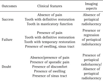
Success of the clinical determination of the endodontically treated teeth includes: absence of pain and swelling; absence of drainage and sinus tract; functional tooth with normal periapical physiology; absence or regression of periapical bone rarefaction (12,16-19). The doubtful cases (transition phase) between RCT success or failure during a follow-up may be associated with limitations of imaging exams (5) or lesions from non-endodontic origin (61-65). Wu et al. (107) reported that the outcomes of RCT should be re-evaluated in long-term longitudinal studies using CBCT and stricter evaluation criteria.
C. Estrela et al.
Table 1. Categorizing common operative procedures errors and clinical factors associated with root canal treatment
Clinical factors / Common operative procedural errors 1. Endodontic treatment planning
The absence of RCT planning contributes to disagreeable surprises.
Procedural errors characterize risk factors that may contribute to failure of RCT. 2. Pulp and periapical disease diagnosis
The decision making for RCT shows high success rate when established based on correct diagnosis.
The differential diagnosis of diseases from endodontic or non-endodontic origin should be prudently made, avoiding misdiagnosis.
3. Anesthesia
Tooth pain is a strong reason why patients look for dental care (69). Many patients consider pain and dentistry as synonymous (70);
The worst remembrance of a patient is the experience and memory of pain during RCT. 4. Access cavity preparation
The presence of nodules in the pulp chamber, calcification, resorptions, dislocated tooth (incorrect inclination in the arc) or a restoration covering the crown may difficult the access cavity preparation and induce accidents.
An incomplete access cavity reduces the quality of emptying and may alter the ideal shape of RCP. Exaggerate access cavity favors root perforation, and makes the tooth susceptible to coronal /radicular fracture.
5. Isolation with rubber dam
To guarantee sanitization process during the operative procedures, an isolation of tooth with rubber dam is recommended, which minimizes cross infection and prevents accidents, like aspiration and deglutition of endodontic instruments, and cytotoxic action of irritant substances on vital tissues.
Sodium hypochlorite is the most indicated irrigant solution, but outside the root canal produces many complications, including severe pain.
6. Root canal preparation
Selection of the technique used for RCP and endodontic instruments, as well as the operator’s experience are important aspects to consider in view of the new strategies and instruments;
Operative procedural errors during RCP (ledge formation, cervical root perforations, foramen transportations, loss of the working length, missed root canal, overinstrumentation, overirrigation, fracture of instruments) may occur due the lack of attention by the professional.
7. Root canal filling and retreatment
The elimination of empty spaces inside the pulp cavity constitutes the main purpose of root canal filling (RCF). The quality of RCP has direct influence on RCF.
A viable option for failure treatment is the retreatment, which presents doubtful prognoses. The new access is influenced by various factors: to find a missed root canal, filling material, pulp cavity anatomy, iatrogenesis (loss of working length -ledge formation, perforation, fractured instrument), the presence of intraradicular post (type, length, diameter and sealer).
8. Restoration of endodontically treated teeth
Perfect selection of tooth restoration has direct influence on successful RCT.
Conservation of the remaining root dentin and crown is fundamental to improve the resistance of endodontically treated teeth.
The best option to recover the tooth integrity is to reduce the time elapsed between root canal treatment and final restoration, and a bonding restoration associated or not with post.
Several teeth are lost due to fracture or perforation associated to inappropriate selection of intraradicular post. 9.Postoperative pain
An extremely displeasing occurrence for the patient and professional is the surprise of pain immediately after RCT, which compromises all the competence of professional.
The common causes of periapical pain include occlusal trauma, over-instrumentation, over-irrigation, over-medication, over-fillings or remains of extruded materials and invasion by microorganisms (dissemination of toxins and enzymes).
10. Follow up of endodontically treated teeth
The limitations in longitudinal studies are due to small recall of patients to RCT follow-up, lack of well-documented records with periapical radiographies of the initial treatment that allows to compare with the actual ones. These factors are essential to determine the quality of the adopted therapeutic protocol.
Operative Errors and Clinical Factors in endodontics success. Wu et al. (129) considered that the extraction
and retreatments were rarely recorded as failures and the recall rate was often small. Other studies reported the same problem in the recall of patients to quality control of the therapeutic protocol and clinical studies (60,129,130).
Knowledge of the previous status of pulp and periapical tissue when the RCT was made, the used resources to determine the periapical status, the time of treatment conclusion and its correct restoration are essential to follow-up. Quality control may be best defined by alternatives like CBCT to evaluate the multidimensional tooth structure using map-reading strategy in complex or doubtful cases (12). Table 1 categorizes the common operative procedural errors and the clinical factors associated with RCT. The RCT success is associated with regression of periapical inflammation, good endodontic and coronal sealing and functional tooth (Table 2).
The limitations regarding the small recall of patients to RCT follow-up, a well-documented record, with periapical radiographies of initial treatment that allow comparing with the actual ones, interfere in longitudinal studies. These factors are essential to determine the quality of the adopted therapeutic protocol. The biological and mechanical characteristics of an endodontically treated tooth implies in understanding the outcome as a multifactorial event for the lifetime of the individual.
Professionals must have in mind that in every phase of RCT an operative error may have adverse implication on
prognosis and may be risk factor to failure. The RCT success is not influenced by isolate factors, but the combination of a rigorous sequence of the protocols associated with the knowledge of probable operative procedural errors and its consequences is essential to avoid future problems with the tooth health.
Resumo
Erros de procedimentos operatórios devem ser bem analisados e evitados em função de influenciar negativamente o prognóstico do tratamento do canal radicular. Tratamento do canal radicular bem sucedido previne a perda do dente, evita dor pulpar e lesão periapical. Esta revisão objetiva categorizar erros de procedimentos operatórios comuns e os fatores clínicos associados ao tratamento do canal radicular. Neste intuito, serão abordados os erros mais comuns e os fatores clínicos dentro da seguinte sequência operatória: planejamento do tratamento endodôntico, diagnóstico da doença pulpar e periapical, anestesia, preparo do acesso cavitário, isolamento do campo operatório, preparo do canal radicular, obturação e retratamento do canal radicular, restauração do dente tratado endodonticamente, dor pós-tratamento do canal radicular, e acompanhamento do dente tratado endodonticamente. O profissional deve estar consciente de que em cada fase operatória um erro pode ter implicação no prognóstico, e ser fator de risco ao fracasso. O conhecimento dos prováveis erros de procedimentos operatórios e suas consequências é essencial para evitar futuros problemas com a saúde do dente.
Acknowledgements
The authors deny any conflicts of interest related to this study. This study was supported in part by grants from the National Council for Scientific and Technological Development (CNPq grants 306394/2011-1 to C.E.).
References
1. Arai Y, Tammisalo E, Iwai K, Hashimoto K, Shinoda K. Development of a compact computed tomographic apparatus for dental use. Dent Maxillofac Radiol 1999;28:245-248.
2. Mozzo P, Procacci C, Taccoci A, Martini PT, Andreis IA. A new volumetric CT machine for dental imaging based on the cone-beam technique: preliminary results. Eur Radiol 1998;8:1558-1564.
3. Cotton TP, Geisler TM, Holden DT, Schwartz SA, Schindler WG. Endodontic applications of cone beam volumetric tomography. J Endod 2007;33:1121-1132.
4. Patel S, Dawood A, Pitt Ford T, Whaites E. The potential applications of cone beam computed tomography in the management of endodontic problems. Int Endod J 2007;40:818-830.
5. Estrela C, Bueno MR, Leles CR, Azevedo B, Azevedo JR. Accuracy of cone beam computed tomography and panoramic and periapical radiography for detection of apical periodontitis. J Endod 2008;34:273-279. 6. Peters OA. Current challenges and concepts in the preparation of root
canal systems: a review. J Endod 2004;30:559-567.
7. Peters OA, Laib A, Gohring TN, Barbakow F. Changes in root canal geometry after preparation assessed by high-resolution computed tomography. J Endod 2001;27:1-6.
8. Spångberg LSW, Haapasalo M. Rationale and efficacy of root canals medicaments and root filling materials with emphasis on treatment outcome. Endod Topics 2002;2:35-58.
9. Ørstavik D. Materials used for root canal obturation: technical, biological and clinical testing. Endod Topics 2005;12:25-38.
10. Shrestha A, Kishen A. Antibacterial nanoparticles in endodontics: A Review J Endod 2016;42:1417-1426.
11. Sousa-Neto MD, Marchesan MA, Pécora JD, Brugnera-Júnior A, Silva-Sousa YTC, Saquy PC. Effect of Er:YAG laser on adhesion of root canal sealers. J Endod 2002;28:185-187.
12. Estrela C, Holland R, Estrela CR, Alencar AH, Sousa-Neto MD, Pécora JD.
Table 2. Characteristics of clinical and imaging outcomes in RCT
Outcomes Clinical features Imaging aspects
Success
Absence of pain Tooth with definitive restoration
Tooth in masticatory function
Absence of periapical radiolucency
Failure
Presence of pain Tooth with definitive restoration Tooth with temporary restoration Presence of swelling, sinus tract
Presence or regression of periapical radiolucency
Doubt
Absence/presence of pain Presence of sporadic pain Presence of discomfort
Presence of swelling Presence of sinus tract
Presence of periapical radiolucency/
C. Estrela et al.
Characterization of successful root canal treatment. Braz Dent J 2014; 25:3-11.
13. Silva JA, Alencar AHG, Rocha SS, Lopes LG, Estrela C. Three-dimensional image contribution for evaluation of operative procedural errors in endodontic therapy and dental implants. Braz Dent J 2012:23:127-134. 14. Alencar AHG, Dummer PMH, Oliveira HCM, Pecora JD, Estrela C.
Procedural errors during root canal preparation using rotary NiTi instruments detected by periapical radiography and cone beam computed tomography. Braz Dent J 2010;21:543-549.
15. Alves RAA, Souza JB, Alencar AHG, Pécora JD, Estrela C. Detection of procedural errors with stainless steel and NiTi instruments by undergraduate students using conventional radiograph and cone beam computed tomography. Iran Endod J 2013;8:161-165.
16. Strindberg LZ. The dependence of the results of pulp therapy on certain factors. An analytical study based on radiographic and clinical follow-up examinations. Acta Odontol Scand 1956;14 (sfollow-uppl 21):1-174. 17. Grossman LI, Shephard LI, Pearson LA. Roentgenologic and clinical
evaluation of endodontically treated teeth. Oral Surg Oral Med Oral Pathol 1964;17:368-374.
18. Seltzer S, Bender IB, Smith J, Freedman I, Nazimor H. Endodontic failures - an analysis based on clinical, roentgenographic and histologic findings. Oral Surg Oral Med Oral Pathol 1967;23:500-530.
19. Bender IB, Seltzer S, Soltanoff W. Endodontic success - a reappraisal of criteria. Oral Surg Oral Med Oral Pathol 1966;22:780-801. 20. Ray HA, Trope M. Periapical status of endodontically treated teeth in
relation to the technical quality of the root filling and the coronal restoration. Int Endod J 1995;28:12-18.
21. Tronstad L, Asbjornsen K, Doving L, Pedersen I, Eriksen HM. Influence of coronal restorations on the periapical health of endodontically treated teeth. Endod Dent Traumatol 2000;16:218-221.
22. Kirkevang LL, Ørstavik D, Horsted-Bindslev P, Wenzel A. Periapical status and quality of root fillings and coronal restorations in a Danish population. Int Endod J 2000;33:509-515.
23. Eriksen HM, Kirkevang LL, Petersson K. Endodontic epidemiology and treatment outcome: general considerations. Endod Topics 2002;2:1-9. 24. Hollanda ACB, Alencar AHG, Estrela CRA, Bueno MR, Estrela C.
Prevalence of endodontically treated teeth in a Brazilian adult population. Braz Dent J 2008;19:313-317.
25. Estrela C, Leles CR, Hollanda ACB, Moura MS, Pécora JD. Prevalence and risk factors of apical periodontitis in endodontically treated teeth in a selected population of Brazilian adults. Braz Dent J 2008;19:34-39. 26. Estrela, Guedes AO, Silva JA, Leles CR, Estrela CRA, Pécora JD. Diagnostic
and clinical factors associated with pulpal and periapical pain. Braz Dent J 2011;22:306-311.
27. Marending M, Peters OA, Zehnder M. Factors affecting the outcome of orthograde root canal therapy in a general dentistry hospital practice. Oral Surg Oral Med Oral Pathol Oral Radiol Endod 2005;99:119-124. 28. Nair PNR. Biology and pathology of apical periodontitis. In: Estrela C.
Endodontic Science. 2 ed. São Paulo-SP, Brasil: Artes Médicas, 2009, v. 1, p. 285-347.
29. Nair PNR. Non-microbial etiology: Foreign body reaction maintaining post-treatment apical periodontitis. Endod Topics 2003;6:96-113. 30. Nair PNR. Non-microbial etiology: Periapical cysts sustain
post-treatment apical periodontitis. Endod Topics 2003;6:114-134. 31. Nair PNR, Sjögren U, Figdor D, Sundqvist G. Persistent periapical
radiolucencies of root filled human teeth, failed endodontic treatments and periapical scars. Oral Surg Oral Med Oral Pathol 1999;87:617-627. 32. Sundqvist G, Figdor D. Life as an endodontic pathogen: Ecological
differences between the untreated and root-filled root canals. Endod Topics 2003;6:3-28.
33. Nair PNR, Henry S, Cano V, Vera J. Microbial status of apical root canal system of human mandibular first molars with primary apical periodontitis after ‘one-visit’ endodontic treatment. Oral Surg Oral Med Oral Pathol Oral Radiol Endod 2005;99:231-252.
34. Bergenholtz G, Lekholm U, Milthon R, Engström B. Influence of apical overinstrumentation and overfilling on re-treated root canals. J Endod 1979;5:310-314.
35. Holland R, Nery MJ, Mello W, Souza V, Bernabé PFE, Otoboni-Filho JA. Root canal treatment with calcium hydroxide I: effect of overfilling
and refilling. Oral Surg Oral Med Oral Pathol 1979;47:87-92. 36. Holland R, Nery MJ, Mello W, Souza V, Bernabé PFE, Otoboni-Filho
JA. Root canal treatment with calcium hydroxide II: effect of instrumentation beyond the apices. Oral Surg Oral Med Oral Pathol 1979;47:93-96.
37. Wu M-K, Wesselink P, Walton R. Apical terminus location of root canal treatment procedures. Oral Surg Oral Med Oral Pathol Oral Radiol Endod 2000;89:99-103.
38. Ricucci D. Apical limit of root canal instrumentation and obturation, part 1: literature review. Int Endod J 1998;31:384-393.
39. Ricucci D, Langeland K. Apical limit of root canal instrumentation and obturation, part 2: a histologic study. Int Endod J 1998;31:394-409. 40. Kojima K, Inamoto K, Nagamatsu K, et al. Success rate of endodontic
treatment of teeth with vital and nonvital pulps: a meta-analysis. Oral Surg Oral Med Oral Pathol Oral Radiol Endod 2004;97:95-99. 41. Schaeffer MA, White RR, Walton RE. Determining the optimal
obturation length: a meta-analysis of literature. J Endod 2005;31:271-274.
42. Moura MS, Guedes OA, Alencar AHG, Azevedo B, Estrela C. Influence of length of root canal obturation on apical periodontitis detected by periapical radiography and cone beam computed tomography. J Endod 2009;35:805-809.
43. Bueno MR, Estrela C, Figueiredo JAP, Azevedo BC. Map-reading strategy to diagnose root perforations near metallic intracanal posts by using cone beam computed tomography. J Endod 2011;37:85-90.
44. Bender IB, Seltzer S. Roentgenographic and direct observation of experimental lesions in bone I. J Amer Dent Ass1961;62:152-160. 45. Bender IB. Factors influencing the radiographic appearance of bony
lesions. J Endod 1982,8:161-170.
46. Estrela C, Bueno MR, Azevedo B, Azevedo JR, Pécora JD. A new periapical index based on cone beam computed tomography. J Endod 2008;34:1325-1331.
47. Estrela C, Bueno MR, Alencar AH, et al. Method to evaluate inflammatory root resorption by using cone beam computed tomography. J Endod 2009;35:1491-1497.
48. Pécora JD, Woefel JB, Sousa-Neto MD, Issa EP. Morphology study of the maxillary molars. Part II: Internal anatomy. Braz Dent J 1992;3:53-57. 49. Vertucci FJ. Root canal morphology and its relationship to endodontic
procedures. Endod Topics 2005;10:3-29.
50. Cantatori G, Berutti E, Castellucci A. Missed anatomy: frequency and clinical impact. Endod Topics 2009;15:3-31.
51. Versiani MA, Pecora JD, Sousa-Neto MD. Microcomputed tomography analysis of the root canal morphology of single-rooted mandibular canines. Int Endod J 2013;46:800-807.
52. Estrela C, Bueno MR, Couto GS, Rabelo LEG, Alencar AHG, Silva RG, et al.. Study of root canal anatomy in human permanent teeth in a subpopulation of Brazil’s center region using cone-beam computed tomography - Part 1. Braz Dent J 2015;26:530-536.
53. Estrela C, Nunes CABCM, Guedes AO, Alencar AHG, Estrela CRA, Silva RG, et al.. Study of anatomical relationship between posterior teeth and maxillary sinus floor in a subpopulation of the Brazilian central region using cone-beam computed tomography - Part 2. Braz Dent J 2016;27:9-15.
54. Estrela C, Rabelo LEG, Souza JB, Alencar AHG, Estrela CRA, Sousa- Neto MD, et al.. Frequency of root canal isthmi in human permanent teeth determined by cone-beam computed tomography. J Endod 2015;41:1535-1539.
55. Haumman CHJ, Chandler NP, Tong DC. Endodontic implications of the maxillary sinus: a review. Int Endod J 2002;35:127-141.
56. Maillet M, Bowles WR, McClanahan SL, John MT, Ashmad M. Cone-beam computed tomography evaluation of maxillary sinusitis. J Endod 2011;37:753-757.
57. Obayashi N, Ariji Y, Goto M, Izumi M, Naitoh M, Kurita K, et al.. Spread of odontogenic infection originating in the maxillary teeth: Computerized tomographic assessment. Oral Surg Oral Med Oral Pathol Oral Radiol Endod 2004;98:223-231.
Operative Errors and Clinical Factors in endodontics
Endod 2016;42:42-46.
59. Estrela C, Porto OCL, Costa NL, Garrote MS, Decurcio DA, Bueno MR, et al.. Large reactional osteogenesis in maxillary sinus associated with secondary root canal infection detected using cone-beam computed tomography. J Endod 2015;41:2068-2078.
60. Estrela C, Silva JA, Decurcio DA, Alencar AHG, Estrela CRA, Faitaroni LA, et al.. Monitoring nonsurgical and surgical root canal treatment of teeth with primary and secondary infections. Braz Dent J 2014;25:494-501.
61. Bueno MR, Carvalhosa AAC, Castro PHS, Pereira KC, Borges FT, Estrela C. Mesenchymal chondrosarcoma mimicking apical periodontitis. J Endod 2008;34:1415-1419.
62. Faitaroni LA, Bueno MR, Carvalhosa AA, Ale KAB, Estrela C. Ameloblastoma suggesting large apical periodontitis. J Endod 2008;34:216-219.
63. Carvalhosa AA, Estrela CRA, Borges AH, Guedes OA, Estrela C. 10-year follow-up of calcifying odontogenic cyst in the periapical region of vital maxillary central incisor. J Endod 2014;40:1695-1697.
64. Carvalhosa AA, Zandonade RM, Castro PHS, Estrela CRA, Borges AH, Estrela C. 8-year follow-up of central giant cell lesion mimicking apical periodontitis. J Endod 2014;40:1708-1712.
65. Yamamoto-Silva FP, Silva BSF, Batista AC, Mendonça EF, Pinto-Júnior DS, Estrela C. Chondroblastic osteosarcoma mimicking periapical abscess. J Appl Oral Sci 2017, [in press].
66. Estrela CRA, Estrela C. Endodontic infection control. In: Estrela C. Endodontic Science. 2 ed. São Paulo-SP, Brasil: Artes Médicas, 2009, v. 1, p. 497-530.
67. Estrela C, Alencar AHG, Silva JA, Estrela CRA. Endodontic treatment planning. In: Estrela C. Endodontic Science. 2 ed. São Paulo-SP, Brazil: Artes Médicas, 2009, v. 1, p. 49-80.
68. Bruno KF, Silva JA, Silva TA, Batista AC, Alencar AHG, Estrela C. Characterization of inflammatory cell infiltrate in human dental pulpitis. Int Endod J 2010;43:1013-1021.
69. Seltzer S. Pain. In: Seltzer S (Editor). Endodontology: biologic considerations in endodontic procedures. 2nd ed. Philadelphia:Lea & Febiger; 1998. p. 471-499.
70. Hargreaves KM. Pain mechanisms of the pulpodentin complex.In: Seltzer and Bender’s dental pulp. Hargreaves KM, Goodis HE (Editors). Chicago: Quintessence Publishing; 2002. p.181-203.
71. Sessle BJ. Recent developments in pain research: central mechanisms of orofacial pain and its control. J Endod 1986;2:435-444.
72. Trowbridge HO. Review of dental pain - histology and physiology. J Endod 1986;12:445-452.
73. Estrela C, Sydney GB, Bammann LL, Felippe-Jr O. Mechanism of action of calcium and hydroxyl ions of calcium hydroxide on tissue and bacteria. Braz Dent J 1995;6:85-90.
74. Gomes BPFA, Drucker DB, Lilley JD. Clinical significance of dental root canal microflora. J Endod 1996;24:47-55.
75. Estrela C, Estrela CRA, Barbin EL, Spanó JCE, Marchesan MA, Pécora JD. Mechanism of action of sodium hypochlorite. Braz Dent J 2002;13:113-117.
76. Siqueira-Jr JF. Endodontic infections: concepts, paradigms, and perspectives. Oral Surg Oral Med Oral Pathol Oral Radiol Endod 2002;94:281-293.
77. Estrela C, Sydney GB, Figueiredo JA, Estrela CR. Antibacterial efficacy of intracanal medicaments on bacterial biofilm: a critical review. J Appl Oral Scie 2009; 17:1-7.
78. Estrela CRA, Estrela C, Reis C, Bammann LL, Pécora JD. Control of microorganisms in vitro by endodontic irrigants. Braz Dent J 2003;14:187-192.
79. Estrela C, Estrela CRA, Decurcio DA, Hollanda ACB, Silva JA. Antimicrobial efficacy of ozonated water, gaseous ozone, sodium hypochlorite and chlorhexidine in infected human root canals. Int Endod J 2007;40:85-93.
80. Zehnder M. Root canal irrigants. J Endod 2006;32:389-398. 81. Zhu WC, Gyamfi J, Niu LN, Schoeffel GJ, Liu SY, Santarcangelo F, et al..
Anatomy of sodium hypochlorite accidents involving facial ecchymosis - A review. J Dent 2013;4:935-948.
82. Farreras DCR, Puente CG, Estrela C. Sodium hypochlorite chemical
burn in an endodontist’s eye during canal treatment using operating microscope. J Endod 2014;40:1275-1279.
83. Varise TG, Estrela C, Guedes DF, Sousa-Neto MD, Pécora JD. Detection of organochlorine compounds formed during the contact of sodium hypochlorite with dentin and dental pulp. Braz Dent J 2014;25:109-116.
84. Ahmad IA. Rubber dam usage for endodontic treatment: a review. Int Endod J 2009;42:963-972.
85. Roahen JO, Lento CA. Using cyanoacrylate to facilitate rubber dam isolation of teeth. J Endod 1992;18:517-519.
86. Yared GM. Canal preparation using only one Ni-Ti rotary instrument: preliminary observations. Int Endod J 2008;41:339-344.
87. Peters OA, Arias A, Paqué F. A micro-computed tomography assessment of root canal preparation with a novel instrument, TRUShape, in mesial roots of mandibular molars. J Endod 2015;41:1545-1550.
88. Gagliardi J, Versiani MA, Sousa-Neto MD, Plazas-Garzon A, Basrani B. Evaluation of the shaping characteristics of ProTaper Gold, ProTaper Next, and ProTaper Universal in curved canals. J Endod 2015;41:1718-1724.
89. Mesgouez C, Rilliard F, Matossian L, Nassiri K, Mandel E. Influence of operator experience on canal preparation time when using the rotary Ni-Ti ProFile system in simulated curved canals. Int Endod J 2003;36:161-165.
90. Pettiette MT, Delano EO, Trope M. Evaluation of success rate of endodontic treatment performed by students with stainless-steel K-files and nickel-titanium hand files. J Endod 2001;27:124-127. 91. Sonntag D, Delschen S, Stachniss V. Root-canal shaping with manual
and rotary Ni-Ti files performed by students. Int Endod J 2003;36:715-723.
92. Hanni S, Schonenberger K, Peters OA, Barbakow F. Teaching an engine-driven preparation technique to undergraduates: initial observations. Int Endod J 2003;36:476-482.
93. Gekelman D, Ramamurthy R, Mirfarsi S, Paque F, Peters OA. Rotary nickel-titanium GT and ProTaper files for root canal shaping by novice operators: a radiographic and microcomputed tomography evaluation. J Endod 2009;35:1584-1588.
94. Pecora JD, Capelli A. Shock of paradigms on the instrumentation of curved root canals. Braz Dent J 2006;17:3-5.
95. Lopes HP, Elias CN. Fracture of endodontic files type K. theoretical and practical fundamentals. Rev Bras Odontol 2001;58:406-410.
96. Lopes HP, Elias CN. Fracture of motor-driven nickel-titanium endodontic instruments. Theoretical and practical fundamentals. Rev Bras Odontol 2001;58:207-210.
97. Estrela C, Bueno MR, Sousa-Neto MD, Pécora JD. Method for determination of root curvature radius using cone beam computed tomography images. Braz Dent J 2008;19:114-118.
98. Estrela C, Bueno MR, Barletta FB, Guedes OA, Porto OCL, Estrela CRA, et al.. JD. Identification of apical and cervical curvature radius of human molars. Braz Dent J 2015;26:351-356.
99. Pécora JD, Capelli A, Guerisoli DMZ, Spanó JCE, Estrela C. Influence of cervical preflaring on apical file size determination. Int Endod J 2005;38:430-435.
100. Schilder H. Cleaning and shaping the root canal. Dent Clin North Amer 1974;8:269-296.
101. Nair PNR. On the causes of persistent apical periodontitis: a review. Int Endod J 2006;39:249-281.
102. Sundqvist G, Figdor D, Persson S, Sjögren U. Microbiologic analysis of teeth with failed endodontic treatment and the outcome of conservative retreatment. Oral Surg Oral Med Oral Pathol 1998;85:86-93.
103. Pecora JD, Estrela C, Bueno MR, Porto OC, Alencar AHG, Sousa-Neto MD, et al.. Detection of root canal isthmuses in molars by map reading dynamic using CBCT images. Braz Dent J 2013:24:569-574.
104. Estrela C, Silva JA, Decurcio DA, Alencar AHG,Leles CR. Efficacy of sodium hypochlorite and chlorhexidine on Enterococcus faecalis - a systematic review and meta-analysis. J Appl Oral Sci 2008;16:364-368. 105. Grossman LI. Physical properties of root canal cements. J Endod
1976;2:166-175.
C. Estrela et al.
AM, Pécora JD, Saquy PC. Evaluation of the effect of EDTA, EGTA and CDTA on dentin adhesiveness and microleakage with different root canal sealers. Braz Dent J 2002;13:123-128.
107. Wu M-K, Shemesh H, Wesselink PR. Limitations of previously published systematic reviews evaluating the outcome of endodontic treatment. Int Endod J 2009;42:656-666.
108. Ørstavik D. Physical properties of root canal sealers: measurement of flow, working time, and compressive strength. Int Endod J 1983;16:99-107.
109. Dietschi D, Duc O, Krejci I, Sadan A. Biomechanical considerations for the restoration of endodontically treated teeth: a systematic review of the literature - Part 1. Composition and micro and macrostructure alterations. Quintesence Int 2007;38:733-743.
110. Soares CJ, Santana FR, Silva NR, Preira JC, Pereira CA. Influence of the endodontic treatment on mechanical properties of root dentin. J Endod 2007;33:603-606.
111. Soares PV, Santos-Filho PCF, Martins LRM, Soares CJ. Influence of restorative technique on the biomechanical behavior of endodontically treated maxillary premolars. Part I: Fracture resistance and fracture mode. J Prosthet Dent 2008;99:30-37.
112. Soares PV, Santos-Filho PC, Gomide HA, Araujo CA, Martins LR, Soares CJ. Influence of restorative technique on the biomechanical behavior of endodontically treated maxillary premolars. Part II: strain measurement and stress distribution. J Prosthet Dent 2008;99:114-122. 113. Soares CJ, Valdivia AD, Silva GR, Santana FR, Menezes MS. Longitudinal clinical evaluation of post systems: a literature review. Braz Dent J 2012;23:135-140.
114. Silva NR, Raposo LH, Versluis A, Fernandes-Neto AJ, Soares CJ. The effect post, core, crown type, and ferrule presence on the biomechanical behavior of endodontically treated bovine anterior teeth. J Prosthet Dent 2010;104:306-317.
115. Santos-Filho PCF, Soares PV, Verissimo C, Reis BR, Soares CJ. Effects of threaded post placement on strain and stress distribution of endodontically treated teeth. Braz Oral Res 2013;27:305-310. 116. Estrela C, Bueno MR. Epidemiology and therapy of apical periodontitis.
In: Estrela C. Endodontic Science. 2 ed. São Paulo-SP, Brasil: Artes Médicas, 2009, v. 1, p. 297-368.
117. Seltzer S, Naidorf IS. Flare-ups in Endodontics. I. Etiological factors. J Endod 1985;11:472-476.
118. Sundqvist G. Associations between microbial species in dental root
canal infections. Oral Microbiol Immunol 1992;7:257-262.
119. Balaban FS, Skidmore AE, Griffin JA. Acute exacerbations following initial treatment of necrotic pulps. J Endod 1984;10:78-81.
120. Harrison JW, Baumgartner JC, Svek TA. Incidence of pain associated with clinical factors during and after root canal therapy. Part 1. Interappointment Pain. J Endod 1983;9:384-387.
121. Harrison JW, Baumgartner C, Svec TA. Incidence of pain associated with clinical factors during and after root canal therapy. Part 2. Postobturation pain. J Endod 1983;9:434-438.
122. Yoshida M, Fukushima H, Yamamoto K, Ogawa K, Toda T, Sagawa H. Correlation between clinical symptoms and microorganisms isolated from root canals of teeth with periapical pathosis. J Endod 1987; 13:24-28.
123. Barnett F, Tronstad L. The prevalence of flare-ups following endodontic treatment. J Dent Res 1989;68:1253.
124. Gomes BPFA, Drucker DB, Lilley JD. Association of endodontic signs and symptoms with particular combinations of specific bacteria. Int Endod J 1996;29:69-75.
125. Hashioka K, Yamasaki M, Nakane A, Horiba N, Nakamura H. et al.. The relationship between clinical symptoms and anaerobic bacteria from infected root canals. J Endod 1992;18:558-561.
126. Siqueira Jr JF, Rôças IN, Favieri, Machado AG, Gahyva, Oliveira JCM, et al.. Incidence of postoperative pain after intracanal procedures based on an antimicrobial strategy. J Endod 2002 28:457-460.
127. Estrela C, Decurcio DA, Silva JA, et al. Immune-inflammatory cell profile and receptor activator of nuclear factor kappa b ligand/ osteoprotegerin expression in persistent apical periodontitis after root canal retreatment failure. J Endod 2016;42:439-446.
128. Estrela C, Freitas Silva BS, Silva JA, Yamamoto-Silva FP, Pinto-Júnior DD, Gomez RS. Stem cell marker expression in persistent apical periodontitis. J Endod 2017;43:63-68.
129. Wu MK, Wesselink P, Shemesh H. New terms for categorizing the outcome of root canal treatment. Int Endod J 2011;44:1079-1080. 130. Liang YH, Li G, Wesselink PR, Wu MK. Endodontic outcome predictors
identified with periapical radiographs and cone-beam computed tomography scans. J Endod 2011;37:326-331.