This clinical study investigated the effects of endodontic treatment by using different irrigants (limewater + NaOCl and polymyxin B + NaOCl) and intracanal medication on endotoxins in teeth with primary endodontic infection and radiographically visible apical periodontitis. Thirty-three teeth with necrotic pulp and periapical lesions from different patients were selected for this study. Samples were collected after the coronal opening (S1) and after instrumentation (S2). Root canals were divided in 3 groups (n = 11) according to the irrigant combination used: NaOCl + LW: 2.5% NaOCl + calcium hydroxide solution (0.14%, limewater); NaOCl + PmB: 2.5% NaOCl + 10.000 UI/mL polymyxin B; 2.5% NaOCl (control). The third sampling (S3) was performed after ethylenediaminetetraacetic acid and the fourth (S4) after samples got 14 days with intracanal medication with 2% chlorhexidine gel + calcium hydroxide. Endotoxins (lipopolysaccharide) were quantified by chromogenic Limulus amebocyte lysate (LAL). Endotoxins were detected in all root canals after the coronal opening (S1). NaOCl + PmB group presented the greatest endotoxin reduction after instrumentation (76.17%), similar to NaOCl + LW group (67.64%, p<0.05) and different from NaOCl group (42.17%, p<0.05). After intracanal medication period (S4), there was significant increase of endotoxins neutralization. It was concluded that NaOCl + PmB promoted the greatest reduction of endotoxin levels, followed by NaOCl + LW. Intracanal medications had no significant complementary role in the reduction of endotoxins at the end of the treatment
Limewater and Polymyxin B Associated
with NaOCl for Endotoxin Detoxification
in Root Canal with Necrotic Pulp
Alessandra Sverberi Carvalho1, Luciane Dias de Oliveira2, Flávia Goulart da Rosa Cardoso1,3, Felipe Eduardo de Oliveira2, Marcia Carneiro Valera1, Cláudio Antônio Talge Carvalho1
1Department of Restorative Dentistry, Endodontics Division, Institute of Science and Technology, UNESP - Univ Estadual Paulista,, São José dos Campos, SP, Brazil 2Department of Biosciences, Institute of Science and Technology, UNESP - Univ Estadual Paulista, São José dos Campos, SP, Brazil 3Department of Odontology, Endodontics Division, UNITAU - Universidade de Taubaté, Taubaté, SP, Brazil
Correspondence: Flávia G R Cardoso, Avenida Francisco José Longo, 777, Jardim São Dimas, 12245-000 São José dos Campos, SP, Brasil. Tel: +55+12-3941-6404. e-mail: [email protected]
Key Words: Endotoxins, limewater, polymyxin B, sodium hypochlorite.
Introduction
Cases of pulp necrosis and chronic periapical lesions are very frequent in endodontics, with predominance of anaerobic microorganisms, especially Gram-negatives as they release toxic products and also contain endotoxins (LPS) on their cell wall (1-3). The release of lipid-A during bacterial death or multiplication assigns toxicity to Gram-negative bacteria (4).
Endotoxins of live bacteria, complete or fragmented, stimulate macrophages, neutrophils and fibroblasts to produce several inflammatory chemical mediators. Thus, endodontic treatment in teeth with pulp necrosis and chronic periapical reaction should aim not only the elimination of microorganisms, but also the inactivation of their endotoxins and other toxic products (5,6).
The main objective of instrumentation is promoting the cleaning and disinfection of root canal system, associated to maintenance of asepsis obtained by the use of auxiliary chemical substances, intracanal medications and endodontic sealers.
Biomechanical preparation is one of the most important steps for elimination of microorganisms and endotoxins. Aiming this elimination, irrigants with bactericidal
properties can be used, such as sodium hypochlorite (NaOCl) and chlorhexidine. NaOCl is the most frequently employed irrigant in endodontics, especially in the treatment of teeth with infected pulp, due to its low surface tension, ability to dissolve organic material and deodorizing, bleaching, lubricating and antimicrobial characteristics (7). However, it is toxic to periapical tissues (8), especially at high concentrations, and it does not prevent significant endotoxic activity (9).
A.S. Carvalho et al.
on dentin for up to 12 weeks and biocompatibility to the periapical tissues (15).
Polymyxin B, often used in patients with endotoxic shock, is a cationic polypeptide antibiotic that acts primarily on the cell wall of Gram-negative bacteria, causing fast changes in the permeability of the cytoplasmic membrane and causing cell death (16). In addition to the antimicrobial activity, this substance is able to neutralize endotoxins (5, 17) and may be used in endodontics because of its effectiveness in the neutralization of LPS, present in root canals (13).
Therefore, this clinical study evaluated the endotoxin levels in root canals with necrotic pulp and radiographically visible periapical lesion, before endodontic treatment, after biomechanical preparation with different combinations of irrigants (NaOCl + limewater, NaOCl + polymyxin B) and after intracanal medication (calcium hydroxide + chlorhexidine gel).
Material and Methods
Patient Selection
After Ethics Committee’s approval, 33 patients attending the Dental School of São José dos Campos (UNESP), São José dos Campos (SP), Brazil, for primary endodontic treatment of radiographically visible apical periodontitis were included in this study. A detailed dental history was obtained from each patient. Those who had received antibiotic treatment during the last three months or who had any general disease were excluded. The Human Research Ethics Committee of the Dental School of São José dos Campos (UNESP) approved the protocol (023/2008-PH/CEP) describing the samples collection for this investigation and the patients were all volunteer who signed an informed consent form. The selected teeth were single-rooted with primary endodontic infection, showing presence of one root canal and absence of periodontal pockets deeper than 4 mm.
Sampling Procedures
All materials and instruments used in this study were previously sterilized by Co60 gamma radiation at 20 kGy during 6 hours (EMBRARAD, Empresa Brasileira de Radiação, Cotia, SP, Brazil), for elimination of preexisting endotoxins (18).
Teeth were isolated with rubber dam, crown and surrounding structures were disinfected with 30% H2O2 (Terapêutica Farmácia de Manipulação, São José dos Campos, SP, Brazil) for 30 seconds, followed by 2.5% NaOCl (Terapêutica Farmácia de Manipulação, São José dos Campos, SP, Brazil) for 30 seconds. Then, 5% sodium thiosulphate (Terapêutica Farmácia de Manipulação, São José dos Campos, SP, Brazil) was used to inactivate the irrigant (3,6,15). The sterility of the external surfaces of the crown was checked by
taking a swab sample from the crown surface and streaking it on blood agar plates, which were then incubated both aerobically and anaerobically (6). Coronal opening was performed with apyrogenic burs in high-speed handpiece and irrigation with apyrogenic saline solution. The first sample of each root canal was collected immediately after coronal opening (S1).
For collection of the first root canal sample, the canals were filled with apyrogenic saline for 1 minute. The method used for collection has been previously described (5). The content was then aspirated with an insulin syringe and needle (Injex, São Paulo, SP, Brazil). This procedure was repeated until a final volume of 100 µL was obtained.
After the first collection, the cervical and middle thirds of the root canals were prepared by the crown-down technique, using Endo-Eze oscillating files (Ultradent Products, South Jordan, UT, USA), according to the manufacturer’s instructions.
The lumen of the canal was identified by using a K-file size 10 (Dentsply/Maillefer Instruments SA, Ballaigues, Switzerland). Next, cervical interferences were eliminated with Endo-Eze instrument 13/.060 by using the same principles of the crown-down pressureless technique. Instrumentation was continued by using oscillating file 13/.045, K-file (#15), oscillating file 13/.035, K-file (#15), and oscillating file 10/.025, until achieving a depth 3 mm shorter than the full length of the root canal calculated from preoperative radiographs.
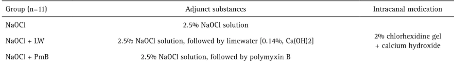
During the preparation of cervical and middle root canal thirds, 5 mL of 2.5% NaOCl solution were used as irrigant and renewed at each change of instrument. The working length (1 mm shorter than the radiographic apex length) was calculated, taking into consideration the tooth radiograph after introduction of a file into the root canal. Apical preparation (0.5-1 mm shorter than the radiographic apex) was performed with 4 K-files. Then, teeth were then divided into 3 groups (n = 11), according to the combination of adjunct substances used during apical root canal preparation (Table 1): NaOCl + LW: 2.5% NaOCl + calcium hydroxide solution (0.14%, limewater); NaOCl + PmB: 2.5% NaOCl + 10.000 UI/mL polymyxin B, and 2.5% NaOCl (control).
In group NaOCl + LW, 5 mL of 2.5% NaOCl was used for the two first files for apical preparation. Next, 5 mL of limewater was used as irrigation agent for the two last files. In group NaOCl + PmB, 5 mL of 2.5% NaOCl was used for the two first files for apical preparation. Next, polymyxin B (Ophtalmos Fórmulas Oficiais Ltda, São Paulo, SP, Brazil) was applied during the two last files, followed by irrigation with 5 mL of apyrogenic saline at each file change.
Limewater and P
olymyxin B in root canals
At the end of biomechanical preparation, all root canals were irrigated with 5 mL of apyrogenic saline and the second sample was collected (S2). Root canals were then filled with 17% EDTA (Asfer Indústria Química Ltda, São Caetano do Sul, SP, Brazil), which was mixed with a file for 3 minutes and then removed with 5 mL of apyrogenic saline before third sample collection (S3).
The root canals were dried with sterile and apyrogenic absorbent paper points of compatible size and filled with intracanal medication consisting of a paste of 2% chlorhexidine gel (Terapêutica Farmácia de Manipulação, São José dos Campos, SP, Brazil) associated with pro-analysis calcium hydroxide (prepared in equal proportions in volume). The medication was introduced into the root canals with a lentulo spiral run at low speed, and a provisional restoration of glass ionomer cement was placed. The intracanal medication was maintained for 14 days.
After this period, the patients returned. The teeth were isolated and disinfected, according to the protocol described above, and the temporary restoration was removed. Root canals were irrigated with 10 mL of apyrogenic saline to remove intracanal medication, and then, the fourth sample collection (S4) was performed. In order to finish the endodontic treatment, root canals were obturated with gutta-percha points and AH plus cement (Dentsply/ Maillefer SA, Ballaigues, Switzerland) by using the active lateral condensation technique.
LAL assay (KQCL test) - Quantification of Endotoxins
All samples collected (S1, S2, S3 and S4) during endodontic treatment (100 µL) were transferred to polypropylene Eppendorf tubes, containing 900 µL of apyrogenic and sterile water, which were used for quantification of endotoxins.
The kinetic chromogenic limulus amebocyte lysate (LAL) (Lonza, Walkersville, MD, USA) assay was performed in order to quantify endotoxins. Escherichia coli endotoxin was used as standard. A positive control (root canal sample contaminated with a known amount of endotoxin) was included for each sample to determine the presence or absence of interfering agents. For the test, 100 μL of apyrogenic water (reaction blank), 5 standard endotoxin solutions [0.005-50 endotoxin units (EU)/mL], root canal samples, and positive controls [each root canal sample
contaminated with a known concentration of endotoxin (10 EU/mL)] were added to a 96-well apyrogenic microplate. Tests were carried out in quadruplicate. The plate was incubated at 37°C ± 1°C for 10 minutes in the Kinetic-QCL reader, which was coupled to a microcomputer with WinKQCL software (Lonza). Next, 100 μL of the chromogenic reagent was added to each well. After the beginning of the kinetic test, the software continuously monitored the absorbance at 405 nm in each microplate well and automatically calculated the log/log linear correlation between reaction time of each standard and the corresponding endotoxin concentration (3,5,6).
Statistical Analysis
The results were analyzed statistically by Kruskal-Wallis and Dunn tests, adopting a level of significance of 5%.
Results
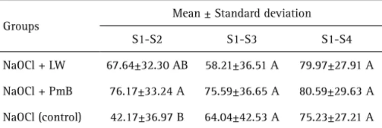
All the samples from first collection (S1) showed presence of endotoxins (33/33 - baseline). It was possible to verify a progressive reduction in endotoxin levels according to the collection (Table 2). The main objective of the endotoxin test was to assess the effectiveness of different irrigants (NaOCl, NaOCl + LW, NaOCl + PmB). The comparison between the reduction percentages observed for each patient after instrumentation (S1 to S2) allowed a better screening of the results obtained for the irrigants analyzed. Descriptive statistics of reduction from S1 to S2, S3 and S4 is presented in Table 3. Kruskal-Wallis test revealed a value for P = 0.0228 (P <.05), which confirms the presence of statistically significant differences between the groups. Application of Dunn’s test identified the difference between groups NaOCl and NaOCl + PmB (Table 3).
Application of EDTA was also evaluated but no difference between the groups was found, which was confirmed by Kruskal-Wallis test for P = 0.1829 (P >.05). In order to evaluate the endodontic treatment by analyzing the effectiveness of intracanal medications for 14 days, it was also considered the percentage of reduction between S1 and S4 for each patient in the study. Kruskal-Wallis test revealed a value of P = 0.4475 (P >.05) for the percentage of reduction between S1 and S4, which no statistically significant difference between the groups. Descriptive
Table 1. Groups treated with different combinations of adjunct substances during root canal preparation
Group (n=11) Adjunct substances Intracanal medication
NaOCl 2.5% NaOCl solution
2% chlorhexidine gel + calcium hydroxide NaOCl + LW 2.5% NaOCl solution, followed by limewater [0.14%, Ca(OH)2]
A.S. Carvalho et al.
statistics of the values and reductions from S1 to S2, S3 and S4 is also presented in Table 3.
Discussion
In this study, the mean reduction from S1 to S2 (after biomechanical preparation) was 42.17% for the group NaOCl; 67.64% for the group NaOCl + LW; and 76.17% for the group NaOCl + PmB. The mean reduction found for the group NaOCl in this study was slightly lower than the values reported by Gomes et al. (19), who observed a reduction percentage of 57.98% of endotoxins after using the same irrigant, and by Martinho and Gomes (20), who found a percentage of reduction of 59.99%. It is possible that the difference in the reduction percentage found in other studies may occur due to the different technical instrumentation used. In those studies, rotary techniques were used, whereas we have used oscillatory system during the endodontic treatment.
These data reveal that 2.5% NaOCl used alone during biomechanical preparation is not very effective against endotoxins present in infected root canals, suggesting that 2.5% NaOCl is not successful for elimination of endotoxins, which remained in the root canal system. Aiming to enhance the effectiveness of the biomechanical preparation, we decided to associate NaOCl to calcium hydroxide solution and polymyxin B, whose action against endotoxins has been demonstrated elsewhere (5,9,21).
The results found for the association NaOCl + LW revealed a reduction percentage of 67.64% from S1 to S2 and 76.17% for the association of NaOCl + PmB, demonstrating the improved effectiveness of irrigation for both associations, even though only the group NaOCl + PmB was significantly different
from NaOCl used alone. Oliveira et al. (5) used limewater and polymyxin B in association with chlorhexidine as irrigants, since the associations showed better results in comparison with chlorhexidine alone. Moreover, the combination of chlorhexidine with limewater was more effective in reducing endotoxins from root canals, indicating that these associations presented the best results.
The use of calcium hydroxide in a solution of apyrogenic water (calcium hydroxide solution) and polymyxin B, used as irrigants, was reported by Oliveira et al. in in vitro (13) and in vivo (5). The authors evaluated the detoxification of endotoxin by using LAL test and production of polyclonal antibodies by B cells. The results revealed that calcium hydroxide and polymyxin B presented an excellent effect on the elimination of endotoxins. Even though a reduction in endotoxin levels was found with the use of associations compared to NaOCl used alone, a small quantity of endotoxin remained in the root canals after biomechanical preparation. The remaining endotoxin probably persisted because calcium hydroxide and polymyxin B, though effective against the LPS, did not penetrate sufficiently into the dentinal tubules to eliminate the remnants present in deepest regions. Safavi and Nichols (11) demonstrated that treatment of LPS with calcium hydroxide releases a high quantity of fatty acids by the hydrolysis of ester bonds in lipid chains of bacterial LPS. Thus, treatment with calcium hydroxide would detoxify LPS, inhibiting its pathological effects (2, 11).
The effectiveness of polymyxin B against endotoxins observed in this study is supported by previous works in the literature (9,13,17,21). Hong et al. (21) reported that the systemic administration of polymyxin B in rats reduced the size of periapical lesions by approximately 80%. According to the author, polymyxin B is a cationic peptide with powerful antioxidant activity. Its molecule bonds to lipid A, negatively charged, altering the three-dimensional conformation of the LPS molecule. In the present study, the polymyxin B associated with 2.5% NaOCl presented the highest percentage of reduction of endotoxin levels (76.17%) from S1 to S2, with statistically significant difference from NaOCl used alone. On the basis of the results currently available for polymyxin B group, it seems fair to suggest that the combined use of NaOCl with this drug may be a viable option for use in root canals in order to obtain greater endotoxin neutralization.
Data gathered in this study demonstrated that Table 3. Descriptive statistics of the percentages of reduction of endotoxin
levels regarding first, second, third and fourth samples found in different experimental groups (NaOCl, NaOCl + LW, NaOCl + PmB)
Groups
Mean ± Standard deviation
S1-S2 S1-S3 S1-S4
NaOCl + LW 67.64±32.30 AB 58.21±36.51 A 79.97±27.91 A
NaOCl + PmB 76.17±33.24 A 75.59±36.65 A 80.59±29.63 A
NaOCl (control) 42.17±36.97 B 64.04±42.53 A 75.23±27.21 A
Different letters indicate statistically significant differences (p<0.05).
Table 2. Levels of endotoxins (mean ± standard deviation, EU/mL) in the root canal samples obtained from different experimental groups
Group S1 S2 S3 S4
NaOCl + LW 1551.26 ± 2726.08 1669.30 ± 168.11 169.75 ± 209.78 85.19 ± 146.64
NaOCl + PmB 1162.69 ± 1319.77 48.23 ± 82.17 37.80 ± 39.34 77.05 ± 174.13
Limewater and P
olymyxin B in root canals
the use of intracanal medication (calcium hydroxide paste associated with 2% chlorhexidine gel) did not increase the neutralization of endotoxins. This medication was selected based on studies that demonstrate its effectiveness against microorganisms and endotoxins (5, 14). This way, in the end of the endodontic treatment (irrigation + intracanal medication), the mean percentages of endotoxin reduction were 75.23% for the group using only NaOCl solution during instrumentation + medication; 79.97% for NaOCl + LW and medication and 80.59% for NaOCl + PmB and medication. It may be noticed that, comparing the percentages obtained after the second collection (S2), the mean reduction obtained after using intracanal medication was greater but not statistically different. It is believed that the use of intracanal medication has adequate antimicrobial action because it remains in contact with dentin and diffuses through the tissues (22). These results show that there is still no technique considered ideal for elimination of infection from the root canal system. In conclusion, biomechanical preparation with the combined use of 2.5% NaOCl + polymyxin B as irrigant presented the greatest reduction of endotoxin levels, followed by 2.5% NaOCl + limewater. Intracanal medication had no significant complementary role in the reduction of endotoxins at the end of the treatment.
Resumo
Este estudo clínico investigou os efeitos do tratamento endodôntico com uso de diferentes irrigantes (NaOCl + água de cal e NaOCl + polimixina B) e medicação intracanal sobre endotoxinas em dentes com infecção endodôntica primária e presença de lesão periapical visível radiograficamente. Foram selecionados para o estudo trinta e três dentes de pacientes que apresentavam necrose pulpar e presença de lesão periapical. As amostras foram coletadas após a abertura coronária (S1) e após a instrumentação (S2). Os canais radiculares foram divididos em 3 grupos (n = 11) de acordo com a combinação de irrigantes utilizada: NaOCl + LW:- hipoclorito de sódio 2,5% + solução de hidróxido de cálcio (água de cal 0,14%); NaOCl + PmB: hipoclorito de sódio a 2,5% + polimixina B 10.000 UI/mL; NaOCl (controle): hipoclorito de sódio a 2,5%. A terceira coleta (S3) foi realizada após aplicação do ácido etilenodiamino tetra acético (EDTA) e a quarta coleta (S4) após 14 dias de medicação intracanal de hidróxido de cálcio + clorexidina gel 2%. Endotoxinas (lipopolissacarídeos) foram quantificadas pelo ensaio cromogênico do lisado de amebócitos de Limulus (LAL). Endotoxinas foram detectadas em todos os canais radiculares após abertura coronária (S1). Grupo NaOCl + PmB apresentou a maior redução de endotoxinas após a instrumentação (76,17%), sendo similar ao grupo NaOCl + LW (67,64%, P >.05) e diferente do grupo NaOCl (42,17%, P <.05). Após o período de medicação intracanal, houve aumento significativo da neutralização de endotoxinas. Concluiu-se que NaOCl + PmB promoveu a maior redução dos níveis de endotoxinas, seguido de NaOCl + LW. A medicação intracanal não teve um papel complementar significativo na redução de endotoxinas no final do tratamento.
References
1. Dahlén G, Bergenholtz G. Endotoxic activity in teeth with necrotic pulps. J Dent Res 1980;59:1033-1040.
2. Nelson-Filho P, Leonardo MR, Silva LA, Assed S. Radiographic evaluation of the effect of endotoxin (LPS) plus calcium hydroxide on apical and periapical tissues of dogs. J Endod 2002;28:694-696.
3. Cardoso FG, Ferreira NS, Martinho FC, Nascimento GG, Manhães LR Jr, Rocco MA, et al. Correlation between volume of apical periodontitis determined by cone-beam computed tomography analysis and endotoxin levels found in primary root canal infection. J Endod 2015;41:1015-1019.
4. Siqueira JR JF, Rôças IN. Bacterial pathogenesis and mediators in apical periodontitis. Braz Dent J 2007;18:267-80.
5. Oliveira LD, Carvalho CA, Carvalho AS, Alves Jde S, Valera MC, Jorge AO. Efficacy of endodontic treatment for endotoxin reduction in primarily infected root canals and evaluation of cytotoxic effects. J Endod 2012;38:1053-1057.
6. Xavier AC, Martinho FC, Chung A, Oliveira LD, Jorge AO, Valera MC, et al. One-visit versus two-visit root canal treatment: effectiveness in the removal of endotoxins and cultivable bacteria. J Endod 2013;39:959-964.
7. Menezes MM, Valera MC, Jorge AO, Koga-ito CY, Camargo CH, Mancini MN. In vitro evaluation of the effectiveness of irrigants and intracanal medicaments on microorganisms within root canals. Int Endod J 2004;37:311-319.
8. Onçag O, Hosgor M, Hilmioglu S, Zekilglu O, Eronat C, Burhanoglu D. Comparison of antibacterial and toxic effects of various root canal irrigants. Int Endod J 2003;36:423-432.
9. Oliveira LD, Jorge AOC, Carvalho CA, Koga-Ito CY, Valera MC. In vitro effects of endodontic irrigants on endotoxins in root canals. Oral Surg Oral Med Oral Pathol Oral Radiol Endod 2007;104:135-142.
10. Leonardo MR, Silva RAB, Assed S, Nelson-Filho P. Importance of bacterial endotoxin (LPS) in endodontics. J Appl Oral Sci 2004;12:93-98.
11. Safavi KE, Nichols FC. Effects of calcium hydroxide on bacterial lipopolysaccharide. J Endod 1993;19:76-79.
12. Silva LAB, Leonardo MR, Assed S, Tanomaru Filho M. Histological study of the effect of some irrigating solutions on bacterial endotoxin in dogs. Braz Dent J 2004;15:109-114.
13. Oliveira LD, Leão MVP, Carvalho CAT, Camargo CH, Valera MC, Jorge AO, et al.. In vitro effects of calcium hydroxide and polymix B on endotoxins in root canals. J Dent 2005;33:107-114.
14. Soares JA, Leonardo MR, Silva LAB, Tanomaru Filho M, Ito IY. Elimination of intracanal infection in dogs’ teeth with induced periapical lesions after rotatory instrumentation: influence of different calcium hydroxide pastes. J Appl Oral Sci 2006;14:172-177.
15. Ferreira NS, Martinho FC, Cardoso FG, Nascimento GG, Carvalho CA, Valera MC. Microbiological profile resistant to different intracanal medications in primary endodontic infections. J Endod 2015;41:824-830.
16. Evans ME, Feola DJ, Rapp RP. Polymyxin B sulfate and a colistin: old antibiotics for emerging multiresistant gram-negative bacteria. Ann Pharmacother 1999;33:960-967.
17. Morrison DC, Jacobs DM. Inhibition of lipopolysaccharide-initiated activation of serum complement by polymyxin B. Infect Immun 1976;13:298-301.
18. Csako G, Elin RJ, Hochstein HD, Tsai CM. Physical and biological properties of U.S. standard endotoxin EC after exposure to ionizing radiation. Infect Immun 1983;41:190-196.
19. Gomes BPFA, Martinho FC, Vianna ME. Comparison of 2.5% sodium hypochlorite and 2% chlorhexidine gel on oral bacterial lipopolysaccharide reduction from primarily infected root canals. J Endod 2009;35:1350-1353.
20. Martinho FC, Gomes BP. Quantification of endotoxins and cultivable bacteria in root canal infection before and after chemomechanical preparation with 2.5% sodium hypochlorite. J Endod 2008;34:268-272. 21. Hong CY, Lin SK, Kok SH, Cheng SJ, Lee MS, Wang TM, et al.. The role of
lipopolysaccharide in infectious bone resorption of periapical lesion. J Oral Pathol Med 2004;33:162-169.
22. Basrani B, Ghanem A, Tjäderhane L. Physical and chemical properties of chlorhexidine and calcium hydroxide-containing medications. J Endod 2004;30:413-417.