www.bjorl.org
Brazilian Journal of
OtOrhinOlaryngOlOgy
1808-8694/$ - see front matter © 2014 Associação Brasileira de Otorrinolaringologia e Cirurgia Cérvico-Facial. Published by Elsevier Editora Ltda. All rights reserved.
http://dx.doi.org/10.1016/j.bjorl.2013.08.001
ORIGINAL ARTICLE
Middle ear packing materials: comparison between absorbable
hemostatic gelatine sponge and sugarcane biopolymer sponge
in rats
Débora Lopes Bunzen
a,*, Nathalia Lins
b, Mariana de Carvalho Leal
a,
Mariana Montenegro de Melo Lira
c, Silvio da Silva Caldas Neto
aa Otorhinolaryngology Service, Universidade Federal de Pernambuco (UFPE), Recife, PE, Brazil b Centre of Experimental Research, Universidade Federal de Pernambuco (UFPE), Recife, PE, Brazil
c Department of Pathology, Hospital das Clínicas, Universidade Federal de Pernambuco (UFPE), Recife, PE, Brazil
Received 15 October 2013; accepted 14 January 2014
KEYWORDS
Biocompatible materials; Otologic surgical proce-dures;
Gelatine sponge, absor-bable;
Biopolymers
Abstract
Introduction: Several biomaterials can be used in ear surgery to pack the middle ear or sup-port the graft. The absorbable gelatin sponge is the most widely used, but it may produce
ibrosis and impair ventilation of the middle ear.
Objective: This experimental study aimed to investigate the inlammatory effects of the
sugarcane biopolymer sponge (BP) in the rat middle ear compared with absorbable gelatin sponge (AGS).
Materials and methods: Prospective experimental study design. Thirty adult female Wistar rats were allocated to receive the BP sponge into the right ear and AGS into the left ear. Animals were randomly killed at 4 and 12 weeks post-procedure. Qualitative histological
assessments were performed to evaluate the inlammatory reaction in the tympanic bullae. Results: The BP sponge caused inlammation more intense and persistent than AGS. The BP
was not absorbed during the experiment. Fibrosis was observed only in the ears with AGS. There were thickening of the mucosa and neoangiogenesis in the group of AGS.
Conclusion: Despite inlammation, the BP sponge produced less ibrosis and neoangiogenesis com -pared to AGS. The sponge BP appeared to be a non-absorbable biomaterial in the middle ear. © 2014 Associação Brasileira de Otorrinolaringologia e Cirurgia Cérvico-Facial. Published by Elsevier Editora Ltda. All rights reserved.
PALAVRAS-CHAVE
Materiais biocompatíveis; Prática diária do otorri-nolaringologista; Esponja de gelatina absorvível;
Biopolímeros
Materiais de suporte na orelha média: comparação entre a esponja de gelatina absorvível e a esponja do biopolímero da cana-de-açúcar em ratos
Resumo
Introdução: Existem diversos biomateriais que podem ser utilizados na cirurgia otológica para preencher a cavidade da orelha média ou dar suporte a enxertos. A esponja de gela-tina absorvível é a mais utilizada, mas pode provocar fibrose e prejudicar a ventilação da orelha média.
Please cite this article as: Bunzen DL, Lins N, Leal MC, Lira MMM, Caldas Neto SS. Middle ear packing materials: comparison between absorbable hemostatic gelatine sponge and sugarcane biopolymer sponge in rats. Braz J Otorhinolaryngol. 2014;80:237-44.
* Corresponding author.
Introduction
Tympanoplasty and tympanomastoidectomy are common otologic surgical procedures in ENT practices. In most of the techniques used, besides the graft material for tympanic membrane (TM) perforation closure, it is necessary to use a supporting or packing material in the cavity of the midd-le ear. The support material must be safe for the patient, biocompatible and should not cause any mucosal reaction, which could compromise middle ear ventilation.1 Ideally it should be conformable to the tympanic cavity and maintain the graft stability long enough for healing.1 After healing, the material may or may not be absorbed by the body.
Several types of support materials may be used in tympanoplasty and tympanomastoidectomy procedures. There are nonabsorbable materials (such as silicone) that require revision surgery to be removed. Absorbable mate-rials can be manufactured from hyaluronic acid, synthet-ic materials, alternative materials (plants) or gelatine. Worldwide, the material most commonly used to provide stability to the tympanic graft is a hemostatic absorbable gelatine sponge, derived from pig dermis. However, stud-ies in the medical literature challenge their use in otology. Signiicant submucosal ibrosis of the middle ear of rats was found in contact areas with this hemostatic sponge.2 One study compared three substances: Nasopore® (poly-urethane membrane), Sepragel® (hyaluronic acid polymer) and Gelfoam® (hemostatic absorbable sponge) that were inoculated into the tympanic bullae of rats. After 3 days, an increased inlammatory reaction was noted in the Gel -foam® group, compared to the other groups; and after 20 days there was a greater degree of subepithelial thicken-ing and ibrosis in this group.3
The middle ear mucosa is very reactive. Tonnaer et al. inoculated various substances (bacteria, hemocyanin, char-coal or saline) in the tympanic bulla of rats by a transtym-panic route.4 The authors concluded that they could pro-voke an acute otitis media with any of the tested substances (except saline) by contact of the substance with the middle ear mucosa.4 The dificulty in inding a biocompatible mate -rial that would cause minimal damage to mucosa has stimu-lated the search for new biomaterials.
The sugarcane biopolymer is a macromolecule pro-duced by the bacterium Zoogloea sp. when this organism is grown in a culture medium rich in molasses from sugar-cane.5 It has been shown to be biocompatible in membra-nous form in several studies conducted in different sites.6 Silva et al.7 conducted an experimental study using the
biopolymer as a tympanic graft ixed on the external surface of TM perforation, and noted the closure of the tympanic perforation in most cases. Mayer et al.8 studied
the inlammatory reaction of the sugarcane biopolymer membrane in the middle ear of rats. These authors noted the presence of exudate and mucosal thickening that re-gressed over time.
One form of the sugarcane biopolymer is a non-po-rous laminar sheet that was experimentally evaluated to replace or ix in place the graft in TM perforation re -pair procedures. Another form is a sugarcane biopolymer sponge that is porous and dense. In contact with water, this biomaterial expands only slightly and becomes crum-bly over time. This type of material could be used as sup-port for the graft or even as packing material for the tympanic cavity and ear canal. The present study aims to evaluate the inlammatory reaction of the sponge form of the sugarcane biopolymer when it contacts the mid-dle ear mucosa, and to compare it with that of absorb-able gelatine sponge, marketed as Gelfoam®. The early
inlammatory response will be analyzed through the char -acterization of the exudate and submucosal edema. The delayed inlammatory response will also be assessed by evaluating for neoangiogenesis and ibrosis, as well as any chronic exudate.
Materials and methods
In the present study, 30 healthy Wistar female albino rats (Rattus norvegicus albinus), weighing 211-290 g (mean 247.25 g), about four months old, were used. We excluded from the study any rat with an abnormality of the middle ear or the tympanic membrane (perforation or extensive myringosclerosis).
The selected animals were operated and followed-up at
Objetivo: Investigar os efeitos da reação inflamatória provocada pela esponja do bio-polímero da cana-de-açúcar (BP) comparada a esponja de gelatina absorvível (EGA) na mucosa da orelha média de ratos.
Materiais e métodos: Estudo experimental prospectivo. A esponja do BP foi implantada na orelha direita e a EGA na orelha esquerda de 30 ratos Wistar fêmeas. Os animais foram sacrificados com 4 e 12 semanas após o procedimento. Avaliação histológica qualitativa foi realizada para verificar a reação inflamatória na bula timpânica.
Resultados: A esponja do BP provocou exsudato inflamatório mais intenso e persistente que a EGA. O BP não foi absorvido durante o tempo de observação. Traves de fibrose foram observadas apenas nos ouvidos com a EGA. Houve espessamento da mucosa e neo-angiogênese no grupo da EGA.
Conclusão: Apesar da reação inflamatória, a esponja do BP provocou menos fibrose e neoangiogênese quando comparada a EGA. A esponja do BP comportou-se como um bio-material não-absorvível na orelha média.
the Center of Experimental Surgery. The animals were kept in a vivarium with constantly controlled temperature (22 ± 2°C) in collective cages with sawdust in its loor with ive animals before the experiment, and in individual cages af-ter the experiment. The animals were under illumination for 12 hours/day. The rats were fed ad libitum with indus-trialized ration (Labina®).
Study model
This is a prospective, controlled, analytical, experimental study.
Procedures
For the procedure, the rats underwent general anesthesia, using ketamine chlorydrate (5 mg/100 g body weight) IM, xylazine chlorydrate (2 mg/100 g body weight) IM, and atro-pine (0.16 ml/100 g body weight) SC. Antibiotic prophylaxis was administered with cephalothin (1.3 mg/100 g body wei-ght) IM.
Arbitrarily, all right ears were allocated to the sugarcane sponge biopolymer (BP) group, and all left ears of the same rats formed the absorbable gelatine sponge, Gelfoam® (GF) group.
All animals underwent otomicroscopy with a surgical microscope (MC-M31 column microscope, DF Vasconcelos®).
After otomicroscopy with magniication ×10, the included rats underwent ear surgery.
Ear surgery
The surgical procedure was performed on a surgical tab-le with comptab-lete asepsis and antiseptic precautions. Af-ter anesthesia and shaving the retroauricular and cervical regions, a ventrolateral 2 cm incision was performed with a #15 scalpel blade 0.5 cm from the external ear oriice (Fig. 1). Then, the muscle plane was retracted to avoid unnecessary trauma, thus allowing the visualization of the tendon of insertion of the digastric muscle at the base of the skull. In this position, the posterior portion of the tym-panic bulla is deeply located (Fig. 2). After the retroauricu-lar exposure, the tympanic bulla was perforated gently with the tip of a needle 40×1.20 mm. Then, with the alligator forceps, the corresponding material was introduced. In the same rat, the biopolymer was placed on the right side, and Gelfoam® on the left side. The size of the biomaterials was
standardized (5 mm long × 1 mm thick, Fig. 3). After the in -troduction of the biomaterials, the muscle layer was reposi-tioned, and the skin was sutured with 4-0 nylon. All surgery procedures were performed by the same investigator to avoid variations in technique. After 15 days, the rat skin na-turally expels the suture, obviating the need to remove it.
The operated rats were subdivided in two subgroups by random allotment: T1 time – rats sacriiced at 4 weeks, and T2 time – rats sacriiced 12 weeks after surgery. At these time intervals the animals were euthanized painlessly by in-traperitoneal administration of sodium thiopental followed by a lethal dose of barbiturate by intracardiac injection. After euthanasia, their tympanic bullae were removed for study.
Figure 1 Wistar rat in lateral decubitus. Surgical incision in the retroauricular region of the left ear. A, anterior; P, posterior.
Figure 2 Exposure of the location of the tympanic bulla, deep to the cervical muscles on the left side of the rat. A, anterior; P, posterior; S, superior; I, inferior.
Preparation and histological analysis
The tympanic bullae were isolated and prepared for histo-logical study. The ixation was performed in 10% buffered formalin and decalciication in 5% nitric acid for 24 hours. In order to improve ixation, a 2-mm hole was made in the posterior part of the tympanic bulla for introduction of the buffered formalin. The material was dehydrated in an increasing ethanol series of 70%, 80%, 90% and 100%, one hour each, and then subjected to a diaphanization pro-cess in xylene, and inally embedded in parafin. With the specimens already embedded in blocks of parafin, 5-µm slices were obtained in a microtome (Spencer AO) with a 50-micron interval. The slices were made in a transverse plane to the TM.
Then the material was stained with haematoxylin-eo-sin (HE). After the proceshaematoxylin-eo-sing of all slides, we selected those whose section was situated at the level of the end of the malleus handle, corresponding to mesotympanum, and whose section was situated at the level of the long process of the incus, corresponding to epitympanum. The selected sections were subjected to qualitative analysis by an experienced pathologist. In the analysis, the mor-phology of the mucosa and TM of the bullae, type and in-tensity of the inlammatory reaction, presence of ibrosis, and behavior of biopolymer, compared to Gelfoam, were observed.
The description of histological indings was subdivid -ed according to the following parameters: TM or mucosal thickness, cell type in the inlammatory iniltrate, and sub -mucosal neovascularization and ibrosis.
The TM thickness was rated as absent, mild, moder-ate or severe. The intensity of inlammation was described based on the observation of exudate cellularity and extent of the process through the tympanic bulla cavity. The in-tensity of the inlammatory activity was rated as follows: null – absence of inlammatory signals; mild – exudate with little cell iniltration, with a reaction involving up to 1/3 of the tympanic bulla lumen (epitympanum or mesotym-panum); moderate – exudate with moderate cell iniltra -tion, with a reaction involving between 1/3 and 2/3 of the bulla lumen (epitympanum and mesotympanum); intense – exudate with extensive cell iniltration, with a reaction involving more than 2/3 of the bulla accompanied by signs of necrosis.
The inflammatory infiltrate was also analyzed for cellularity. If there were more lymphocytes and plasma cells, it was rated as chronic and lymphomononuclear (LMN). If there was greater number of neutrophils, the infiltrate was rated as acute and polymorphonuclear (PMN). If there was as much LMN as PLM, it was consid-ered as a subacute exudate.
Neoangiogenesis occurs from pre-existing vessels. In a late phase of inlammation, there is vasodilatation and increased vascular permeability and degradation of the basement membrane with endothelial cell migration to-ward the angiogenic stimulus. The presence or absence of this behaviour of mucosal inlammation was evaluated.
Presence of fibrosis is also part of the late phase of inflammation, and was characterized by presence of fi-broblasts and collagen deposition in the extracellular matrix. The subepithelial fibrosis in the bulla was rated
as mild, moderate or severe, depending on the exten-sion of the process into the tympanic bulla.
In assessing the degree of absorption of the material into the tympanic bulla, we considered whether there was any sign of persistence of the material in the slides investigated.
Statistical analysis
The data obtained were categorized and presented descrip-tively and analytically. All data were grouped into tables. In the comparative statistical analysis, we checked whether there were differences between biomaterials. We also com-pared data between T1 and T2 times for each group sepa-rately. The Fisher’s exact test was used due to the small number of observations. Results whose descriptive levels (p values) were <0.05 were considered statistically signiicant. The statistical calculations were performed using SPSS sof-tware for Windows version 18.0 – Statistical Package for the Social Sciences.
Ethical considerations
This study followed the principles governing the Code of Ethics and the experimental animal protection laws, according to the abiding principles in Brazil, especially Law No. 9,605 – art. 32, and Decree No. 3,179 - art. 17, dated September 21, 1999, which deal with the issue of using animals for scientiic purposes. Furthermore, the study had full approval from the Ethics Committee for Animal Use, and was registered under protocol No. 23076.020776/2010-48, in agreement with the fact that the death of the animals used in this study is justiied, because there are no alternative resources for the rea-lization of our scientiic procedure. The use of general anesthesia before any procedure aimed to avoid pain and reduce animal stress. During the experiment, the experi-mental animals were followed-up by veterinary oficers of the Centre for Experimental Surgery.
Results
Of the total of 30 rats in the study, 15 rats were randomly selected for T1, and 15 for T2 group. In the T1 group, two animals died due to anesthetic complications during surgery. Of the 13 operated rats, 6 were lostdue to pro-blems such as bilateral secondary infection or failure of the histotechnical processing. Of the 7 remaining rats , two had unilateral otitis (left side) and remained in the study. Thus, at the end of the first month of observation of the experiment, we obtained 7 right ears (BP group) and 5 left ears (GF group).
The description of the histological findings was sub-divided according to the following parameters: TM and mucosal thickness, cell type in the inflammatory infil-trate, neovascularization and severity of the submu-cosal fibrosis. To characterize the sample studied, we present in tables 1 and 2 the absolute (N) and relative (percentage) frequencies of classes in each qualitative variable.
To compare the groups with respect to the histolog-ical findings, we applied the Fisher’s exact test, due to the small number of observations. For the statistical analysis, the histological results rated as absent or mild were pooled and compared to the pooled outcome of the data rated as moderate or severe.
Tables 3 and 4 list the statistical analysis of the data. These tables show a comparison between the biomaterials.
There was no statistical difference between the ana-lyzed parameters when comparing the results in T1 and T2 only for BP group, as well as when this comparison were performed in T1 and T2 for GF group.
Regarding the presence of the implant after 3 months of the experiment, BP was seen in all the tympanic bul-lae. GF was seen in only one tympanic bulla. Bone neo-formation was observed in two bullae with GF and in three bullae with BP.
The fibrogenic response pattern was different be-tween the two groups. In BP implants, fibrosis, when ob-served, was mild in most cases. On the other hand, some cases in the GF implants developed, fibrous adhesions, with the air spaces fully occupied. These adhesions were observed in this group both at one and three months af-ter the surgery.
Table 1 Histological indings in the tympanic bulla of rats after one month of the experiment (T1).
Histological
findings Muc. Th. TM. Th. LMN PMN Neoangio Fibrosis BP (n = 7)
Absent 1 (14.28%) 3 (42.86%) - - 1 (14.28%) -Light 3 (42.86%) 4 (57.14%) 1 (14.28%) 1 (14.28%) 6 (85.71%) 5 (71.43%)
Moderate - - 4 (57.14%) 3 (42.86%) - 2 (28.57%)
Intense 3 (42.86%) - 2 (28.57%) 3 (42.86%) - -GF (n = 5)
Absent 2 (40.0%) 3 (60.0%) - 5 (100%) - -Light 1 (20.0%) - 4 (80.0%) - 4 (80.0%) 1 (20.0%)
Moderate 1 (20.0%) 1 (20.0%) 1 (20.0%) - 1 (20.0%) 3 (60.0%)
Intense 1 (20.0%) 1 (20.0%) - - - 1 (20.0%)
BP, biopolymer; GF, Gelfoam®; Muc.Th., mucosal thickening; TM.Th., tympanic membrane thickening; LMN, lymphmononuclear
infiltrate; PMN, polymorphonuclear infiltrate; Neoangio, neoangiogenesis.
Table 2 Histological indings in the tympanic bulla of rats after 3 months of the experiment (T2).
Histological
findings Muc. Th. TM. Th. LMN PMN Neoangio Fibrosis BP (n = 15)
Absent 1 (6.66%) 8 (53.33%) - - 5 (33.33%) 1 (6.66%)
Light 5 (33.33%) 5 (33.33%) - 1 (6.66%) 8 (53.33%) 8 (53.33%)
Moderate 4 (26.66%) 1 (6.66%) 12 (80.0%) 6 (40.0%) 2 (13.33%) 5 (33.33%)
Intense 5 (33.33%) 1 (6.66%) 3 (20.0%) 8 (53.33%) - 1 (6.66%)
GF (n = 11)
Absent 1 (9.09%) 5 (45.45%) - 9 (81.81%) - -Light 1 (9.09%) 3 (27.27%) 7 (63.63%) 1 (9.09%) 3 (27.27%) 4 (36.36%)
Moderate 3 (27.27%) 3 (27.27%) 4 (36.36%) - 7 (63.63%) 3 (27.27%)
Intense 6 (54.54%) - - 1 (9.09%) 1 (9.09%) 4 (36.36%)
BP, biopolymer; GF, Gelfoam®; Muc.Th., mucosal thickening; TM.Th., tympanic membrane thickening; LMN, lymphmononuclear
Discussion
The use of biomaterials in otologic surgery is a common practice in otorhinolaryngology. After the introduction of packing materials into the tympanic cavity in the 50s by Zollner and Wullstein, the use of biomaterials became po-pular.9,10 Depending on the indication and surgical techni-que proposed to the patient, materials were developed to be used as implants in the replacement of the ossicles of the middle ear, oval window sealants, hemostasis materials, ventilation tubes, support materials for graft and packing material for tympanic cavity or external ear canal.1 The su-pport and packing materials are indicated in most tympano-plasty and mastoidectomy procedures.
Currently, Gelfoam® is the absorbable biomaterial most used in otologic surgery. Initially this sponge was used as a hemostatic agent in neurosurgery. The expandability of the material in contact with luid and its absorption by the body in the medium term resulted in Gelfoam being used to occupy space in various surgical areas. In 1951, Blaine11 described its use not only as a hemostatic agent, but also as a replacement of destroyed tissues. Since the 50s, Gel-foam® has been used in various otologic operations, but, since the 1960s, some studies noted deleterious effects on the middle ear. Schuknecht12,13 in two reports, documented the occurrence of hearing loss after stapedectomy surgery and concluded that this failure was secondary to the use of Gelfoam® in the oval window.
The middle ear mucosa and TM, when in contact with some harmful material, suffer subepithelial oedema and hyperplasia, evidenced by histological thickening. In the present study, we observed, both in T1 and T2 of our ex-periment, that none of the biomaterials tested caused a signiicant TM thickening. Laurent et al.14 also observed no changes in TM in experimental surgeries using GF.
The tympanic membrane is composed of three layers, and its innermost layer is contiguous with the tympanic bulla mu-cosa, being subject to the same injuries and with a similar inlammatory response. Mayer et al.8 observed a signiicant TM thickening after inoculation of material through a perfo-rated eardrum. The authors noted that this inding resulted not only from the presence of the material, but also from the handling of TM itself, and concluded that the methodology for application of the material in the experimental surgery of the tympanic bulla may inluence the results.8 Therefore, our study used the retrobullar route, already described by other authors, which causes no damage to TM.
In our study, although not statistically different, the GF group in T2 had the highest percentage of severe mucosal thickening in the tympanic bulla (81.85%). This trend for a more intense mucosal thickening secondary to GF use was expected, since other experimental studies have reported it previously. Krupala et al.15 found a thickening with
mu-cosal inlammation in 9 of 10 animals in which GF was used as support material. Jang et al.16 found signiicant mucosal thickening, when compared GF versus Interceed®. In their Table 3 Comparison between groups of biomaterials in
re-lation to histological indings in the tympanic bulla of rats
after 1 month of experiment (T1). Histological
findings
Group – T1
P value BP (n = 7) GF (n = 5)
Muc. Th.
Absent/Light 4 (57.1%) 3 (60.0%) > 0.999 Moderate/Severe 3 (42.9%) 2 (40.0%)
TM. Th.
Absent/Light 7(100%) 3 (60.0%) 0.152 Moderate/Severe 0(0%) 2 (40.0%)
LMN
Absent/Light 1(14.3%) 4 (80.0%) 0.062 Moderate/Severe 6 (85.7%) 1 (20.0%)
PMN
Absent/Light 1(14.3%) 5 (100%) 0.015a
Moderate/Severe 6 (85.7%) 0 (0%)
Neoangio
Absent/Light 7(100%) 4 (80.0%) 0.417 Moderate/Severe 0 (0%) 1 (20.0%)
Fibrosis
Absent/Light 5 (71.4%) 1 (20.0%) 0.234 Moderate/Severe 2(28.6%) 4 (80.0%)
a Statistically significant (p < 0.05).
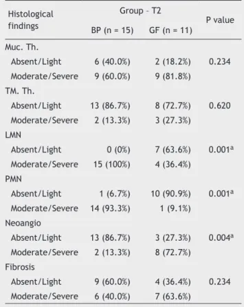
Table 4 Comparison between groups of biomaterials in relation to histological findings in the tympanic bulla of rats after 3 months of experiment (T2).
Histological findings
Group – T2
P value BP (n = 15) GF (n = 11)
Muc. Th.
Absent/Light 6 (40.0%) 2 (18.2%) 0.234 Moderate/Severe 9 (60.0%) 9 (81.8%)
TM. Th.
Absent/Light 13 (86.7%) 8 (72.7%) 0.620 Moderate/Severe 2 (13.3%) 3 (27.3%)
LMN
Absent/Light 0 (0%) 7 (63.6%) 0.001a
Moderate/Severe 15 (100%) 4 (36.4%)
PMN
Absent/Light 1 (6.7%) 10 (90.9%) 0.001a
Moderate/Severe 14 (93.3%) 1 (9.1%)
Neoangio
Absent/Light 13 (86.7%) 3 (27.3%) 0.004a
Moderate/Severe 2 (13.3%) 8 (72.7%)
Fibrosis
Absent/Light 9 (60.0%) 4 (36.4%) 0.234 Moderate/Severe 6 (40.0%) 7 (63.6%)
study, the authors performed myringotomy procedures with scariication of the mucosa before the procedure; and after 3 weeks, the histological indings revealed adhesions and mucosal thickening in 7 of 10 ears with the use of GF.16 After eight weeks of observation, Bahadir et al.17 found moderate mucosal thickening in rats treated with inoculation of Gel-foam per transtympanic route. When a scariication of the mucosa in contact with the GF was performed, the authors found a much more exuberant mucosal thickening; 6 rats exhibited a moderate, and 4 a severe, grade of thickening in a total of 10 animals.16
An inlammatory iniltrate was present in all cases, but with a difference regarding to the cellularity of the process. In ears packed with BP, a greater inlammatory reaction was noted, with the presence of both polymorphonuclear (PMN) and lymphocytic (LMN) iniltration. The PMN iniltrate was present around the BP sponge, and the LMN iniltrate in the underlying mucosa. On the other hand, in the ears packed with GF, the LMN iniltrate represented most cases and there was no PNM exudate, with a statistically signiicant difference between biomaterials.
The amount of exudate was also signiicantly different between groups, since 85.7% of the BP exudate were consid -ered of moderate to severe at the beginning of the experi-ment, unlike GF, which caused mild or absent inlammatory reaction in most cases. The inlammatory reaction caused by BP sponge remained different from GF throughout the experiment. In T2, table 4 shows that in most cases BP main-tained a moderate to intense inlammatory response, both in PMN as in LMN exudate, while the group of ears treated with PG showed mild or no reaction, with statistically signif-icant difference. There was no change in inlammatory in -tensity over time; BP group remained with the most intense reaction and GF group with the lightest reaction.
In other studies, BP also induced an important inlam -matory response. In the form of the membrane used in the tympanic bulla of rats, BP led to the formation of a sub-acute exudate, rated as moderate to severe in 33.3% of cases.8 As vascular replacement, BP induced inlammatory reaction with the presence of neutrophils, lymphocytes, and ibrosis.18 In subaponeurotic tissue, BP was tested as a pubovaginal sling.19 Severe inlammatory reaction with the presence of PMN and giant cells around the biomaterial was reported.19 In the present study, the inlammatory iniltrate proved important, especially in terms of permeating the sponge; in the bulla region, contralateral to BP, there was no exudate. Finally, it remains unknown to what extent this persistent inlammatory response may impair the ventila -tion and the scarring process of the tympanic bulla.
The degradation of BP was slower than GF’s: this also contributed to the maintenance of the immune stimulus. The chemical composition of BP was deined as a cellulose polysaccharide composed of different monomeric bases, namely: glucose 87.57%, xylose 8.58%, ribose 1.68%, glucu -ronic acid 0.83%, mannose 0.82%, arabinose 0.37%, galac -tose 0.13%, fucose 0.01%, and rhamnose 0.01%.5 Although, in theoretical terms, all these elements are easily degraded in the body, the persistence of an intense inlammatory re -action and the continued presence of BP sponge 3 months after surgery in in vivo experiments demonstrate a a level of inlamatory response not observed in the sponge derived from pig dermis. One possibility could be that BP is of plant
origin, in contrast to the animal origin of GF, or perhaps there is some immunological dificulty of the rodent species. The persistence of BP in the body may have applicability in other situations; this product may become a non-absorbable support material. Future biomolecular studies could be per-formed to better assess this property.
Despite the presence of a persistent inlammatory exu -date, the BP sponge does not seem to induce neovascular-ization and ibrosis in the same proportion as the GF sponge. Table 4 shows that neovascularization was absent or mild in 86.7% in the tympanic bullae with BP, but was moderate to severe in 72.7% in the GF group, a statistically signiicant difference. The neovascularization occurs as an endothelial proliferation in the presence of an inlammatory stimulus. The immune reaction caused by GF allowed the vessels to dilate with the formation of new vascular meshes, which probably point to an organization of the inlammatory pro -cess. This intense neovascularization in ears treated with GF may be related to a higher rate of ibrosis at a later time. After all, in our result and in the literature, there is a greater tendency to ibrosis in the middle ear exposed to GF.
In the descriptive results, we noted not only the sub-mucosal ibrosis of the GF group, but also trabeculae of ibrosis crossing the cavity of the tympanic bulla. In the 1980s, otologists in Sweden found adhesions with trabec-ulae of connective tissue in the middle ear in reoperations of tympanoplasty and in programmed revision mastoideco-my surgeries.20 There was a suspicion that the hemostatic sponge used as support could be the cause of these chang-es. In experimental surgery with rats, Swedish researchers found tympanic membrane retraction with synechiae and ibrosis in the middle ear in the presence of Gelfoam after 2 months.20 These authors also observed new bone formation and submucosal thickening in both the promontory and the hypotympanum after 3 months of experiment.20 Hellstrom et al. concluded that Gelfoam® could be involved in the failure of some otologic surgeries.20
The presence of moderate or severe ibrosis in 80% and 63.6% in T1 and T2, respectively, is consistent with studies in theliterature implanted GF.Rates a high as 90% of cases with moderate to severe ibrosis have been reported.3,16,20-22 In
oth-er studies, 60% of the cases exhibited ibrosis, but thoth-ere are also papers that noted this change in only 20% of cases with GF.2,15,17
BP is a polymer derived from sugarcane, a low cost and abundant biomass in Brazil, capable of changing its natu-ral state to gel, membrane or sponge simply through phys-ical phenomena, without need of adding chemphys-ically active products. BP is biocompatible in vitro; but more studies are needed to ensure a satisfactory end result in ear surgery.
Conclusion
The sugarcane biopolymer sponge caused a more intense inlammatory reaction with exudate, compared to absor -bable gelatine sponge. There was little neovascularization and mild ibrosis in the sugarcane biopolymer sponge group, compared to absorbable gelatine sponge group. Further stu-dies may elucidate whether this behaviour of the sugarcane biopolymer may be useful in otologic operations.
Conlicts of interest
The authors declare no conlicts of interest.
Acknowledgements
To Camila Sartechi, for his help in the statistical analysis of this study. To Adriana Amorim, Med. Vet., by her care for the experimental animals.
References
1. Shen Y, Teh BM, Friedland PL, Eikelboom RH, Atlas MD. To pack or not to pack? A contemporary review of middle ear packing agents. Laryngoscope. 2011;121:1040-8.
2. Liening DA, Lundy L, Silberberg B, Finstuen K. A comparison of the biocompatibility of three absorbable hemostatic agents in the rat middle ear. Otolaryngol Head Neck Surg. 1997;116:454-7.
3. Dogru S, Haholu A, Gungor A, Kucukodaci Z, Cincik H, Ozdemir T, et al. Histologic analysis of the effects of three different su-pport materials within rat middle ear. Otolaryngol Head Neck Surg. 2009;140:177-82.
4. Tonnaer ELGM, Ingels KJAO, Rijkers GT, Curfs JHAJ. Antigenic as well as nonantigenic stimuli induce similar middle ear res-ponses in the rat. Laryngoscope. 2003;113:322-7.
5. Paterson-Beedle M, Kennedy JF, Melo FAD, Lloyd LL M V. A cellu-losic exopolysaccharide produced from sugarcane molasses by a Zoogloea sp. Carbohydrate Polymers. 2000;42:375-83. 6. Assis FD, Melo D, Marques E. Citotoxicidade de biopolímero de
cana-de-açúcar. An Fac Med Univ Fed Pernamb. 2004;49:73-7. 7. Silva DB, Aguiar JLA, Marques A, Coelho A, Rolim Filho EL. Mi-ringoplastia com enxerto livre de membrana de biopolímero de cana-de-açúcar e fáscia autóloga em Chinchilla laniger. An Fac Med Univ Fed Pernamb. 2006;51:45-51.
8. Mayer DLB, Araújo JG de, Leal de C, Caldas Neto S da S, Ataíde RF, Mello RJV de. Membrana do biopolímero da cana-de-açúcar: avaliação experimental na orelha média [Su-garcane biopolymer membrane: experimental evaluation in the middle ear]. Braz J Otorhinolaryngol. 2011;77:44-50. 9. Zollner F. The prognosis of the operative improvement of
hearing in chronic middle ear infections. Ann Otol Rhinol Laryngol.1957;66:907-17.
10. Wullstein H. The restoration of the function of the middle ear, in chronic otitis media. Ann Otol Rhinol Laryngol. 1956;65:1021-41. 11. Blaine G. Absorbable gelatin sponge in experimental surgery.
Lancet. 1951;2:427-9.
12. Schuknecht HF. Sensorineural hearing loss following stapedec-tomy. Acta Otolaryngol. 1962; 54: 336-40.
13. Schuknecht HF. Gelfoam as an implant in oval window following stapedectomy. Ann Otol Rhinol Laryngol. 1971;80:415-8. 14. Laurent C, Hellström S, Stenfors LE. Hyaluronic acid reduces
connective tissue formation in middle ears illed with absorba -ble gelatin sponge: an experimental study. Am J Otolaryngol. 1986;7:181-6.
15. Krupala JL, Gianoli GJ, Smith RA. The eficacy of hyaluronic
acid foam as a middle ear packing agent in experimental tym-panoplasty. Am J Otol. 1998;19:546-50.
16. Jang CH, Park H, Cho YB, Choi CH. The effect of Interceed for reducing adhesion as a middle ear packing agent: an experi-mental study. Int J Pediatr Otorhinolaryngol. 2008;72:1517-21. 17. Bahadir O, Aydin S, Caylan R. The effect on the middle-ear
cavity of an absorbable gelatine sponge alone and with corti-costeroids. Eur Arch Otorhinolaryngol. 2003;260:19-23. 18. Aguiar JLA, Lins EM, Marques SRB, Coelho ARB, Rossiter RO,
Melo RJV. Surgarcane biopolymer patch in femoral artery an-gioplasty on dogs. Acta Cir Bras. 2007;22:77-81.
19. Lucena R. Utilização do biopolímero da cana-de-açúcar como novo material para sling pubovaginal: análise estereológica. Recife: Universidade Federal de Pernambuco. Tese. Recife: Universidade Federal de Pernambuco, Centro de Ciências da Saúde; 2007.
20. Hellström S, Salén B, Stenfors LE. Absorbable gelatin sponge (Gelfoam) in otosurgery: one cause of undesirable postoperati-ve results? Acta Otolaryngol. 1983;96:269-75.
21. McGhee MA, Dornhoffer JL. The effect of gelilm in the pre
-vention of ibrosis in the middle ear of the animal model. Am J
Otol. 1999;20:712-6.
22. Huang G, Chen X, Jiang H. Effects of NasoPore packing in the middle ear cavity of the guinea pig. Otolaryngol Head Neck Surg. 2011;145:131-6.
23. Loffroy R, Guiu B, Cercueil J-P, Krausé D. Endovascular thera-peutic embolisation: an overview of occluding agents and their effects on embolised tissues. Curr Vasc Pharmacol. 2009;7:250-63.
24. Park AH, Jackson A, Hunter L, McGill L, Simonsen SE, Alder SC, et al. Cross-linked hydrogels for middle ear packing. Otol Neurotol. 2001;27:1170-5.
25. Li G, Feghali JG, Dinces E, McElveen J, Van de Water TR.
Evaluation of esteriied hyaluronic acid as middle ear-packing