www.bjorl.org.br
Brazilian Journal of
OtOrhinOlaryngOlOgy
1808-8694/$ - see front matter © 2014 Associação Brasileira de Otorrinolaringologia e Cirurgia Cérvico-Facial. Published by Elsevier Editora Ltda. All rights reserved.
http://dx.doi.org/10.1016/j.bjorl.2014.02.003
ORIGINAL ARTICLE
Effect of sugarcane biopolymer gel injected in rabbit vocal fold
Rodrigo Augusto de Souza Leão
a,b,*,Raquel Coelho de Assis
c, Silvio da Silva Caldas Neto
a,d,
Mariana Montenegro de Melo Lira
a,e,f, Silvio José de Vasconcelos
d,ba Universidade Federal de Pernambuco (UFPE), Recife, PE, Brazil
b Service in Otorhinolaryngology, Hospital Agamenom Magalhães, Recife, PE, Brazil c Hospital Agamenon Magalhães, Recife, PE, Brazil
d Universidade de São Paulo (USP), São Paulo, SP, Brazil
e Universidade Estadual de Campinas (UNICAMP), Campinas, SP, Brazil
f Hospital das Clínicas, Universidade Federal de Pernambuco (UFPE), Recife, PE, Brazil
Received 8 March 2013; accepted 9 February 2014
KEYWORDS
Vocal cords; Biopolymers; Larynx
Abstract
Introduction: Alterations in the vocal folds that involve volume reduction and glottal closure failure result in exaggerated air escape during speech. For such situations, the use of implants or grafts of different materials has been proposed.
Objective: To deine the effect of sugarcane biopolymer gel when implanted in the vocal folds of rabbits.
Methods: This was an experimental study. The vocal folds of rabbits injected with sugarcane biopolymer and saline solution were histologically evaluated after 21 and 90 days.
Results: Mild to moderate inlammation and increased volume were observed in all vocal folds in -jected with biopolymer, when compared to controls. There were no cases of necrosis or calciication. Discussion: This study showed higher inlammatory reaction in cases than in controls and bio -polymer biointegration to the vocal fold. This ibrogenic response with absence of epithelial repercussions suggests that the biopolymer in its gel form can be bioactive and preserve the normal vibratory function of the epithelium.
Conclusion: We show that in spite of producing an inlammatory reaction in vocal fold tissues, the material remained in vocal fold throughout the study period.
© 2014 Associação Brasileira de Otorrinolaringologia e Cirurgia Cérvico-Facial. Published by Elsevier Editora Ltda. All rights reserved.
PALAVRAS-CHAVE
Pregas vocais; Biopolímeros; Laringe
Comportamento do biopolímero da cana-de-açúcar em gel injetado em prega vocal de coelho
Resumo
Introdução: As alterações das pregas vocais que envolvem redução do seu volume e falha no fe-chamento glótico causam um escape exagerado de ar durante a fonação. Para essas situações, tem sido proposta a utilização de implantes ou enxertos de materiais diversos.
Objetivo: Deinir o comportamento do biopolímero de cana-de-açúcar implantado nas pregas vocais de coelhos.
Please cite this article as: Leão RAS, Assis RC, Caldas Neto SS, Lira MMM, Vasconcelos SJ. Effect os sugarcane biopolymer gel injected in rabbit vocal fold. Braz J Otorhinolaryngol. 2014;80:220-5.
* Corresponding author.
Introduction
When affected by congenital or acquired anomalies the vo-cal folds can upset glottal closure during phonation; in these circumstances the persistence of a glottal gap of variable size and shape of results in an exaggerated leak of
exha-led air, which alters or prevents vocal fold vibration and al -ters the voice.1-4 In these situations, the use of implants or grafts of different materials have been proposed to increa-se the affected vocal fold volume and thus improve glottal coaptation during phonation.5
The choice and use of the ideal material depends on
several factors, such whether it is easy to obtain, quantity available, low donor morbidity, immunogenic and mutagen -ic characterist-ics, susceptibility to infection, malleability, absorption rate and cost.6
Currently there is a growing interest in the production through fermentation processes, as well as the scientiic
and clinical use of polysaccharides obtained byusing mi-croorganisms capable of converting certain substances into gels, viscous solutions or membranes (biopolymers).7
An exopolysaccharide biopolymer obtained by
fermenta-tion of sugarcane molasses by a bacterium (Zoogloea SP) was
synthesized after 1990.8 Its low cytotoxicity, biocompatibility
and low production cost stimulated research on this material.9
Its use has been widely studied in several research projects in
the reconstruction of different tissues of the human body.9-15 No study had yet been performed to evaluate the possi-bility of using sugarcane biopolymer (SCB) as graft material in the vocal folds.
Objectives
This study aimed to analyze tissue reaction 21 and 90 days after SCB injection in the vocal folds of rabbits, describing
the presence of the material and the inlammatory process, as well as vocal cord thickness.
Methods
This is a prospective randomized case-control experimental study.
A total of 12 adult male California rabbits, ranging
in weight from 1.80 kg to 3.7 kg, with good nutritional
status and median weight of 2.45 kg were selected. The rabbit was chosen because the vocal folds of rabbits are
similar to human vocal folds. The vivisection of the animals
complied with the requirements of Federal Law No. 9605
art. 32 and Decree No. 3179 art. 17 dated from 09/21/1999. The Ethics Committee on Animal Experimentation of the institution recommended full approval, under number 23076.003670/2009-46.
Surgical procedures were performed under general an
-esthesia without tracheal intubation with laryngeal expo
-sure of rabbits by oral route under direct view. One vocal
fold received an injection of 0.1 mL of SCB in gel form (1%
polysaccharide hydrogel). The contralateral vocal fold was submitted to the same procedure with 0.1 mL of saline solu
-tion. There was no technical dificulty in performing the
procedures in rabbits.
For the histological analysis, the larynx was excised and divided into two hemi-larynges. The animals were divided into two temporal groups, each with ive rabbits, randomly selected. Group deinition was carried out by drawing lots after the surgical procedure. The irst group was euthanized
21 days after the procedure (group I) and the second after 90 days (group II).
The rabbits were euthanized with an overdose of the
anesthetic drugs xylazine and ketamine.
The choice of the vocal fold to be submitted to SCB
im-plantation was randomly assigned before the surgery. The biopolymer was inserted between the vocal muscle and la -ryngeal cartilage (Fig. 1). The vocal fold submitted to saline Métodos: Trata-se de um estudo experimental. Avaliaram-se histologicamente as pregas vocais de coelhos injetadas com biopolímero de cana-de-açúcar e solução isiológica após 21 e 90 dias. Resultados: Foi observada a presença do biopolímero, reação inlamatória leve a moderada e aumento de volume em todas as pregas vocais injetadas em relação às de controle. Não houve casos de necrose ou calciicação.
Discussão: Este trabalho mostrou maior reação inlamatória nos casos que os controles além de biointegração do material na prega vocal. Essa resposta ibrogênica com ausência de reper -cussões epiteliais pode nos sugerir que o biopolímero em sua forma de gel pode ser bioativo e preservar as funções vibratórias normais do epitélio.
Conclusão: Neste trabalho, mostramos que apesar de produzir uma reação inlamatória nos
tecidos das pregas vocais o material perdurou na prega vocal durante todo o período de estudo. © 2014 Associação Brasileira de Otorrinolaringologia e Cirurgia Cérvico-Facial. Publicado por Elsevier Editora Ltda. Todos os direitos reservados.
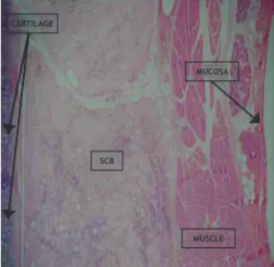
Figure 1 Photomicroscopy of rabbit right vocal fold 3 weeks
after SCB implantation. Note the biopolymer deposits among the skeletal muscle ibers, with no mucosal alterations. (HE staining, 40×)
CARTILAGE
SCB
MUCOSA
solution injection only was considered the control vocal fold. Two animals died; the irst in the immediate postoperative
period and the second during surgery, probably due to deep
anesthesia. Neither of these animals was used in the study. The two hemi-larynges were evaluated by a single exam
-iner, blinded to the group to which they belonged. The sur
-gical specimens were ixed in 10% formalin for 24 hours and three-micrometers thick coronal sections were performed,
going through the membranous portion of the vocal fold.
The sections were stained with hematoxylin-eosin (HE). The inlammatory process intensity in the vocal fold sub
-mitted to biopolymer injection was compared to the con -tralateral vocal fold in the different study periods: 21 days (group I) and 90 days (group II) after the procedure.
A comparative study was performed for the presence of ibrosis, lymphocytic inlammatory iniltrate, polymorpho
-nuclear iniltrate, giant cell predominance, cell types (neu -trophils, mast cells, lymphocytes, plasma cells, eosinophils,
and histiocytes), presence of biopolymers, calciication, ne -crosis and angiogenesis and thickness measurements in its
greatest width in the vocal fold with SCB in relation to the
control fold in both study periods.
The histological variables used for lymphomononuclear
inlammatory iniltrate were absent (0), grade I (< 10% of the area occupied by inlammatory cells), grade II (11%-50%), grade III (> 50%). For polymorphonuclear inlamma
-tory iniltrate: absent (0), grade I (< 10% of the area occu
-pied by inlammatory cells), grade II (11%-50%) and grade III
(> 50%). For giant cells, the mean number of giant cells per
high-power ield was used (magniication × 40).
Qualitative variables for ibrosis were: score 0 (absent), score 1 (mild, rare ibers in up to 10% of the area) score 2
Table 1 Histological characteristics found in Group I - 21 days – Vocal fold with biopolymer.
Rabbit Thickness Polymer LMNII PMNII GC Fibrosis Angiogenesis
1 2.2 + 1 0 13.4 1 1,5
2 3.1 + 2 Eos 1 1.7 0 3,9
4 4.0 + 2 Eos 3 2.6 1 3,7
5 4.0 + 1 0 13.6 1 3,4
10 2.2 + 1 0 64.0 0 2,0
LMNII, lymphomononuclear inflammatory infiltrate (0-3); PMNII, polymorphonuclear inflammatory infiltrate (0-3), Eos, cellular predominance of eosinophils, GC (giant cells), mean number of giant cells per high-power field (40 ×); Fibrosis, intensity of fibrosis observed; Polymer, presence or absence of the biopolymer; Angiogenesis, mean number of vascular spaces per high-power field (40 ×); thickness, measurement of the vocal fold thickness at its point of greatest width.
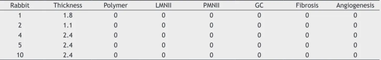
Table 2 Histological characteristics found in Group I - 21 days – Control vocal fold.
Rabbit Thickness Polymer LMNII PMNII GC Fibrosis Angiogenesis
1 1.8 0 0 0 0 0 0
2 1.1 0 0 0 0 0 0
4 2.4 0 0 0 0 0 0
5 2.4 0 0 0 0 0 0
10 2.4 0 0 0 0 0 0
LMNII, lymphomononuclear inflammatory infiltrate (0-3); PMNII, polymorphonuclear inflammatory infiltrate (0-3), Eos, cellular predominance of eosinophils, GC (giant cells), mean number of giant cells per high-power field (40 ×); Fibrosis, intensity of fibrosis observed; Polymer, presence or absence of the biopolymer; Angiogenesis, mean number of vascular spaces per high-power field (40 ×); thickness, measurement of the vocal fold thickness at its point of greatest width.
(moderate, dispersed ibers, young, not modeled, between 10% and 75%) and score 3 (intense, mature ibers, mod
-eled, 75% to 100%). Biopolymer persistence was present or absent; calciication (present or absent), necrosis (present or absent); angiogenesis was evaluated for the mean num
-ber of vascular spaces per high-power ield (magniication
× 40).
Vocal fold thickness was measured from the point of greatest width of the vocal fold from the cartilage to the
epithelial surface, using a ruler overlapping the lamina. To compare the qualitative variables (intensity of the
iniltrate, cell predominance, ibrosis) between the groups, Fisher exact test was used, which tests the homogeneity of the two groups in relation to data distribution of responses
at the different valuated levels. To compare the quantita-tive variables (angiogenesis, giant cells and vocal fold
thick-ness), the Mann-Whitney U-Wilcoxon test was used, which is a nonparametric test for the evaluation of two independent populations when one cannot classify the sample as having
normal distribution.
Results
Tables 1 and 2 show the indings of Group I (21 days). Tables 3 and 4 show the indings in group II (90 days).
All rabbits showed the presence of SCB. No specimen ex
-hibited rejection of the material, necrosis or calciication. The vocal folds injected with saline solution showed no in
When comparing group I (21 days) and group II (90 days)
regarding the presence of ibrosis in the vocal folds with biopolymer, lymphomononuclear inlammatory iniltrate, polymorphonuclear inlammatory iniltrate and the analysis between the predominance of giant cells and angiogenesis between the biopolymer injection groups, there was no sta
-tistically signiicant difference between groups.
Regarding differences in thickness between the implant
-ed and control vocal folds,the difference was statistically signiicant, as shown in Table 5.
Table 3 Histological characteristics found in Group II - 90 days - Vocal fold with biopolymer.
Rabbit Thickness Polymer LMNII PMNII GC Fibrosis Angiogenesis
3 1.3 + 2 Eos 1 0.7 2 5.6
7 2.9 + 1 Eos 1 0.6 2 1.7
9 5.0 + 1 0 32.0 2 3.7
11 2.8 + 1 0 6.2 1 6.3
12 3.0 + 1 0 27.0 1 5.9
LMNII, lymphomononuclear inflammatory infiltrate (0-3); PMNII, polymorphonuclear inflammatory infiltrate (0-3), Eos, cellular predominance of eosinophils, GC (giant cells), mean number of giant cells per high-power field (40 ×); Fibrosis, intensity of fibrosis observed; Polymer, presence or absence of the biopolymer; Angiogenesis, mean number of vascular spaces per high-power field (40 ×); thickness, measurement of the vocal fold thickness at its point of greatest width.
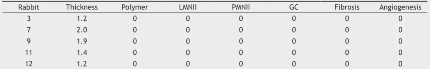
Table 4 Histological characteristics found in Group II - 90 days - Control vocal fold.
Rabbit Thickness Polymer LMNII PMNII GC Fibrosis Angiogenesis
3 1.2 0 0 0 0 0 0
7 2.0 0 0 0 0 0 0
9 1.9 0 0 0 0 0 0
11 1.4 0 0 0 0 0 0
12 1.2 0 0 0 0 0 0
LMNII, lymphomononuclear inflammatory infiltrate (0-3); PMNII, polymorphonuclear inflammatory infiltrate (0-3), Eos, cellular predominance of eosinophils, GC (giant cells), mean number of giant cells per high-power field (40 ×); Fibrosis, intensity of fibrosis observed; Polymer, presence or absence of the biopolymer; Angiogenesis, mean number of vascular spaces per high-power field (40 ×); thickness, measurement of the vocal fold thickness at its point of greatest width.
Table 5 Comparative analysis of thickness, measured at its
greatest width in millimeters, of rabbit vocal folds injected with sugarcane biopolymer and control vocal folds.
Rabbit (n)
SCB / Thickness
(mm)
Control/ Thickness
(mm)
1 2.2 1.3
p = 0.0058
2 3.1 2.9
4 4.0 5.0
5 4.0 2.8
10 2.2 3.0
3 1.3 1.2
7 2.9 2.0
9 5.0 1.9
11 2.8 1.4
12 3.0 1.2
Group I, 21 days and Group II, 90 days.
Significant for comparison. (Mann-Whitney U, WilcoxonTest).
Discussion
Vocal fold alterations that involve a reduction of volume and failure of glottal closure result in an exaggerated air
escape during speech, which makes the voice excessively
breathy.
For some of these cases, surgical procedures resulting in an increase of the affected vocal fold volume to reduce the
There were no epithelial layer lesions in the vocal folds evaluated by microscopy, as shown in Fig. 2.
Figure 2 Photomicroscopy of rabbit right vocal fold 3 months
after SCB implantation. Note the biopolymer deposits betwe -en the vocal muscle ibers, with no mucosal alterations. (HE staining, 40×)
MUCOSA MUSCLE
glottal gap may be indicated. However, placing a material
inside the fold can alter its vibrational properties, either by
the presence of the material itself, or by the inlammatory changes it will produce in the subepithelial space, resulting
in voice hoarseness.
To date, there has been no consensus in the literature regarding the type of material to be used in vocal folds.
Sev-eral materials have been tested, including fat, which seems
to be the most used in Brazil, mainly due to its prompt
avail-ability and low cost and that can be inserted through a sub -mucosal pouch in the vocal fold under general anesthesia.10 Others, such as hydroxyapatite have had positive liter-ature references but are expensive. One of the advantages
of injectable substances is that they can be applied without
the need for general anesthesia, but this is not the case for
hydroxyapatite, fascia and areolar tissue, which need to be
introduced after the creation of a submucosal pouch in the vocal fold.5,6,11,16-18
The sugarcane biopolymer has been the subject of nu-merous studies. Several studies have assessed its properties in different systems, such as the restoration of vascular and
abdominal walls, as well as the middle ear inlammatory
response.9-15
The use of the SCB gel on the vocal folds has never been
described. This material was chosen because it is injectable
and meets certain criteria, such as the fact that it is easy to
handle, its biocompatibility, low cost and low probability of infection, which makes it a good candidate for correction of
pathological vocal fold alterations.6,18
In our study, we found a mild to moderate inlamma
-tory response in both temporal groups of vocal folds with SCB. There was no signiicant inlammatory reaction caused
by the surgical procedure in the control vocal folds. Other
substances studied in the literature showed similar results in relation to the inlammatory response between the cases
and their controls. Duprat, in 20015 studied the behavior of
fat grafts in rabbits and obtained similar inlammatory re
-sponses between cases and controls. The author states that the fat did not appear to interfere with the inlammatory response in the surgical wound of the control vocal fold. However, the surgery performed by this author is different than what we did. He created a pouch to receive the fat graft in the vocal fold and the control was the similar sur
-gical trauma without graft placement. Autologous fat obvi
-ously has a lower cost than the SCB.18
The trauma of saline solution injection was not suficient to trigger inlammatory activity or a ibrogenic responses in
our observations and in the literature using a similar meth-odology. In 2007, Perazzo et al.19 used a surgical technique similar to ours to inject hyaluronic acid in vocal folds of rabbits. This author obtained similar results to those of our
study in control vocal folds, in which the injection of saline solution was used. We observed virtually no inlammatory response in the vocal folds injected with saline solution. Hyaluronic acid, as well as the SCB, can be injected under
local anesthesia in humans, but it has a higher cost.
The use of SCB in gel form resulted in greater inlam
-matory responses than their controls and this inlam-matory activity persisted throughout the study period. The 3-week and 3-month cases were statistically equivalent for all in
-lammatory variables studied. The in-lammatory response basically consisted of lymphomononuclear iniltrate of mild
to moderate intensity and some rabbits showed polymor
-phonuclear iniltrate with eosinophil predominance. This inding was not described in other studies per
-formed with other substances. There has been no descrip
-tion of the presence of eosinophils in literature with SCB use elsewhere. These cells are responsible for allergic reactions
and their presence should be further studied, as the possi-bility of contaminating substances such as dust used in sur-gical gloves may have contributed to their appearance, as
well as the fact that the iniltration site or gel formulation may be related to this inding.
The gel SCB-induced ibrogenesis was mild to moder
-ate in intensity. The observed pattern showed a peripheral predominance with apparent formation of ibrous capsules. Some specimens, such as rabbit 2 vocal fold, with SCB, showed a nodular and non-homogeneous pattern. Even in cases where the biopolymer location was more supericial, there was no alterations in the epithelial layer or Reinke space. As reported in the works of Flinck C,20 Thibeault SL21,22 and Longaker MT23 the preservation of the subepithe-lial layers is extremely important to preserve the mucosal
wave and voice quality.
This ibrogenic response with absence of epithelial reper
-cussions – no retractions, no pus formation or extrusion – sug -gests that the gel biopolymer can be bioactive and preserve
normal epithelial vibratory function. However, the presence of an inlammatory process in the subepithelial layers may
suggest a change in the physical properties of Reinke space,
which also has major importance in voice production. Any reduction in the amplitude of the mucosal wave can cause voice hoarseness. However, this inlammatory response also occurred with all the other materials studied before.
Although there are foreign body giant cells, we observed
elements that suggested good integration of the biopolymer
with tissues, such as the constant presence of angiogenesis and ibrous cords that passed through the biopolymer. Be
-cause of the SCB bio-integration, with cells invading its in
-timacy, it was not possible to easily identify in the studied slides the precise limits of the material within the vocal fold
and sometimes the biopolymer seemed to be disseminated
into the tissue, which prevented the measurement of total
area occupied by the SCB. For this reason, a more detailed analysis of the degree of biopolymer absorption has not been
possible. However, sinceit was more dificult to determine
the biopolymer region in the 3-month group, there may be
some biopolymer absorption over time, even if it was partial. On the other hand, we analyzed vocal fold thickness in its greatest cross-sectional dimension with the purpose of assessing the increase in volume. The larynges were not
sectioned according to a standard in all rabbits, and so the
comparison between temporal groups (21 days and 90 days) would have a signiicant bias. However, as the vocal cords of the each larynx were sectioned in the same way, it was
possible to compare the thickness of those that received
SCB with the control ones.
The vocal fold increase was signiicant and this is ex -tremely important, as one of the main objectives of bioma-terial injection in the vocal folds is precisely to increase its
volume and the consequent glottal closure, allowing im -proved vocal production.
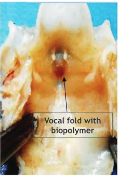
complete absorbtion in the present study. The SCB remained present in all the injected vocal folds. During the
macro-scopic analysis, the vocal fold with SCB was sometimes easily identiied by the difference in volume caused by the
presence of the biopolymer (Fig. 3).
3. Morris MJ, Christopher KL. Diagnostic criteria for the classiica -tion of vocal cord dysfunc-tion. Chest. 2010;138:1213-23. 4. Allen J Cause of vocal fold scar. Curr Opin Otolaryngol Head
Neck Surg. 2010;18:475-80.
5. Duprat A, Costa H, Eckley C, Pupo D, Rossi H. Implante de gordura no espaço de Reinke para a correção de alterações histoestruturais das pregas vocais. Braz J Otorhinolaryngol. 2001;67:78-83.
6. Constantino P, Friedman, C. Soft tissue augmentation and re-placement in head and neck general consideration. Otolaryn-gol Clin North Am.1994;27:1-12.
7. Berwanger AL, Scamparini AR, Domingues NM, Vanzo LT, Trei -chel H, Padilha FF. Produção de biopolímero sintetizado por Sphingomonas capsulata a partir de meios industriais. Ciênc Agrotec. 2007;31:177-83.
8. Melo FAD. Contribuição ao Estudo Cinético da Produção de Polissa-carídeo Extracelulares por Zoogloeasp em Melaço de Cana-de-Açú -car [tese]. Recife: Universidade Federal de Pernambuco; 2003. 9. Castro CMMB, Aguiar JLA, Melo FAD, Silva WTF, Marques E, Silva
DB. Citotoxicidade de biopolímero de cana-de-açúcar. Fac Med Univ Fed Pernamb. 2004;49:119-23.
10. Sato K, Umeno H, Nakashima T. Histological investigation of li -possuctioned fat for injeccion laryngoplasty. Am J Otolaryngol. 2005;26:219-25.
11. Coelho MCOC, Carrazoni PG, Monteiro VLC, Melo FAD, Mota RA, Tenório Filho F. Biopolímero produzido a partir da cana de açú -car para a cicatrização cutânea. Acta Bras Cir. 2001;17:11-3. 12. Vilar FO, Vasconcelos GB, Lima RFB, Lima SVC, Aguiar JL. Um
novo material para tratamento da incontinência urinária: estu -do em ratas. Acta Bras Cir. 2005;20:13-9.
13. Aguiar JL, Lins EM, Marques SRB, Barros Coelho AR, Rossiter RO, Melo RJV. Sugarcane biopolymer patch in femoral ar-teryangioplasty on dogs. Acta Bras Cir. 2007;22:77-81. 14. Silva DB, Aguiar JL, Marques A, Coelho ARB, Rolim Folho EL.
Miringoplastia com enxerto livre de membrana de biopolímero de cana de açúcar e fáscia autóloga em Chinchillalangier. An Fac Med Univ Fed Pernamb. 2006;51:45-51.
15. Mayer DBL, Araújo J, Leal M, Caldas Neto S. Sugarcane bio -polymermembrane: experimental evaluation in the middle ear. Braz J Otorhinolaryngol. 2011;77:44-50.
16. Brandenburg J, Kikhan W, Koshkee D. Vocal cord augmentation with autógenous fat. Laryngoscope. 1992;102:468-95.
17. Shindo M, Zaretsky L, Rice D. Autologous fat injection for unilateral vocal fold paralysis. Ann Otol Rhinol Laryn-gol.1996;105:602-6.
18. Duprat A. Comportamento histológico do enxerto autólogo de gordura na prega vocal de coelho [tese]. São Paulo: Faculdade de Ciências Médicas da Santa Casa de São Paulo; 2001. 19. Perazzo PL, Duprat A, Lancelotti C, Donati F. Estudo
prelimi-nar do comportamento histológico da prega vocal do coelho após injeção de ácido hialurônico. Braz J Otorrinolaryngol. 2007;73:171-8.
20. Flinck C. Structure cordaleet pathologies vocals. Rev Laryngol Otol Rhinol. 2005;126:195-300.
21. Thibeault SL, Rousseau B, Welham NV, Hirano S, Bless DM. Hyaluro -nan levels in acute vocal cord scar. Laryngoscope. 2004;114:760-4 22. Thibeault SL, Bless DM, Gray SD. Interstitial protein alterations
in rabbit vocal folds with scar. J Voice. 2003;17:377-83 23. Longaker MT, Chiu ES, Adzick NS, Stern M, Harrisson MR, Stern
R. Studies in fetal wound healing. Ann Surg. 1991;213:292-6.
Figure 3 Photograph of rabbit larynx after 3 weeks of SCB implantation in the right vocal fold. Macroscopically, we obser -ve the deletion of the groo-ve between the vocal fold and the ipsilateral ventricular band.
Conclusion
Despite causing an inlammatory reaction in vocal fold tis -sues that persisted throughout the study period, injected SCB remained in the vocal fold after three months consi-derably increasing vocal fold volume for up to 90 days in .
Conlicts of interest
The authors declare no conlicts of interest.
References
1. Gimenez LM, Zafra H. Vocal Cord Dysfunction: Anupdate. Ann Allergy Asthma Immunol. 2011;106:267-74.
2. Kenn K, Balkissoon R. Vocal cord dysfunction: what do we know? Eur Respir J. 2011;37:194-200.