*Correspondence: Swatantra Kumar Singh Kushwaha. Department of Phar-macy, Pranveer Singh Institute of Technology, 208020 - Kanpur, India. E-mail: [email protected]
A
vol. 49, n. 4, oct./dec., 2013
Carbon nanotubes as a novel drug delivery system for anticancer
therapy: a review
Swatantra Kumar Singh Kushwaha
1,*, Saurav Ghoshal
1, Awani Kumar Rai
1, Satyawan Singh
21Pranveer Singh Institute of Technology, Kanpur, India, 2Saroj Institute of Technology & Management, Lucknow, India
Carbon nanotubes (CNTs) were discovered in 1991 and shown to have certain unique physicochemical properties, attracting considerable interest in their application in various ields including drug delivery. The unique properties of CNTs such as ease of cellular uptake, high drug loading, thermal ablation, among others, render them useful for cancer therapy. Cancer is one of the most challenging diseases of modern times because its therapy involves distinguishing normal healthy cells from affected cells. Here, CNTs play a major role because phenomena such as EPR, allow CNTs to distinguish normal cells from affected ones, the Holy Grail in cancer therapy. Considerable work has been done on CNTs as drug delivery systems over the last two decades. However, concerns over certain issues such as biocompatibility and toxicity have been raised and warrant extensive research in this ield.
Uniterms: Carbon nanotubes/properties. Carbon nanotubes/use/drugs delivery. Single-Walled Carbon Nanotube. Multiwalled Carbon Nanotube. Anticancer drugs/delivery. Cancer/therapy. Drugs/delivery.
Os nanotubos de carbono foram descobertos em 1991 e suas propriedades físico-químicas únicas demonstradas, despertando interesse em sua aplicação em vários campos, incluindo a entrega liberação de fármacos. As propriedades únicas dos nanotubos de carbono, tais como a facilidade de captação pela célula, carga alta de fármaco, ablação térmica, entre outras, tornaram-nos úteis para terapia de câncer, uma das doenças mais difíceis dos tempos modernos, pois sua terapia envolve a distinção entre as células normais saudáveis e as afetadas pela doença. Os nanotubos de carbono têm um papel importante nessa área porque fenômenos como EPR permitem que estes possam distinguir as células normais das afetadas, que é o Santo Graal na terapia do câncer. Trabalho considerável tem sido feito ao longo das duas últimas década com nanotubos de carbono, como sistemas de liberação de fármacos. No entanto, preocupações sobre algumas questões, como biocompatibilidade e toxicidade, surgiram ao longo do tempo, demandando extensas pesquisa nesse campo.
Unitermos: Nanotubos de carbono/propriedades. Nanotubos de carbono/uso/liberação de fármacos. Nanotubo de carbono de parede única. Nanotubo de parede múltipla. Fármacos anticancer/liberação. Cancer/tratamento. Fármacos/liberação.
INTRODUCTION
Cancer ranks amongst the top three killers in modern society, next to heart and cerebrovascular diseases. In 2009, approximately eight million people died from cancer worldwide according to the WHO. The chemotherapeutic agents used for the treatment of a range of cancers are always associated with severe, sometimes fatal, toxicity due to a lack of target speciicity (Alderton et al.,1992).
advantages over other nano-sized delivery systems, such as an exceptionally high drug loading capacity due to their high surface area and the possibility for incorporating additional therapeutic and diagnostic moieties, either on the surface or their inner cavity. In addition, they interact with cellular membranes in a unique way: some types of carbon nanotubes (CNTs) have been reported to enter mammalian cells by an endocytosis-independent, “needle-like” penetration mechanism, which allows for direct cytoplasmic delivery of therapeutic payloads (Kostarelos
et al., 2007; Mu et al., 2009). A number of studies have already reported successful delivery of anti-cancer drugs to human cancer cells or tumor xenografts by means of carbon nanotubes (Heister et al., 2009; Liu et al., 2007; Ali-Boucetta et al., 2008; Zhang et al., 2009; Liu et al.,
2009; Li et al., 2010).
PROPERTIES OF CARBON NANOTUBES
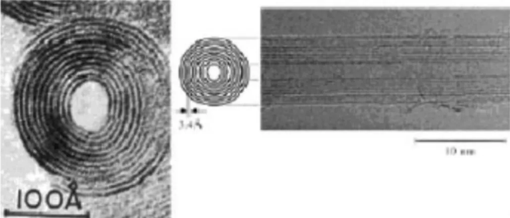
A number of properties result from the regular formation of carbon atoms in graphene cylinders. Carbon nanotubes are a huge cylindrical large molecule consisting of a hexagonal arrangement of sp2 hybridized carbon atoms (C-C distance is about 1.4 Ǻ). The wall of CNTs consists of single or multiple layers of graphene sheets, of which those formed by rolling up of single sheet are called single-walled carbon nanotubes (SWCNTs) and those formed by rolling up of more than one sheet are called multi-walled CNTs (MWCNTs). Both SWCNTs and MWCNTs are capped at both ends of the tubes in a hemispherical arrangement of carbon networks called fullerenes warped up by the graphene sheet (Figure 1). The interlayer separation of the graphene layers of MWCNTs measures approximately 0.34 nm on average, each forming an individual tube, with all the tubes having a larger outer diameter (2.5 to 100 nm) than SWCNTs (0.6 to 2.4 nm). SWCNTs have a better deined wall, whereas MWCNTs are more likely to have structural defects, resulting in a less stable nanostructure. In the medical ield, three main attributes of CNTs have been exploited:
• Their small size.
• Their high surface area to volume ratio. • Their ability to contain chemicals.
Carbon nanotubes can be produced small enough to pass through holes in tumours or to transport DNA (Singh
et al.,2005) The large surface to volume ratio provides a good platform for eficient transportation of chemicals and for the reactions needed for ultra-sensitive glucose detection (Muguruma et al., 2007).
CARBON NANOTUBES AND THEIR
IMPOR-TANCE IN ANTICANCER DRUG DELIVERY
Carbon nanotubes (CNTs) are allotropes of carbon with a cylindrical nanostructure. The discovery and subsequent widespread characterization of carbon nanotubes (CNTs) have opened up a class of materials with unexpected electrical, mechanical, and thermal properties (Alderton et al., 1992). The structure of CNTs can be imagined as the cylindrical roll-up of one or more graphene sheets containing only sp2 hybridized carbon atoms in a honeycomb arrangement (Tasis et al., 2006). Whether in the form of single-walled carbon nanotubes (SWCNTs) or multi-walled carbon nanotubes (MWCNTs), CNTs present several remarkable properties such as high aspect-ratio, ultra-light weight, tremendous strength, high thermal conductivity and signiicant electronic properties ranging from metallic to semiconducting (Ezzati et al., 2011; Lüer et al., 2009). CNTs may have different lengths ranging from several hundred nanometers to several millimeters, but their diameters depend on their class: SWCNTs are 0.4-3 nm in diameter and MWCNTs are 2-500 nm in diameter, depending on the method of synthesis (Kim et al.,2007). MWCNTs also consist of several cylinders of graphitic shells with a spacing layer of 0.3–0.4 nm (Dolatabadi et al., 2011). CNTs are currently one of the most popular nanomaterials used in a number of applications including electronics, energy storage, solar cells, molecular separation, sensing, biosensing, and drug delivery (Dolatabadi et al., 2011; Alpatova et
FIGURE 1 - The structure of SWCNTs with the two ends closed
al., 2010). CNTs can be used as drug-delivery vehicles or ‘nanocarriers’ in cancer therapy and other areas of medicine without causing toxicity to healthy tissue while allowing prolonged release of the drug (Tasis et al.,2006).
In recent years, a wide range of different nanoscale drug delivery vectors have been evaluated (Pan et al.,
2004; Chithrani et al., 2006; Endo et al., 2008). Notably, single-walled carbon nanotubes (SWCNTs) have attracted considerable interest in this regard, as they offer potential advantages over the more widely studied metal nanoparticle systems. These advantages include their ability to carry a high cargo loading, intrinsic stability, and structural lexibility, which can prolong circulation time and hence the bioavailability of the drug molecules carried (Liu et al., 2008; Singh et al., 2006; Worle-Knirsch et al., 2006; Wang et al., 2004). Moreover, SWCNTs have been shown to enter mammalian cells (Cai et al., 2007; Chin et al.,
2007; Kam et al., 2004; Kostarelos et al., 2007) and due to their promising properties, SWCNT-based materials have already been investigated as potential delivery vehicles for intracellular transport of nucleic acids (Cai et al., 2007; Liu
et al., 2007; Pantarotto et al., 2004), proteins (Kam et al.,
2004; Kam et al., 2005) and drug molecules (Prato et al.,
2008; Liu et al., 2007; Chen et al., 2008; Liu, Sun et al.,
2007). SWCNTs have been functionalized with antibodies and low-molecular-weight targeting agents, providing high eficiency for nanotube internalization into cells (Chen et al., 2008; Liu et al., 2007; Bottini et al., 2006; Cato et al.,
2008, Singh et al., 2005; Kang et al., 2008; Shao et al.,
2007; Welsher et al., 2008; Zeineldin et al., 2009).These systems also have higher loading capacity and allow for the incorporation of targeting agents and stealth molecules may also be added to evade the immune system.(Mu et al., 2009; Heister et al.,2009) Such systems have been loaded with drug molecules such as doxorubicin (DOX) via p-p stacking interactions, methotrexate, Paclitaxel and quercetin.(Modi et al., 2011; Dolatabadi et al., 2011; Wang
et al., 2009; Yuan et al., 2006; Abarrategi et al., 2008; Fu
et al., 2007). Nevertheless, the irst generation SWCNTs
proved to be unsuitable as drug delivery vectors as they tended to form bundles that disperse poorly in aqueous solutions and were therefore unsuitable for pharmacological use. To overcome this drawback, synthetic polymers such as poly(phenylacetylene) (Zeineldin et al., 2009, Yuan et al., 2006) and natural polymers such as polysaccharides (Kam et al., 2005; Yuan et al., 2006; Abarrategi et al., 2008; Fu et al., 2007; Hasegawa et al., 2004; Long et al., 2008; Numata et al., 2004; Zhang et al., 2008) have been used to encase SWCNTs via non-covalent interactions, thereby improving their compatibility with water and physiological environments.
ADVANTAGES OF CARBON NANOTUBES
OVER CONVENTIONAL CANCER THERAPY
Conventional treatment such as surgery is hindered by accessibility to tumorous cells and the risk of operating near or on vital organs. Also, selective treatment in chemotherapy and radiation is limited. On the whole, present treatment methods are not very effective at stopping the spread or recurrence of cancer. Nanomedicine provides a means of targeted delivery of drugs. Since the cancerous cells are on the nanoscale, there is a potential for highly eficient drug delivery (Misra et al., 2010). This has two major beneits. First, the total quantity of drug required is less, a concern primarily associated with the more costly drugs. Additionally, no solvent is required for delivery of the drug, which means that undesired health effects from the solvent can be prevented. Second, a lower concentration of the toxin is delivered to other parts of the body, without the risk of the protective nanocarrier degrading. Thus, fewer health side effects are suffered by the patient undergoing treatment. A further advantage of nanocarriers is that a range of drugs can be attached for a variety of purposes including; therapeutic, diagnostic, targeting and barrier avoidance, effectively allowing a toolkit to enable treatment speciic to each patient’s cancer.
METHODS FOR OPENING, FILLING AND
CAPPING CARBON NANOTUBES
As mentioned previously, carbon nanotubes are end-capped and thus for drug loading there are essentially two approaches which include the illing of carbon nanotubes during synthesis or after synthesis. Adding the contents of the nanotubes in-situ tends to be a less eficient approach,
producing a yield of around 10% whereas the post-synthesis process can be better controlled and yields of 50-100% are achievable (Monthioux, 2002). The appropriate method depends on the material that is to be inserted into the CNT. The criteria include melting temperature, reactiveness, surface tension and sensitivity of the material.
functional group is either bonded to the inside or outside of the walls. The most common mechanism for filling CNTs is capillarity. The limiting factor in capillarity is the diameter of the CNT and the surface tension of the material (the threshold material surface tension is approximately 200mN/m). However, according to (Gao et al., 2003), hydrophobic and Van der Waals forces also play a role in aqueous solutions. For chemicals with higher surface tensions it is possible to lower this tension by creating a suitable composite, which can be chemically reduced to the original substance once the CNT has been illed. The CNTs are washed using a solution which has been chosen to offer only limited solubility to the impregnating luid and thus can dissolve only deposits left outside the CNT (Qiang et al., 2008). After illing, the CNTs are capped by
passing a current which fuses the ends closed (de Jonge et al., 2005). The loading of CNTs remains an area requiring further research and more frequently mathematical methods are used rather than laboratory experiments due to the comparatively lower cost (Hilder et al., 2009).
Drug loading
The location of the drug to be delivered by the CNT can be internal or external. Internalisation or encapsulation relies on Van der Waals forces for insertion into the CNT and is best used for drugs that are sensitive to external environments and easily broken down (Hillebrenner et al., 2006).
Drug targeting
I n t r ad i ti o n a l c a n c e r tr e a t me n ts in v o lv in g chemotherapeutic agents, drug delivery is unavoidably indiscriminate. The toxic drug treatment is therefore applied to both tumour and regular cells which has the effect of, at best, causing numerous unpleasant side-effects in the patient and, at worst, leading to the death of the patient due to the toxicity of the drugs themselves. If it were possible to target the delivery of chemotherapeutic agents to only the tumour cells then this would both decrease the adverse side effects and also allow more effective, concentrated doses/agents that would be too toxic for traditional chemotherapy.
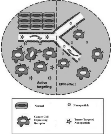
There are two basic techniques used for targeted nanoscale drug delivery. The first is passive (or size-mediated) targeting, where this relies upon the unique size of nanoparticles and the growth behavior of tumours. As the tumour grows it requires greater and greater amounts of oxygen and nutrients, engaging new blood vessels by a process called angiogenesis. Unlike in regular blood
vessels, the endothelial cells (which regulate the transfer of molecules across the vessel) in tumours can be spaced as far apart as 600 nm from each other. This defect allows increased permeability of nanoparticles into the interstitial space. In addition, there is poor lymphatic drainage in these tumorous areas.
These combined effects lead to a phenomenon known as enhanced permeability and retention (EPR) (Figure 2). By proper design of nanoparticles, this effect can be harnessed to locally increase density of nanoparticles (and their therapeutic agents) in the tumour, up to at least 10 times that of drugs not transported via nanoparticles (Misra et al., 2010). Another effect of angiogenesis and the desire to increase supply of nutrients to the tumour cell is the process of glycolysis used to increase energy level. This has the resulting effect of a locally decreased pH. This could potentially be utilised as an effective means of controlled drug release within the cancerous tumour, given a nanotube capped with a substance biodegradable by an acidic environment (Misra
et al., 2010). The second technique is active targeting - this involves using antibody- or ligand-targeted binding as a means of selective delivery to cancer cells or tumours. This technique requires knowledge of the target receptor or antigen on the cancer cells, preferably with a number of properties that are unique to the cancer cells and that are
expressed with high enough density to distinguish them from surrounding healthy cells (Wang et al., 2009).
Research has shown that nanoscale drug delivery devices can be targeted speciically at cancer cells using the latter method, albeit mostly in laboratory tests. Applications for diagnosis and therapeutic treatment by other imaging agents and drug delivery vehicles such as repeatedly-branched, spherical dendrimers (typically by covalent bonding to the molecules periphery or outer functional groups (Morgan et al., 2006)) or robust silica-coated micelles for delivery of hydrophobic anti-cancer agents (Huo et al., 2006) could also be employed using carbon nanotubes as a potential delivery vehicle. Central to a nanoparticle’s eficacy when it enters the body is its ability to bypass the immune system for at least as long as it takes to reach and react to the cancer cells. For example, polymer-coated liposomes of the irst generation were cleared by the mononuclear phagocytic system (MPS) within minutes. The second generation has a modiied liposome surface that, unlike the unmodiied phospholipid liposome surface, does not attract plasma proteins and hence the MPS. This allows the liposomes to remain circulating within the blood for much longer and are hence more likely to reach their target, either passively or actively (Papahadjopoulos et al., 1990). Similar techniques are required to ensure high circulation periods when nanotubes are used.
Thermal ablation is a unique property attached with nanotubes to cause an increase of 10 0C, the temperature
rise required to induce a form of hyperthermia in the cells, ultimately causing their death (Burke et al., 2009). It is a universal means of destroying any type of cancer. Currently thermal ablation of tumour cells is done by radiofrequency-based heating of an ablation catheter which is inserted into the tumour with the assistance of an ultrasound transducer for imaging, When the catheter is in place, radiofrequency energy is applied via the catheter into the tissue which is then absorbed as thermal energy, destroying the cells. The success of this technique depends heavily on the surgeon’s accuracy when inserting the catheter. By using targeted nanotubes and the aforementioned properties of these when exposed to near infra-red (NIR) light, more accurate, reliable thermal ablation is achievable without requiring an incision (Liu
et al., 2009; Chakravarty et al., 2008).
Another phenomenon and potential use of NIR light in conjunction with CNTs is in the triggered release of the drug payload. It was found that six 10 second pulses at 1.4W/cm2 intensity released a DNA cargo into the occupied cell (Kam et al., 2005). The release of the payload was not accompanied by cellular destruction, enabling precise control of the therapeutic treatment. An additional beneit of
the increased local temperature is the enhanced permeability of the tumour vasculature. This improves drug uptake into the tumour resulting in more effective chemotherapeutic treatment. It is conceivable that CNTs could be utilised as both a drug-delivery vehicle and as a means of thermal ablation, improving the efficacy of treatment yet with reduced intensity (Burke et al., 2009).
Passive and active targeting
Previous attempts at antibody-mediated drug delivery have been largely unsuccessful due to the loss of specificity of the antibodies on binding with drug molecules. It was found that using nanotubes to support antibodies did not change their properties and so did not inhibit their targeting abilities. Targeting methods such as active or passive targeting are a direct result of functionalisation. Passive targeting is a result of inertness and physical size of the macromolecule, “hiding” it from the immune system. CNTs must be nanosized to prevent cellular opsonisation (the susceptibility of the macromolecule to ingestion by phagocytes resulting in its destruction) by the innate immune system but also functionalised with molecules/polymer chains such as PEG which do not promote an adaptive immune response. The CNT must also be of sufficient size to utilise the EPR effects and so a trade-off is required. PEG is useful in determining the degree of optimal functionalisation as it is an easily controllable variable. This passive targeting can cause problems; microspheres can lead to chemoembolism-type problems in the lymphatic nodes. For such cases, functionalisation with nanomagnetic particles (e.g. iron oxide) and placing of a magnet at the desired location for extended periods of time allows for drug release over an extended period. Active targeting requires functionalisation with tumour-speciic binding sites to selectively bind to tumour cells. Many cells of various cancers are known to overexpress certain receptors, such as brain tumours showing typically 100 k to 900 k LDL (low density lipoprotein) receptors. Functionalising CNTs with LDL not only increases uptake dramatically in the cancer cells, but reduces uptake in other cells that have far fewer LDL receptors.
Crossing the blood-brain barrier
Drug delivery targeted to lymphatic system
Many cancers metastasize through the lymphatic canal. Drug delivery systems targeted to the lymphatic system can block the metastasis of cancers effectively. Using radical polymerization, polyacrylic acid (PAA) can be appended onto CNTs, making them highly hydrophilic. Through coprecipitation, Fe3O4-based magnetic nanoparticles can be adsorbed on the PAA-CNT surface.
Through the interaction with COOH groups of grafted PAA, the nanoparticles can be stabilized from clustering. By stirring the solution containing PAA-CNT, Fe3O4-based magnetic nanoparticles, and gemcitabine for 24 h, gemcitabine was loaded into the nanosystem with a loading eficiency of 62%. It was found that CNTs were seen only in the local lymphatic nodes and were absent in the major organs, such as liver, kidney, heart, spleen, and lungs, after 3 h of subcutaneous injection. Without the help of such a nanostructures (Yang et al., 2010), gemcitabine cannot preferentially distribute in the lymphatic system.
FUNCTIONALISATION
Raw carbon nanotubes have highly hydrophobic surfaces and are not soluble in aqueous solutions whereas pristine CNTs are not soluble in any solution. A solution to this problem is Functionalisation. Functionalisation of CNTs is a process of chemical synthesis where desired functional groups can be introduced onto the walls of CNTs for various applications producing functionalised carbon nanotubes (f-CNT). The aim of this process in cancer treatment is the enhancement of biocompatibility within the body, enhancement of encapsulation tendency and solubility, multimodal drug delivery, and imaging with the speciic properties imparted related to the desired function. Modiications to CNTs can be divided into two categories; covalent and noncovalently bonded.
Covalent bonding
Covalent chemical bonding of polymer chains to CNTs results in strong chemical bonds between nanotubes and the attached molecule. Various covalent reactions have been developed to graft molecules based on their varying properties and can be further classiied as Grafting to or Grafting from reactions, which involve the addition of preformed polymer chains or the polymerisation of monomers from surface derived initiators on CNTs, respectively. Both to and from methods involve reaction to the surface of CNT by functionalisation reactions. Molecules or polymer chains reacting with the surface
of pristine, pre-functionalised or oxidised CNTs are the three main methods used to attach molecules covalently. Oxidation of CNTs, representing one of the most common modiications, uses oxidising agents such as concentrated nitric acid to form carboxyl groups at the most reactive sites; i.e. the ends, which are more reactive, and on any defects on the walls, such as 5-membered rings (Bianco
et al., 2005; Singh et al., 2005; Prato et al., 2008). The curvature of the CNT places a strain on the sp2 hybridised
carbon atoms, reducing the energy barrier required to convert the sp2 hybridized bonds to sp3 compared with lat
graphene. This results in pristine CNT being susceptible to various addition reactions such as the Bingel reaction (Bianco et al., 2005; Singh et al., 2005; Prato et al., 2008). Covalent bonding gives a robust attachment which is generally stable in a bioenvironment. However, intrinsic physical properties of CNTs such as photoluminescence and Raman scattering are drastically reduced due to the disruption to the CNT structure associated with covalent bonding. Due to this, covalent bonding cannot be used to functionalise CNTs used for photothermal ablation or for imaging purposes. A number of covalent functionalisation reactions have been used to modify carbon nanotubes to suit various applications (Spitalsky et al., 2010).
Non-covalent bonding
Non-covalent bonding of molecules to CNTs is generally the more widely used method of drug delivery according to the literature. An ideal non-covalently functionalised CNT should have specific properties; the more closely matched, the greater the usefulness in biological roles. This can be carried out by creating micelle-type structures where amphiphilic molecules are coated to the CNT. Another common form of functionalisation is π-π bonding achieved by stacking pyrene molecules onto the surface of the CNT. This type of bonding can also be applied to single strands of DNA by virtue of the aromatic DNA base units. This was shown to be unstable as it is cleaved by nucleases and consequently the biological applications are so far limited. Non-covalent bonding does not disrupt the π – network where, except for a shortening of length, the physical properties of the CNTs are essentially preserved, showing great promise for imaging and photothermal ablation (Liu et al., 2009).
ATTACHMENT OF DRUG TO CNT AND ITS
RELEASE
This is in order to employ a bond that is biologically cleaved either near the cell or more usefully, within the cell, releasing the payload. In a recent study, it was found that the lack of enhanced eficacy between the delivery of methotrexate, an anti-cancer drug, and the nonconjugated drug was from the amide bond, attaching the drug to the f-CNT. The bond was found to be too stable and not biologically cleaved. Improved delivery can result from a bond that is biologically stable, so as not to breakdown before it has reached the required location, but can be enzymatically cleaved within the cell (Prato et al., 2008). The use of NIR to release CNT-encapsulated drugs is another promising release mechanism, as living organisms are generally transparent to NIR. This is particularly applicable to polar drugs which do not readily cross the lipid bilayer. The NIR heats the CNT and can be used to accelerate the diffusion of the molecule inside the tube to the cell. Extremely low diffusion coefficients trap molecules of high polarity. When heated, the diffusion coeficient increases up to 7-fold, facilitating the release of polar drugs (Chaban et al., 2010).
Kinetics of CNTs
As drug carriers, the administration, absorption, and transportation of CNTs must be considered for obtaining the desired treatment effects. The studied routes of CNT administration include oral and injections FIGURE 3 - Modiication of SWCNTs. (1 Dhar et al., 2008, 2 Jia et al., 2007, 3 Georgakilas et al., 2005, 4 Chen et al., 2010, 5 Liu et al., 2008, 6 Meng et al., 2008, 7 Zheng et al., 2003, 8
Hong et al., 2010).
such as subcutaneous injection, abdominal injection, and intravenous injection. There are different ways of absorption and transportation when CNTs are administered by different routes. The absorbed CNTs are transported from the administration sites to the effect-relevant sites by blood or lymphatic circulation. After administration, absorption is the irst key step for drug carriers to complete their drug-delivering mission. Studies have suggested that CNTs themselves are capable of being absorbed. It has also been established that physically shortened CNTs that are orally administered can be absorbed through the columnar cells of intestinal mucous membrane, where this was conirmed by transmission electron microscopy (Liu et al., 2011).
Distribution indicates the sites or places the absorbed CNTs can arrive and exist, of great importance in clinical pharmacology and toxicology of CNTs as drug carriers. There have been experiments to investigate in vivo and
ex vivo biodistributions, as well as tumor targeting ability of radiolabeled SWCNTs (diameter, approximately 1 to 5 nm; length, approximately 100 to 300 nm) noncovalently functionalized with phospholipids(PL)- PEG in mice using positron emission tomography and Raman spectroscopy, respectively. It was interesting to note that the PEG chain lengths determine the biodistribution and circulation of CNTs.
The nonbiodegradability in the body and non-eliminability from the body raise questions on the possibility of their successful use in clinical practice, factors which have always been a concern. Functionalized SWCNTs seem to be metabolizable in the animal body. For example, SWCNTs with carboxylated surfaces have demonstrated their unique ability to undergo 90-day degradation in a phagolysosomal simulant, resulting in shortening of length and accumulation of ultraine solid carbonaceous debris. Unmodified, ozonolyzed, aryl-sulfonated SWCNTs exhibit no degradation under similar conditions. The observed metabolism phenomenon may be accredited to the unique chemistry of acid carboxylation, which, in addition to introducing the reactive, modiiable COOH groups onto CNT surfaces, also induces collateral damage to the tubular graphenic backbone in the form of neighboring active sites that provide points of attack for further oxidative degradation (Raffa et al., 2010).
BIOCOMPATIBILITY OF CNTS AND ITS
ENHANCEMENT
• Coating should be suficiently stable to resist de -tachment from nanotube surface under biological conditions
• Amphiphilic coating molecules should have a low critical micelle concentration so CNT is stable once removed from solution
• Coating should have functional groups which are available for bioconjugation with antibodies or other molecules to create various CNT conjugates for various applications.
Producing biocompatible CNT requires low levels of toxicity and to ease-of-processing by the body. Addition of polyethylene glycol (PEG) by attachment via phospholipids allows for this, as both constituents are easily removed from the body over time. The toxicity is greatly reduced by functionalisation (Bianco et al.,
2005). A problematic feature of many current drugs is the circulation time in vivo which in vitro testing cannot determine. Low circulation times greatly reduce effectiveness as the drug is removed from the body too quickly, whereas long circulation times have a detrimental effect on healthy tissue increasing the side effects. f-CNT can tailor the blood circulation half-life so as to maximize absorbance by tumours, but minimise the accumulation in skin dermis, for example. The extent of functionalisation with polymers such as PEG and the length of these chains play an in important role in biodistribution. Increasing the extent (i.e. the density) of PEGylation (PEGylation is the process of covalent attachment of polyethylene glycol polymer chains) and the lengths of polymer chains, increased the circulation time in in vivo studies on mice. Densities of 10% PEGylation and chain lengths of 5k monomer units were found to have ideal half-life circulation times of 12-13 hours with high uptakes in tumours but low uptakes in other cells such as skin dermis. This is a good example of how functionalisation can be
inely tuned to optimise the in vivo behaviour of f-CNT as well as applications in general (Raffa et al., 2010).
Cell uptake is partially dependent on the length of CNTs and is another criteria to be taken into account when attempting to ind design criteria for CNT carrier systems. CNTs having lengths within the submicron range were found to readily accumulate in cells (Shvedova et al.,
2010); further suggesting surface chemistry has a minor role in the uptake. Most studies carried out use varying lengths of CNT, but with more speciic manufacturing techniques available, future work should look in more detail at the optimal length for varying applications.
Toxicity of carbon nanotubes
As has been mentioned in this paper, carbon nanotubes are a high-profile, nano-scale technology that is being considered in many technological fields. Increasingly however, concerns have been raised over potential toxicity issues with carbon nanotubes (Firme et al., 2010; Pacurari
et al., 2008) and there is presently a lack of data and understanding about their impact on biological systems. Given the probable wide-spread use of CNTs in the future, it is imperative to understand their impacts on biological systems before they can be used in mainstream drug delivery. The most attractive properties of nano-materials for biomedical applications i.e. their small size, large surface area, high reactivity, and high aspect ratio, are also the main factors of potential cytotoxicity. It is thought that, although there may be several mechanisms of causing cell damage, the main way would be due to DNA damage (Ji et al.,
2010). The study suggests that SWCNTs can induce adverse cellular responses through activation of molecular signaling associated with oxidative stresses (cancer inducing).
Several groups have already observed that CNTs can exhibit behaviour similar to that of asbestos ibres when conducting experiments on mice (Thurnherr et al., 2011).
This concern becomes understandable when the structure of both compounds is compared. Structures of chrysotile asbestos (left) and MWCNT (right) are shown above. The main issue with asbestos (and the concern with CNTs) is that due to their nano-scale and light weight, they easily become airborne and are carried into the lungs. Asbestos is known to have caused scarring of the lungs (pulmonary fibrosis). This leads to a host of health problems and diseases due to the reduced surface area within the lung to transfer oxygen into the blood stream. CNTs resemblance to asbestos ibres, in terms of aspect ratio, bio-persistence and reactivity contribute to this concern. Whilst there is good reason to be concerned about the potential similarities to asbestos ibres, there is evidence to suggest that industrially produced MWCNTs in high doses do not result in cell death in lung epithelial (tissue) in the way that asbestos ibres do. Additionally, long term exposure to pristine MWCNTs at low concentrations did not cause any major adverse effects (Lindberg et al., 2009).
Factors found to affect CNT toxicity
A list of factors that have been found to have an inluence on the degree of toxicity of CNTs (Thurnherr et al., 2011) follows below:
• Concentration / dose of CNTs.
• SWCNTs or MWCNTs
• Length of the tubes
• Catalyst residues left over during synthesis or func-tionalization
• Degree of aggregation • Oxidisation
• Functionalisation.
Whilst many studies show conflicting results on some of these properties, two seem to yield the most concurrent results; concentration and functionalisation. Various studies have been conducted with regard to the effect of dose concentration on cell viability. The two parameters used to monitor this test are concentration of dose and incubation time. It has been shown using rat erythrocytes (red blood cells) that at MWCNT concentrations of 25 µg/mL no adverse effects to the cells were observed. At concentrations of 50 µg/mL however, erythrocyte haemolysis (breaking of the cell membrane) was increased. One likely explanation is that at these higher concentrations the MWCNTs agglomerate, which appears to accelerate the haemolysis process (Bottini et al., 2006).
Several papers agree that high dose concentrations and prolonged incubation times both increase the induced toxicity and thus decrease cell viability. Research has shown cell viability decreases signiicantly in human bronchial epithelial cells. (Kalaugher et al., 2005) The trend shows how DNA damage increases considerably with dose concentration of SWCNTs (non-functionalised SWCNT). The concentration and incubation time of a dose is an area of nanotechnology in cancer treatment which requires much further study, as it will be important to optimise these for the treatment and eradication of cancerous growths as well as to minimise the body’s exposure to the drug (should it prove to have a degree of toxicity). The focus of a large body of research has been the degree to which functionalisation affects CNT toxicity. This is also likely to be one of the areas of research that receives most attention because active and passive targeting is directly related to the
TABLE I - List of anticancer drugs delivered using CNTs
S.N. Name of Drug Modiication /
Functionalization SWCNT / MWCNT Advantage
1 Doxorubicin (Heister et al., 2006;
Xiaoke et al.,2009)
PEG conjugation SWCNT Reduced toxicity
2 Mitoxantrone (Heister et al., 2006) PEG conjugation SWCNT Reduced toxicity
3 Paclitaxel (Tian et al., 2011) PEG conjugation SWCNT Increased circulation period
4 Cisplatin (Liu et al., 2007) Non functionalised SWCNT Decreased toxicity
5 Carboplatin (Hampel et al., 2008) SWCNT
6 Doxorubicin (Li et al., 2011) Conjugated with folate MWCNT Active targeting
7 Paclitaxel (Tian et al., 2011) Folate conjugate MWCNT Increased circulation period
8 Methotrexate (Modi et al., 2011) PEGlyated MWCNT Controlled toxicity
9 Quercitin (Dolatabadi, et al., 2011) PEGylated SWCNT Reduced side effect
10 Folic Acid (Reddy et al., 2006) – MWCNT Active targeting, longer
type and degree of functionalisation of the CNT. It has been demonstrated that increasing the degree of functionalisation of a SWCNT can dramatically decrease its cytotoxicity. (Azizian et al., 2010) The executive director for the Centre for Biological and Environmental Nanotechnology (CBEN) has stated regarding this study “...it’s the same answer: change the surfaces. This is an important demonstration that there are general trends in biological responses to nano-particles”. Long side-chain functional groups on SWCNTs can lower toxicity and have been shown to increase the CNTs biocompatibility with cells. This property of CNTs for cancer treatment appears to be particularly promising, as the functionalisation of CNTs is essential for passive and active cancer treatment (Yang et al., 2010).
CONCLUSION
In the ield of CNT technology for cancer treatment, the issues surrounding CNT toxicity remain inconclusive, as there are numerous conlicting studies demonstrating both toxic and non-toxic behavior, in spite of a number of drugs which have been delivered using carbon nanotubes. [Table 1] This is in part; it seems, due to the nature of the research being conducted. That is, there is no real benchmark for comparing results. Due to the range of parameters listed earlier which have been shown to affect CNTs toxicity, this is an area which will require the continual attention of toxicologists in the future. Using just one example, some evidence (Thurnherr et al., 2011) points to Fe (iron) impurities on CNTs at high concentrations increasing the observed cytotoxic response, while other research (Kolosnjaj-Tabi et al., 2010) claims the opposite. Clearly, much further study is required in this area before CNT technology can be applied to cancer treatment. That said, it is an extremely promising application of nano-technology and is definitely worth further research, as the current methods for cancer treatment are indiscriminately harmful and only partially effective.
REFERENCES
ABARRATEGI, A.; GUTIERREZ, M.C.; MORENO-VICENTE, C.; HORTIGUELA, M.J.; RAMOS, V.; LOPEZ-LACOMBA, J.L. Multiwall carbon nanotube
scaffolds for tissue engineering purposes. Biomaterials,
v.29, n.1, p.94-102, 2008.
AJAYAN, P.M.; EBBESEN, T.W.; ICHIHASHI, T.; IIJIMA, S.; TANIGAKI, K.; HIURA, H. Opening carbon nanotubes
with oxygen and implications for filling. Nature, v.362,
n.6420, p.522-525, 1993.
ALDERTON, P.M.; GROSS, J.; GREEN, M.D. Comparative study of doxorubicin, mitoxantrone, and epirubicin in combination with ICRF-187 (ADR-529) in a chronic
cardiotoxicity animal model. Cancer Res., v.52, n.1,
p.194-201, 1992.
ALI-BOUCETTA, H.; AL-JAMAL, K.T.; MCCARTHY, D.; PRATO, M.; BIANCO, A.; KOSTARELOS, K. Multiwalled carbon nanotube-doxorubicin supramolecular complexes
for cancer therapeutics. Chem. Commun, v.28, n.4,
p.459-461, 2008.
ALPATOVA, A.L.; SHAN, W.; BABICA, W. Single-walled carbon nanotubes dispersed in aqueous media via non-covalent functionalization: effect of dispersant on the stability, cytotoxicity, and epigenetic toxicity of nanotube
suspensions. Water Res., v.44, n.2, p.505-520, 2010.
AZIZIAN, J.; TAHERMANSOURI, H.; BIAZAR, E.; HEIDARI, S.; KHOEI, D.C. Functionalization of carboxylated multiwall nanotubes with imidazole
derivatives and their toxicity investigations. Int. J.
Nanomedicine, v.5, p.907-914, 2010.
BEG, S.; RIZWAN, M.; SHEIKH, A.M.; HASNAIN, M.S.; ANWER, K.; KOHLI, K. Advancement in carbon
nanotubes: basics, biomedical applications and toxicity, J.
Pharm. Pharmacol., v.63,n.2, p.141-163, 2011.
BIANCO, A.; KOSTARELOS, K.; PRATO, M. Applications of
carbon nanotubes in drug delivery. Curr. Opin.Chem. Biol.,
v.9,n.6, p.674-679, 2005.
BOTTINI, M.; CERIGNOLI, F.; DAWSON, M.I.; MAGRINI, A.; ROSATO, N.; MUSTELIN, T. Full-length single-walled carbon nanotubes decorated with streptavidin-conjugated quantum dots as multivalent intracellular fluorescent
nanoprobes. Biomacromolecules, v.7, n.8, p.2259-2263,
2006.
BURKE, A.; DING, X.; SINGH, R. Long-term survival following a single treatment of kidney tumors with multiwalled carbon nanotubes and near-infrared radiation.
Proc. Natl. Acad. Sci., v.106, n.31, p.12897-12902, 2009.
CAI SHAO‐YU; KONG JI‐LIE. Advance in research on carbon
canotubes as diagnostic and therapeutic agents for tumour.
CAI, D.; DOUGHTY, C.A.; POTOCKY, T.B.; DUFORT, F.J.; HUANG, Z.; BLAIR, D. Carbon nanotube-mediated delivery of nucleic acids does not result in non-specific
activation of B lymphocytes. Nanotechnology, v.18, n.365,
p.101-110, 2007.
CATO, M.H.; D’ANNIBALE, F.; MILLS, D.M.; CERIGNOLI, F.; DAWSON, M.I.; BERGAMASCHI, E. Cell-type
speciic and cytoplasmic targeting of PEGylated carbon
nanotubebased nanoassemblies. J. Nanosci. Nanotechnol.,
v.8, n.5, p.2259-2269, 2008.
CHABAN, V.V.; SAVCHENKO, T.I.; KOVALENKO, S.M.; PREZHDO, O.V. Heat-driven release of a drug molecule
from carbon nanotubes: a molecular dynamics study. J.
Phys. Chem., v.114, n.42, p.13481-13486, 2010.
CHAKRAVARTY, P.; MARCHES, R.; ZIMMERMAN, N.; SWAFFORD, A.; BAJAJ, P.; MUSSELMAN, I.; PANTANO, P.; DRAPER, R.; VITETTA, E. Thermal ablation of tumor cells with antibody-functionalized
single-walled carbon nanotubes. Proc. Natl. Acad. Sci., v.105, n.25,
p.8697-8702, 2008.
CHEN, D.; WU, X.; WANG, J.; HAN, B.; ZHU, P.; PENG, C.H. Morphological observation of interaction between PAMAM
dendrimer modiied single walled carbon nanotubes and
pancreatic cancer cells. Nano Biomed. Eng., v.2, n.4,
p.60-65, 2010.
CHEN, J.Y.; CHEN, S.Y.; ZHAO, X.R.; KUZNETSOVA, L.V.; WONG, S.S.; OJIMA, I. Functionalized single-walled carbon nanotubes as rationally designed vehicles
for tumortargeted drug delivery. J. Am. Chem. Soc., v.130,
n.49, p.16778-16785, 2008.
C H I N , S . F. ; B A U G H M A N , R . H . ; D A LT O N , A . B . ; DIECKMANN, G.R.; DRAPER, R.K.; MIKORYAK, C. Amphiphilic helical peptide enhances the uptake of
single-walled carbon nanotubes by living cells. Exp. Biol. Med.,
v.232, n.9, p.1236-1244, 2007.
CHITHRANI, B.D.; GHAZANI, A.A.; CHAN, W.C.W. Determining the size and shape dependence of gold
nanoparticle uptake into mammalian cells. Nano Lett., v.6,
n.4, p.662-668, 2006.
CHONN, A.; CULLIS, P.R. Recent advances in lipo-somal
drug-delivery systems. Curr. Opin. Biotechnol., v.6, n.6,
p.698-708, 1995.
DEJONGE, N.; DOYTCHEVA, M.; ALLIOUX, M.; KAISER, M.; MENTINK, S.; TEO, K.; LACERDA, R.; MILNE, W.
Cap closing of thin carbon nanotubes. Adv. Mater., v.17,
n.4, p.451-455, 2005.
DHAR, S.; LIU, Z.; THOMALE, J.; DAI, H.; LIPPARD, S.J. Targeted single-wall carbon nanotube mediated Pt (IV)
prodrug delivery using folate as a homing device. J. Am.
Chem. Soc., v.130, n.34, p.11467-11476, 2008.
DOLATABADI, J.E.N.; JAMALI, A.A.; HASANZADEH, M. Quercetin delivery into cancer cells with single walled
carbon nanotubes. Int. J. Biosci. Biochem. Bioinforma., v.1,
n.1, p.21-25, 2011.
DOLATABADI, J.E.N.; MASHINCHIAN, O.; AYOUBI, B.; JAMALI, A.A.; MOBED A.; LOSIC, D.; OMIDI, Y.; GUARDIA, M. Optical and electrochemical DNA
nano-biosensors. Trend Anal. Chem., v.30, n.3, p.459-472, 2011.
EBBESEN, T.W. Wetting, filling and decorating carbon
nanotubes. J. Phys. Chem. Solids, v.57 n.6-8, p.951-955,
1996.
ENDO, M.; STRANO, M.S.; AJAYAN, P.M. Potential applications of carbon nanotubes. In: JORIO, A.;
DRESSELHAUS, G.; DRESSELHAUS, M.S. (Ed.). Topics
in applied physics. Berlin: Springer; Heidelberg: Verlag, 2008. v.21, p.13-61.
FANG, J.; NAKAMURA, H.; MAEDA, H. The EPR effect: unique features of tumor blood vessels for drug delivery, factors involved, and limitations and augmentation of the
effect. Adv. Drug Delivery Rev., v.63, n.3, p.136-151, 2011.
FIRME, C.P.; BANDARU, P.R. Toxicity issues in the application
of carbon nanotubes to biological systems. Nanomedicine,
v.6, n.2, p.245-256, 2010.
GAO, Y.K.; DX, C.; CS, O. Spontaneous insertion of dna
oligonucleotides into carbon nanotubes. Nano Letters, v.3,
n.4, p.471-473, 2003.
GEORGAKILAS, V.; TZITZIOS, V.; GOURNIS, D.; PETRIDIS, D. Attachment of magnetic nanoparticles on
carbon nanotubes and their soluble derivatives. Chem.
HAMPEL, S.; KUNZE, D.; HAASE, D. Carbon nanotubes
illed with a chemotherapeutic agent: a nanocarrier mediates
inhibition of tumor cell growth. Nanomedicine, v.3, n.2,
p.175-182, 2008.
HASEGAWA, T.; FUJISAWA, T.; NUMATA, M.; UMEDA, M.; MATSUMOTO, T.; KIMURA, T. Single-walled carbon nanotubes acquire a specific lectin-affinity through supramolecular wrapping with lactose-appended
schizophyllan. Chem. Commun., v.1, n.19, p.2150-2151,
2004.
HEISTER, E.; NEVES, V.; TÎLMACIU, C.; LIPERT, K.; SANZ BELTRÁN, V.; COLEY, H. Triple functionalisation of single-walled carbon nanotubes with doxorubicin, a
monoclonal antibody, and a luorescent marker for targeted
cancer therapy. Carbon, v.47, n.9, p.2152-2160, 2009.
HEISTER, E.; NEVES, V. Drug loading, dispersion stability, and therapeutic efficacy in targeted drug delivery with
carbon nanotubes. Carbon, v.128, n.2, p.10568-10571,
2006.
HILLEBRENNER, H.; BUYUKSERIN, F.; KANG, M.; MOTA, M.O.; STEWART, J.D.; MARTIN, C.R. Corking nano test
tubes by chemical self-assembly. J. Am. Chem. Soc., v.128,
n.13, p.4236-4237, 2006.
HONG, S.Y.; TOBIAS, G.; AL-JAMAL, K.T.; BALLESTEROS, B.; ALI-BOUCETTA, H.; LOZANO-PEREZ, S.; NELLIST, P.D.; SIM, R.B.; FINUCANE, C.; MATHER, S.J.; GREEN, M.L.; KOSTARELOS, K.; DAVIS, B.G.
Filled and glycosylated carbon nanotubes for in vivo
radioemitter localization and imaging. Nat. Mat., v.9, n.6,
p.485-490, 2010.
HUO, Q.; LIU, J.; WANG, L.-Q.; JIANG, Y.; LAMBERT, T.N.; FANG, E. A new class of silica cross-linked micellar
coreshell nanoparticles. J. Am. Chem. Soc., v.128, n.19,
p.6447-6453, 2006.
IANCU, C.; MOCAN, L. Advances in cancer therapy through the use of carbon nanotubemediated targeted hyperthermia.
Int. J. Nanomedicine, v.6, p.1675-1684, 2011.
IIJIMA, S. Helical microtubules of graphitic carbon. Nature,
v.354, n.6348, p.56–58, 1991.
DOLATABADI, J.E.N.; OMIDI, Y.; LOSIC, D. Carbon nanotubes as an advanced drug and gene delivery
nanosystem. Curr. Nanosci., v.7, n.3, p.297-314, 2011.
JI, S.R.; LIU, C.; ZHANG, B.; YANG, F.; XU, J.; LONG, J.; JIN, C.; FU, D.L.; NI, Q.X.; YU, X.J. Carbon nanotubes
in cancer diagnosis and therapy. Biochim. Biophys. Acta.,
v.1806, n.1, p.29-35, 2010.
JIA, N.; LIAN, Q.; SHEN, H.; WANG, C.; LI, X.; YANG, Z. Intracellular delivery of quantum dots tagged antisense oligodeoxynucleotides by functionalized multiwalled
carbon nanotubes. Nano Lett., v.7, n.10, p.2976-2980, 2007.
KALAUGHER, L. Nanotube toxicity linked to functionalization.
Technology Update (abbreviation), v.3, n.???, p.1-9, 2005.
KAM, N.W.S.; DAI, H.J. Carbon nanotubes as intracellular protein transporters: generality and biological functionality.
J. Am. Chem. Soc., v.127, n.16, p.6021-6026, 2005.
KAM, N.W.S.; JESSOP, T.C.; WENDER, P.A.; DAI, H.J. Nanotube molecular transporters: internalization of carbon
nanotube-protein conjugates into mammalian cells. J. Am.
Chem. Soc., v.126, n.22, p.6850-6851, 2004.
KAM, N.W.S.; O’CONNELL, M.; WISDOM, J.A.; DAI, H.J. Carbon nanotubes as multifunctional biological transporters and near-infrared agents for selective cancer
cell destruction. Proc. Natl. Acad. Sci., v.102, n.33,
p.11600-11605, 2005.
KANG, B.; YU, D.C.; CHANG, S.Q.; CHEN, D.; DAI, Y.D.;
DING, Y.T. Intracellular uptake, traficking and subcellular
distribution of folate conjugated single walled carbon
nanotubes within living cells. Nanotechnology, v.19, n.37,
p.375103-375110, 2008.
KIM, S.N.; RUSLING, J.F.; PAPADIMITRAKOPOULOS, F. Carbon nanotubes for electronic and electrochemical
detection of biomolecules. Adv. Mater., v.19, n.20,
p.3214-3228, 2007.
KOLOSNJAJ-TABI, J.; HARTMAN, K.B.; BOUDJEMAA, S.; ANANTA, J.S.; MORGANT, G.; SZWARC, H.; WILSON,
L.J.; MOUSSA, F. In vivo behavior of large doses of
ultrashort and full-length single-walled carbon nanotubes after oral and intraperitoneal administration to Swiss mice.
ACS Nano, v.4, n.3, p.1481-1492, 2010.
KOSTARELOS, K.; LACERDA, L.; PASTORIN, G.; WU, W.; WIECKOWSKI, S.; LUANGSIVILAY, J. Cellular uptake of functionalized carbon nanotubes is independent
of functional group and cell type. Nat. Nanotechnol., v.2,
LI, R.; WU, R.; ZHAO, L. Folate and iron difunctionalized multiwall carbon nanotubes as dual-targeted drug nano-
carrier to cancer cells. Carbon, v.49, n.5, p.1797-1805,
2011.
LI, R.B.; WU, R.; ZHAO, L.; WU, M.H.; YANG, L.; ZOU, H.F. P-glycoprotein antibody functionalized carbon nanotube overcomes the multidrug resistance of human leukemia
cells. ACS Nano, v.4, n.3, p.1399-1408, 2010.
LINDBERG, H.K.; FALCK, G.C.-M.; SUHONEN, S.; V I P P O L A , M . ; VA N H A L A , E . ; C ATA L ´ A N , J . ; SAVOLAINEN, K.; NORPPA, H. Genotoxicity of nanomaterials: DNA damage and micronuclei induced by carbon nanotubes and graphite nanofibres in human
bronchial epithelial cells in vitro. Toxicol. Lett., v.186, n.3,
p.166-173, 2009.
LIU, S.Y.-W.; LEE, K.-F.; LAI, P.B.-S. Needle track seeding: a real hazard after percutaneous radiofrequency ablation for
colorectal liver metastasis. World J. Gastroenterol., v.15,
n.13, p.1653-1655, 2009.
LIU, X.; TAO, H.; YANG, K.; ZHANG, S.; LEE, S.-T.; LIU, Z. Optimization of surface chemistry on single-walled carbon nanotubes for in vivo photothermal ablation of tumors.
Biomaterials, v.32, n.1, p.144-151, 2011.
LIU, Z.; CHEN, K.; DAVIS, C.; SHERLOCK, S.; CAO, Q.; CHEN, X.; DAI, H. Drug delivery with carbon nanotubes
for in vivo cancer treatment. Cancer Res., v.16, n.???,
p.6652-6660, 2008.
LIU, Z.; DAVIS, C.; CAI, W.B.; HE, L.; CHEN, X.Y.; DAI, H.J. Circulationand long-term fate of functionalized, biocompatible single-walled carbon nanotubes in mice
probed by Raman spectroscopy. Proc. Natl. Acad. Sci.,
v.105, n.5, p.1410-1415, 2008.
LIU, Z.; FAN, A.C.; RAKHRA, K.; SHERLOCK, S.; GOODWIN, A.; CHEN, X. Supramolecular stacking of doxorubicin on carbon nanotubes for in vivo cancer therapy.
Angew Chem. Int. Ed. Engl.,v.48, n.41, p.7668-7772, 2009.
LIU, Z.; SUN, X.M.; NAKAYAMA-RATCHFORD, N.; DAI, H.J. Supramolecular chemistry on watersoluble carbon
nanotubes for drug loading and delivery. ACS Nano, v.1,
n.1, p.50-56, 2007.
LIU, Z.; TABAKMAN, S.; WELSHER, K.; DAI, H. Carbon nanotubes in biology and medicine: in vitro and in vivo
detection, imaging and drug delivery. Nano Res., v.2, n.2,
p.85-120, 2009.
LIU, Z.; WINTERS, M.; HOLODNIY, M.; DAI, H.J. RNA delivery into human T cells and primary cells with
carbon-nanotube transporters. Angew Chem. Int. Ed. Engl., v.46,
n.12, p.2023-2027, 2007.
LIU, Z.; CAI, L. H.E.; NAKAYAMA, N.; CHEN, K.; SUN, X.; CHEN, X.; DAI. H. In vivo Biodistribution and highly
eficient tumour targeting of carbon nanotubes in mice. Nat.
Nanotechnol., v.2, n.1, p.47-52, 2007.
LONG, D.W.; WU, G.Z.; ZHU, G.L. Noncovalently modiied
carbon nanotubes with carboxymethylated chitosan: a
controllable donor-acceptor nanohybrid. Int. J. Mol. Sci.,
v.9, n.2, p.120-130, 2008.
LÜER, L.; HOSEINKHANI, S.; POLLI, D.; CROCHET, J.; HERTEL, J.; LANZANI, G. Size and mobility of excitonsin
(6,5) carbon nanotubes. Nat. Phys., v.5, n.1, p.54-58., 2009.
MENG, J.; JIE, M.; DUAN, J.; KONG, H.; LI, L.; WANG, C.; XIE, S.; CHEN, S.; GU, N.; XU, H.; YANG, X-D. Carbon nanotubes conjugated to tumor lysate protein enhance the
eficacy of an antitumor immunotherapy. Small, v.4, n.9,
p.1364-1370, 2008.
M I S R A , R . ; A C H A RYA , S . ; S A H O O , S . K . C a n c e r nanotechnology: application of nanotechnology in cancer
therapy. Drug Discov. Today, v.15, n.19-20, p.842-850,
2010.
MODI, C.D.; PATEL, S.J.; DESAI, A.B.; MURTHY, R.S.R. Functionalization and evaluation of PEGylated carbon
nanotubes as novel drug delivery for methotrexate. J. Appl.
Pharm. Sci., v.1, n.5, p.103-108, 2011.
MONTHIOUX, M. Filling single-wall carbon nanotubes.
Carbon, v.40, n.10, p.1809-1823, 2002.
MORGAN, M.T.; NAKANISHI, Y.; KROLL, D.J.; GRISET, A.P.; CARNAHAN, M.A.; WATHIER, M.; OBERLIES, N.H.; MANIKUMAR, G.; WANI, M.C.; GRINSTAFF, M.W. Dendrimer-encapsulated camptothecins: increased solubility, cellular uptake, and cellular retention affords
enhanced anticancer activity in vitro. Cancer Res., v.66,
MU, Q.X.; BROUGHTON, D.L.; YAN, B. Endosomal leakage and nuclear translocation of multiwalled carbon nanotubes:
developing a model for cell uptake. Nano Lett., v.9, n.12,
p.4370-4375, 2009.
MUGURUMA, H.; MATSUI, Y.; SHIBAYAMA, Y. Carbon nanotube-plasma polymer-based amperometric biosensors: enzyme-friendly platform for ultrasensitive glucose
detection. Jpn. J. Appl. Phys., v. 46, n.9A, p.6078-6082,
2007.
NANDAKUMAR, A. Consolidated report of the population based cancer registries. National cancer registry programme. Indian Council of Medical Research, v.4, n.2, p.23-24, 2010. (Material not found! Please, see it if it is correct).
NUMATA, M.; ASAI, M.; KANEKO, K.; HASEGAWA, T.; FUJITA, N.; KITADA, Y. Curdlan and schizophyllan (beta-1,3-glucans) can entrap single-wall carbon nanotubes in
their helical superstructure. Chem Lett., v.33, n.3,
p.232-233, 2004.
PACURARI, M.; YIN, X.J.; ZHAO, J.; DING, M.; LEONARD, S.S.; SCHWEGLER-BERRY, D.; DUCATMAN, B.S.; SBARRA, D.; HOOVER, M.D.; CASTRANOVA, V.; VALLYATHAN, V. Raw single-wall carbon nanotubes induce oxidative stress and activate MAPKs, AP-1, NF-kappaB, and Akt in normal and malignant human
mesothelial cells. Environ. Health Perspect,v.116, n.9,
p.1211-1217, 2008.
PAN, D.J.; TURNER, J.L.; WOOLEY, K.L. Shell cross-linked nanoparticles designed to target angiogenic blood vessels via alpha(v)beta(3) receptor-ligand interactions.
Macromolecules, v.37, n.???, p.7109-7115, 2004.
PANTAROTTO, D.; SINGH, R.; MCCARTHY, D.; ERHARDT, M.; BRIAND, J.P.; PRATO, M. Functionalized carbon
nanotubes for plasmid DNA gene delivery. Angew Chem.
Int. Ed. Engl., v.43, n.39, p.5242-5246, 2004.
PAPAHADJOPOULOS, D.G.A. Liposomes designed to avoid
the reticuloendothelial system. Prog. Clin. Biol. Res., v.343,
p.85-93, 1990.
PRATO, M; KOSTARELOS K.A.B. Functionalized carbon
nanotubes in drug design and discovery. Acc. Chem. Res.,
v.41, n.1, p.60-68, 2008.
FU, Q.; WEINBERG, G.; SU, D-S. Selective illing of carbon
nanotubes with metals by selective washing. New Carbon
Mater., v.23, n.1, p.17-20, 2008.
RAFFA, V.; CIOFANI, G.; VITTORIO, O.; RIGGIO, C.; CUSCHIERI, A. Physicochemical properties affecting
cellular uptake of carbon nanotubes. Nanomedicine, v.5,
n.1, p.89-97, 2010.
REDDY, S.T.; REHOR, A.; SCHMOEKEL, H.G.; HUBBELL, J.A.; SWARTZ, M.A. In vivo targeting of dendritic cells in
lymph nodes with poly(propylene sulide) nanoparticles. J.
Control. Release, v.112, n.1, p.26-34, 2006.
SHAO, N.; LU, S.; WICKSTROM, E.; PANCHAPAKESAN, B. Integrated molecular targeting of IGF1R and HER2 surface receptors and destruction of breast cancer cells using
single wall carbon nanotubes. Nanotechnology, v.18, n.31,
p.315101-315109, 2007.
SHVEDOVA, A.A.; KAGAN, V.E. The role of nanotoxicology in realizing the ’helping without harm’ paradigm of nanomedicine: lessons from studies of pulmonary effects
of single-walled carbon nanotubes. J.Intern. Med., v.267,
n.1, p.106-118, 2010.
SINGH, R.; PANTAROTTO, D. Binding and condensation of plasmid DNA onto functionalized carbon nanotubes: toward
the construction of nanotube-based gene delivery vectors. J.
Am. Chem. Soc., v.127, n.12, p.4388-4396, 2005.
SINGH, R.; PANTAROTTO, D.; LACERDA, L.; PASTORIN, G.; KLUMPP, C.; PRATO, M. Tissue biodistribution and blood clearance rates of intravenously administered carbon
nanotube radiotracers. Proc. Natl. Acad. Sci., v.103, n.9,
p.3357-3362, 2006.
SPITALSKY, Z.; TASIS, D.; PAPAGELIS, K.; GALIOTIS, C. Carbon nanotubepolymer composites: chemistry,
processing, mechanical and electrical properties. Prog.
Polym. Sci., v.35, n.3, p.357-401, 2010.
TASIS, D.; TAGMATARCHIS, N.; BIANCO, A.; PRATO, M.
Chemistry of carbon nanotubes. Chem. Rev., v.106, n.3,
p.1105-1136, 2006.
TAYLOR, A.; TURNBULL, B. The potential use of carbon
Nanotubes for cancer treatment. Nature, v.321, n.2,
THURNHERR, T.; BRANDENBERGER, C.; FISCHER, K.; DIENER, L.; MANSER, P.; MAEDER-ALTHAUS, X.; KAISER, J.P.; KRUG, H.F.; ROTHEN-RUTISHAUSER, B.; WICK, P.A. comparison of acute and long-term effects of industrial multiwalled carbonnanotubes on human lung
and immune cells invitro. Toxicol. Lett., v.200, n.3,
p.176-186, 2011.
TSANG, S.C.; CHEN, Y.K.; HARRIS, P.J.F.; GREEN, M.L.H.
A simple chemical method of opening and illing carbon
nanotubes. Nature, v.372, n.6502, p.159-162, 1994.
WANG, H.F.; WANG, J.; DENG, X.Y.; SUN, H.F.; SHI, Z.J.; GU, Z.N. Biodistribution of carbon single-wall carbon
nanotubes in mice. J. Nanosci. Nanotechnol., v.4, n.8,
p.1019-1024, 2004.
WANG, X. Fabrication of ultralong and electrically uniform
single-walled carbon nanotubes on clean substrates. Nano
Lett., v.9, n.9, p.3137-3141, 2009.
WANG, X.; WANG, Y. Advances of cancer therapy by
nanotechnology. Cancer Res. Treat.,v.41, n.1, p.1-11, 2009.
WELSHER, K.; LIU, Z.; DARANCIANG, D.; DAI, H. Selective probing and imaging of cells with single walled
carbon nanotubes as near-infrared luorescent molecules.
Nano Lett., v. 8, n.2, p.586-590, 2008.
WORLE-KNIRSCH, J.M.; PULSKAMP, K.; KRUG, H.F. Oops they did it again! Carbon nanotubes hoax scientists
in viability assays. Nano Lett., v.6, n.6, p.1261-1268, 2006.
XIAOKE, Z.; LINGJIE, M. Targeted delivery and controlled
release of doxorubicin to cancer cells using modiied single
wall carbon nanotubes. Biomaterials, v.30, n.30,
p.6041-6047, 2009.
YANG, D.; YANG, F.; HU, J.; LONG, J.; WANG, C.; FU, D.; NI, Q. Hydrophilic multiwalled carbon nanotubes decorated with magnetite nanoparticles as lymphatic targeted drug
delivery vehicles. Chem. Commun., n.29, p.4447-4449,
2009.
YANG, Z.; ZHANG, Y.; YANG, Y.; SUN, L.; HAN, D.; HONG, L.I.; WANG, C. Pharmacological and toxicological target organelles and safe use of single-walled carbon nanotubes as
drug carriers in treating Alzheimer disease. Nanomedicine,
v.6, n.3, p.427-441, 2010.
YUAN, W.Z.; SUN, J.Z.; DONG, Y.Q.; HAUSSLER, M., YANG, F.; XU, H.P. Wrapping carbon nanotubes in pyrene-containing poly(phenylacetylene) chains: solubility, stability, light emission, and surface photovoltaic properties.
Macromolecules, v.39, n.23, p.8011-8020, 2006.
ZEINELDIN, R.; AL-HAIK, M.; HUDSON, L.G. Role of
polyethyleneglycol integrity in speciic receptor targeting
of carbon nanotubes to cancer cells. Nano Lett., v.9, n.2,
p.751-757, 2009.
ZHANG, X.K.; MENG, L.J.; LU, Q.G.; FEI, Z.F.; DYSON, P.J. Targeted delivery and controlled release of doxorubicin to
cancer cells using modiied single wall carbon nanotubes.
Biomaterials, v.30, n.30, p.6041-6047, 2009.
ZHANG, X.K.; WANG, X.F.; LU, Q.H.; FU, C.L. Inluence of
carbon nanotube scaffolds on human cervical carcinoma HeLa cell viability and focal adhesion kinase expression.
Carbon, v.46, n.3, p.453-460, 2008.
ZHENG, M.; JAGOTA, A.; SEMKE, E.D.; DINER, B.A.; MCLEAN, R.S.; LUSTIG, S.R.; RICHARDSON, R.E.; TASSI, N.G. DNA-assisted dispersion and separation of
carbon nanotubes. Nat. Mater., v.2, n.5, p.338-342, 2003.
ZHONG TIAN; YINFENG SHI; MIN YIN; HEBEI SHEN; NENGQIN JIA. Functionalized multiwalled carbon nanotubes-anticancer drug carriers: synthesis, targeting
ability and antitumor activity. Nano Biomed. Eng., v.3, n.3,
p.157-162, 2011.
Received for publication on 27th August 2012