Ginsenoside Rg1 protects human renal tubular
epithelial cells from lipopolysaccharide-induced
apoptosis and in
fl
ammation damage
X.J. Ni, Z.Q. Xu, H. Jin, S.L. Zheng, Y. Cai and J.J. Wang
Transplantation Center, The First Affiliated Hospital of Wenzhou Medical University, Wenzhou, China
Abstract
Ginsenoside Rg1, one of the most notable active components ofPanax ginseng, has been widely reported to exert anti-inflammatory actions. This study aimed to reveal whether ginsenoside Rg1 also exhibits beneficial roles against lipo-polysaccharide (LPS)-induced apoptosis and inflammation in human renal tubular epithelial cells, and to evaluate the potential role of the component on tubulointerstitial nephritis treatment. HK-2 cells were treated with various doses of ginsenoside Rg1 (0, 50, 100, 150, and 200mM) in the absence or presence of 5mg/mL LPS. Thereafter, CCK-8 assay,flow cytometry, western blot, migration assay, reactive oxygen species (ROS) assay, and ELISA were carried out to respectively assess cell viability, apoptosis, migration, ROS activity, and the release of inflammatory cytokines. As a result, ginsenoside Rg1 protected HK-2 cells from LPS-induced injury, as cell viability was increased, cell apoptosis was decreased, and the release of MCP-1, IL-1b, IL-6, and TNF-awas reduced. Ginsenoside Rg1 functioned to HK-2 cells in a dose-dependent manner, and the 150mM dose exhibited the most protective functions. Ginsenoside Rg1 had no significant impact on cell migration and ROS activity, while it alleviated LPS-induced ROS release and migration impairment. Furthermore, the down-regulations of p-PI3K, p-AKT, and up-down-regulations of PTEN, p-IkBa, p-p65, Bcl-3 induced by LPS were recovered to some extent after ginsenoside Rg1 treatment. In conclusion, ginsenoside Rg1 protects HK-2 cells against LPS-induced inflammation and apoptosis via activation of the PI3K/AKT pathway and suppression of NF-kB pathway.
Key words: Ginsenoside Rg1; LPS; Apoptosis; Inflammation; PI3K/AKT pathway; NF-kB pathway
Introduction
Ginseng, the root of the widely popular plant Panax ginseng, has been used as Chinese traditional medicine for at least 1000 years (1). Currently, it is one of the most extensively researched and prescribed alternative medicines worldwide (1). This drug exerts pharmacological actions for several diseases including diabetes, neurological condi-tion, cardiovascular disease, and even cancer (1–4). The molecular components responsible for the beneficial effect of ginseng include ginsenosides, which are triterpene saponins that consist of rigid steroid skeleton and sugar moieties. Among the 30 different varieties of ginsenosides, Rg1 is one of the major pharmacologically active and abundant ingredient (1). Several studies have demonstrated the different beneficial effects of ginsenoside Rg1, which include anti-inflammatory and anti-apoptotic effects (1–7). An early in vivostudy has reported that intraperitoneal injection with ginsenoside Rg1 in rats resulted in a prevention of urinary protein excretion, an elevation of serum cholesterol content, as well as histopathological changes such as hypercellularity
and adhesion (8), indicating the therapeutic potential of ginsenoside Rg1 in nephritis. However, to our knowledge, the detailed functions of ginsenoside Rg1 on tubulointerstitial nephritis and its underlying molecular mechanisms have not been uncovered.
PI3K/AKT pathway has been shown to play a central role in different biological processes such as angiogenesis, tumorigenesis, and inflammation (3). This pathway is also known to play an important role in inflammation by taking part in cell growth modulation, and maturation of different cellular components of the inflammation pathway (3). Similarly, NF-kB pathway is responsible for modulating multiple cellular processes especially in inflammation and apoptosis (4). Several chronic inflammation diseases like inflammatory bowel disease, sepsis, arthritis, and athero-sclerosis have been revealed to be associated with chronic activation of NF-kB (4,7). Recent studies have indicated that ginsenoside Rg1 is able to activate PI3K/AKT signal-ing through a glucocorticoid receptor (GR)-dependent manner,
Correspondence: J.J. Wang:<[email protected]>
and it has been shown to be a functional ligand of GR (9). In contrast, ginsenoside Rg1 suppresses the activation of NF-kB pathway also through a GR-dependent manner (4,10). Based on thesefindings we hypothesize that ginsenoside Rg1 might be a potential anti-inflammatory drug at least in part via modulation of PI3K/AKT and NF-kB pathways.
This study aimed to explore the beneficial role of ginseno-sides Rg1 against lipopolysaccharide (LPS)-induced apop-tosis and inflammation damage in human renal tubular epithelial cells and also investigate its underlying mecha-nism. This study will providein vitroinformation supporting ginsenoside Rg1 as a potential anti-inflammatory drug for tubulointerstitial nephritis treatment.
Material and Methods
Cell culture and treatment
Human renal tubular epithelial cell line HK-2 was obtained from American Type Culture Collection (ATCC, USA), and was cultured in Dulbecco’s Modified Eagle’s Medium/Nutrient Mixture F-12 (DMEM-F12, 3:1, Gibco-BRL, USA) supplemented with 10% fetal bovine serum (FBS, Gibco-BRL) in a humidified 5% CO2atmosphere at 37°C.
Ginsenoside Rg1 with purity greater than 98% (National Institutes for Food and Drug Control, China) were dis-solved in DMSO (Sigma-Aldrich, USA) and mixed with the medium so that the final concentration of the vehicle was less than 0.1%. To analyze the functional impacts of ginsenoside Rg1 following LPS stimulation, cells were incubated with various doses of ginsenoside Rg1 (0, 50, 100, 150, and 200mM) in the presence or absence of 5mg/mL LPS (fromEscherichia coliO111:B4, Sigma-Aldrich) for 24 h (11).
CCK-8 assay
The viability of HK-2 cells was assessed by using a Cell Counting Kit-8 (CCK-8, Dojindo Molecular Technologies, USA). Briefly, cells were seeded on a 96-well plate with a density of 5103cells/well, and then cells were treated with ginsenoside Rg1 and/or LPS for 24 h. The cells were further incubated in fresh medium for 48 h, and then 10mL CCK-8 solution was added to the culture medium. The plates were incubated for 30 min at 37°C in humidified 95% air and 5% CO2. The absorbance was measured at
450 nm using a Microplate Reader (Bio-Rad, USA).
Apoptosis analysis
The FITC-Annexin V/PI detection kit from Beijing Biosea Biotechnology Co., Ltd. (China) was utilized in the present work for detection of cell apoptosis. In brief, cells were grown to about 70% confluence in 6-well plates and treated with ginsenoside Rg1 and/or LPS for 24 h, followed by 48-h incubation in fresh medium at 37°C. Cells (1105) in each sample were then collected and resuspended in 200 mL Annexin-Binding Buffer, and stained with 10 mL FITC-Annexin V and 5mL PI for 30 min in the dark at room
temperature. Flow cytometry analysis was done by using a FACS can (Beckman Coulter, USA).
Western blot
The protein used for western blotting was extracted using RIPA lysis buffer (Beyotime Biotechnology, China) supple-mented with protease inhibitors (Roche, Switzerland). The proteins were quantified using the BCAtProtein Assay Kit (Pierce, USA). The western blot system was established using a Bio-Rad Bis-Tris Gel system according to the manufacturer’s instructions. Primary antibodies were pre-pared in 5% blocking buffer at a dilution of 1:1,000. Primary antibody was incubated with the membrane at 4°C overnight, followed by wash and incubation with secondary antibody marked by horseradish peroxidase for 1 h at room tempera-ture. After rinsing, the polyvinylidene difluoride (PVDF) mem-brane carrying blots and antibodies were transferred into the Bio-Rad ChemiDoct XRS system, and then 200 mL Immobilon Western Chemiluminescent HRP Substrate (Millipore, USA) was added to cover the membrane surface. The signals were captured and the intensity of the bands was quantified using Image LabtSoftware (Bio-Rad, China).
Migration assay
Cell migration was determined by using a modified Transwell chamber with a pore size of 8 mm (Corning, USA). For migration assay, cells suspended in 200 mL of serum-free medium were seeded on the upper compart-ment of 24-well Transwell culture chamber, and 600 mL of complete medium containing 10 mL mitomycin C was added to the lower compartment. After 12 h incubation at 37°C, cells werefixed with methanol. Non-traversed cells were carefully removed from the upper surface of thefilter with a cotton swab. Traversed cells on the lower side of the filter were stained with crystal violet (Sigma-Aldrich) and counted. Relative cell migration was calculated as the number of the treated cells normalized to the number of the control cells adhering to the lower chamber.
Reactive oxygen species (ROS) assay
ROS was measured byflow cytometry using 2,7-dichloro-fluorescein diacetate (DCFH-DA; Nanjing Jiancheng Tech-nology Co., China). The cells were seeded in a 6-well plate, and after treatment and washing twice with phosphate buffer saline (PBS), cells were incubated in serum-free culture medium containing 10mM DCFH-DA for 20 min at 37°C in the dark. Subsequently, the cells were washed with PBS, and a trypsin digestion method was used for sample collec-tion. All samples were centrifuged and the supernatants were removed. The cells were resuspended to 500mL PBS and thefluorescent intensities were measured using aflow cytometer (488 nm excitation, 521 nm emission).
measured by human CCL2/MCP-1 Quantikine ELISA kit, human IL-1 beta/IL-1F2 Quantikine ELISA kit, human IL-6 Quantikine ELISA kit, and human TNF-alpha Quantikine ELISA kit (all from R&D Systems, UK), respectively, accord-ing to the manufacturer’s instructions.
Statistical analysis
All experiments were done in triplicate. The results of multiple experiments are reported as means±SD. Statis-tical analyses were performed using Graphpad statisStatis-tical software (GraphPad Software Inc., USA). P values were calculated using one-way analysis of variance (ANOVA). Po0.05 was considered to be statistically significant.
Results
Ginsenoside Rg1 alleviated LPS-reduced HK-2 cells viability
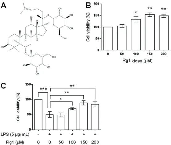
HK-2 cells were treated with increasing doses of ginseno-side Rg1 (structure depicted in Figure 1A) in the presence or absence of 5mg/mL LPS, and then cell viability was mea-sured to evaluate the functional effects of ginsenoside Rg1 on
cell viability. At doses of 100mm (Po0.05), 150mm (Po0.01), and 200mm (Po0.01), ginsenoside Rg1 caused a significant increase in the cell viability compared to control group of HK-2 cells (Figure 1B). In addition, 5 mg/mL of LPS dramatically reduced cell viability (Po0.001), while 100mm (Po0.05), 150mm (Po0.01), and 200mm (Po0.01) ginsenoside Rg1 partially alleviated LPS-reduced cell viability (Figure 1C).
Ginsenoside Rg1 alleviated LPS-induced HK-2 cells apoptosis
Cell apoptosis was also detected following various doses of ginsenoside Rg1 in the presence or absence of LPS. Flow cytometry detection results showed that 5mg/mL of LPS induced a significant increase in apoptotic cell rate (Po0.001, Figure 2A). High doses of ginsenoside Rg1 (100, 150, and 200mM) significantly suppressed LPS-induced apoptosis (Po0.01 or Po0.001). Expression changes of apoptosis-related factors in cells were detected by western blot analysis, as shown in Figure 2B and C. Bax and p53 were up-regulated, Bcl-2 was down-regulated, while caspase-3 was cleaved after LPS stimulation (all Po0.001). More importantly, 100, 150, and 200 mM of
Figure 1.A, Structural formula of ginsenoside Rg1.B, Viability of HK-2 cells treated with ginsenoside Rg1 (0, 50, 100, 150, and 200mM) or pretreated with 5mg/mL lipopolysaccharide (LPS) (C). Data are reported as means±SD (n=3). *Po0.05, **Po0.01, and ***Po0.001
ginsenoside Rg1 could alleviate LPS-induced abnormal expressions of these factors (Po0.05, Po0.01 or Po0.001), and it seemed that the regulatory effects of ginsenoside Rg1 might be in a dose-dependent manner.
Ginsenoside Rg1 alleviated LPS-reduced migration in HK-2 cells
To detect the role of ginsenoside Rg1 in the migra-tion of human renal tubular epithelial cells, HK-2 cells were treated with LPS and/or 150 mm ginsenoside Rg1, and then cell migration was assessed. LPS significantly reduced the relative migration rate (Po0.001), while ginsenoside Rg1 showed no effect on the migration of HK-2 cells. Moreover, ginsenoside Rg1 at a dose of 150 mm significantly (Po0.05) suppressed LPS-induced decrease in migration of HK-2 cells (Figure 3).
Figure 2.Ginsenoside Rg1 inhibited lipopolysaccharide (LPS)-induced HK-2 cells apoptosis. HK-2 cells were treated with ginsenoside Rg1 (0, 50, 100, 150, and 200mM) in the absence or presence of 5mg/mL LPS, and then apoptotic cell rate (A), and protein expression levels of Bax, Bcl-2, p53, cleaved-caspase3, and pro-caspase3 were assessed (BandC). Data are reported as means±SD (n=3). *Po0.05, **Po0.01, and ***Po0.001 (ANOVA).
Figure 3.Ginsenoside Rg1 alleviated lipopolysaccharide (LPS)-induced HK-2 cell migration. HK-2 cells were treated with 150mM of ginsenoside Rg1 and/or 5 mg/mL of LPS, and then relative migration rate was detected. Data are reported as means±SD
Ginsenoside Rg1 decreased LPS-induced ROS Given that overproduction of ROS can induce apop-tosis through both extrinsic and intrinsic pathways (12),
we further detected ROS activity of the HK-2 cells follow-ing by treatment with LPS and/or 150 mm ginsenoside Rg1. As mean fluorescence intensity of ROS showed in Figure 4, LPS significantly (Po0.001) increased the ROS activity of the HK-2 cells compared to the control cells (HK-2 cells in absence of LPS). Ginsenoside Rg1 (150mm) had no impact on ROS activity, while it alleviated the LPS-induced increase of ROS activity (Po0.01).
Ginsenoside Rg1 decreased LPS-induced inflammation response
The concentrations of four inflammatory cytokines, MCP-1, IL-1b, IL-6, and TNF-a, in HK-2 cells culture super-natant were detected following ginsenoside Rg1 and LPS treatment. As shown in Figure 5A–D, LPS exposure significantly elevated these four inflammatory cytokines levels in HK-2 cells culture supernatants (all Po0.001). However, ginsenoside Rg1 decreased LPS-induced pro-duction of these four cytokines (Po0.05, Po0.01 or Po0.001). A high dose of ginsenoside Rg1 exhibited a Figure 4.Ginsenoside Rg1 decreased lipopolysaccharide
(LPS)-induced increase in reactive oxygen species (ROS) activity. HK-2 cells were treated with 150mM of ginsenoside Rg1 and/or 5mg/mL of LPS, and then ROS activity was detected. Data are reported as means±SD (n=3). **Po0.01 and ***Po0.001 (ANOVA).
Figure 5. Ginsenoside Rg1 decreased lipopolysaccharide (LPS)-induced inflammation response. HK-2 cells were treated with ginsenoside Rg1 (0, 50, 100, 150, and 200mM) in the absence or presence of 5mg/mL LPS, and then the concentrations of (A) MCP-1, (B) IL-1b, (C) IL-6, and (D) TNF-ain culture supernatant were measured. Data are reported as means±SD (n=3). *Po0.05, **Po0.01,
stronger down-regulatory impact on these cytokines, and the maximum decrease in the levels of the above-mentioned cytokines of inflammation were seen at a dose of 150mm.
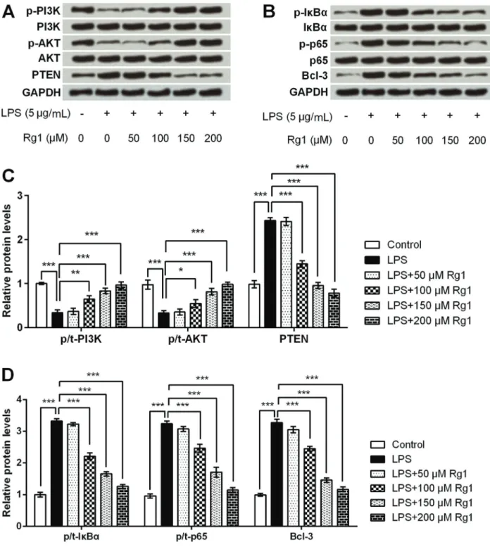
Ginsenoside Rg1 suppressed LPS-induced HK-2 cells injury by activation of PI3K/AKT and inactivation of NF-jB signaling pathways
Further, we focused on PI3K/AKT and NF-kB signal-ing pathways to reveal whether they are implicated in ginsenoside Rg1 modulation of HK-2 cells. Western blot analysis showed that p-PI3K and p-AKT levels were down-regulated, and PTEN (phosphatase and tensin homolog), p-IkBa, p-p65, and Bcl-3 were up-regulated after LPS stimulation (all Po0.001, Figure 6A–D). Mean-while, ginsenoside Rg1 recovered these down-regulations and up-regulations (Po0.05, Po0.01 or Po0.001), in a dose-dependent manner. No impact was observed in the total levels of PI3K, AKT, IkBaand p65 after LPS and/or ginsenoside Rg1 treatment.
Discussion
Ginsenoside Rg1, the principal component of Chinese medicinal plantPanax, is well-known to possess several beneficial effects (1). Studies have described the protec-tive role of ginsenoside Rg1 in diseases associated with cardiovascular system, nervous system, using umbilical cord cells and stem cells (1–7).
The most important beneficial effects of ginsenoside Rg1 include anti-apoptosis and anti-inflammatory actions. Hu and his colleagues reported that ginsenoside Rg1 protects murine stem cells against H2O2-induced
apopto-sis, as evidenced by the down-regulation of cleaved caspase-3 and up-regulation of Bcl-2 (5). Furthermore, it has been demonstrated that ginsenoside Rg1 protects against H2O2-induced apoptosis in a dose-dependent
mech-anism (13). The molecule also can inhibit the production of LPS-stimulated cytokines (10), and protect from ischemia/ reperfusion injury through attenuation of inflammation and apoptosis (14,15). Similar to these studies, we also found anti-apoptotic and anti-inflammatory roles of ginseno-side Rg1 in LPS-stimulated human renal tubular epithelial cells, as HK-2 cells viability was increased, the percent-age of apoptotic cell was reduced, and the production of inflammatory cytokines (MCP-1, IL-1b, IL-6, and TNF-a) were decreased following treatment with increasing doses of ginsenoside Rg1 in the presence of LPS.
It is widely accepted that relatively high levels of ROS cause redox imbalance and induce cell apoptosis or necrosis in a wide variety of physiological and pathological conditions (16,17). On the other hand, the generation of ROS can be promoted by pro-inflammatory cytokines, such as IL-1band TNF-a, and further deteriorate the infl am-mation response (18,19). In the current study, we found that ginsenoside Rg1 had no significant impact on ROS
activity, but significantly reduced LPS-induced ROS. This finding further confirmed that ginsenoside Rg1 exerts anti-apoptotic and anti-inflammatory roles in LPS-stimulated HK-2 cells.
In addition to the importance of cell apoptosis and inflammation, numerous other events such as cell migra-tion play pivotal roles in the structure and funcmigra-tion of renal tubular epithelial cells (20,21). Migration is neces-sary for the renal tubular epithelial cells to acquire normal morphology, cell-cell contact, and transport capacity (22,23). To date, several studies have focused on the role of ginsenoside Rg1 in cell migration, however the mecha-nism of the interaction is controversial. In breast cancer MCF-7 cells, ginsenoside Rg1 treatment suppress phorbol myristate acetate-induced cell migration and invasion (4). In contrast, in endothelial progenitor cells, ginsenoside Rg1 could promote cell migration in a dose- and time-dependent manner (24). The discrepancy might be due to the different cell types and stress stimuli. In this study, we found that ginsenoside Rg1 treatment had no impacts on HK-2 cells migration, while it could alleviate LPS-reduced migration, indicating a protective role in LPS-induced impairment of migration capacity. This fact revealed that ginsenoside Rg1 improves cell migration not via modula-tion of epithelial mesenchymal transimodula-tion (EMT) process, because in that case normal cells (undamaged) treated with ginsenoside Rg1 would also acquire migration capacity enhancement. The exact mechanism of ginsenoside Rg1 effect on human renal tubular epithelial cells migration still needs further investigation.
The PI3K/AKT is an important component of the intra-cellular signaling pathways tightly linked with cell prolifera-tion, apoptosis, and inflammation response (25–27). NF-kB pathway, a family of transcription factors, is respon-sible for various biological processes, especially immune and inflammation responses (28,29). More interestingly, PI3K/AKT can impact on crosstalk with NF-kB pathway (30), as it is known to regulate the transcriptional activity of NF-kB pathway through phosphorylation and facilitation of IkBa degradation (28). In this study, we explored the protein expressions of main factors in this two signaling pathways and found that PI3K/AKT pathway was acti-vated and NF-kB pathway was suppressed following ginsenoside Rg1 treatment. These data indicated the underlying mechanisms of anti-inflammatory and anti-apoptotic actions of ginsenoside Rg1. Ourfindings are similar to several other studies that reported that ginseno-side Rg1 activates PI3K/AKT signaling (3), and suppresses NF-kB pathway (4,7,31,32).
Acknowledgments
This study was supported by Science and Technol-ogy Department of Wenzhou (No. Y20130199), Zhejiang
Provincial Natural Science Foundation of China (No. LQ13 H100003), and National Natural Science Foundation of China (No. 81501382).
References
1. Sun ZG, Chen LP, Wang FW, Xu CY, Miao G. Protective effects of ginsenoside Rg1 against hydrogen peroxide-induced injury in human neuroblastoma cells.Neural Regen Res2016; 11: 1159–1164, doi: 10.4103/1673-5374.187057. 2. Zong Y, Ai QL, Zhong LM, Dai JN, Yang P, He Y, et al. Ginsenoside Rg1 attenuates lipopolysaccharide-induced inflammatory responses via the phospholipase C-g1 signaling pathway in murine BV-2 microglial cells.Current Medl Chem2012; 19: 770–779, doi: 10.2174/0929867127 98992066.
3. Zhang Y, Zhang Z, Wang H, Cai N, Zhou S, Zhao Y, et al. Neuroprotective effect of ginsenoside Rg1 prevents cogni-tive impairment induced by isoflurane anesthesia in aged rats via antioxidant, anti-inflammatory and anti-apoptotic effects mediated by the PI3K/AKT/GSK-3b pathway. Mol Med Rep2016; 14: 2778–2784, doi: 10.3892/mmr.2016.5556. 4. Li LI, Wang Y, Benquan QI, Yuan D, Dong S, Guo D, et al. Suppression of PMA-induced tumor cell invasion and migration by ginsenoside Rg1 via the inhibition of NF-k B-dependent MMP-9 expression.Onco Rep2014; 32: 1779– 1786, doi: 10.3892/or.2014.3422.
5. Hu J, Gu Y, Fan W. Rg1 protects rat bone marrow stem cells against hydrogen peroxide-induced cell apoptosis through the PI3K/Akt pathway.Mol Med Rep2016; 14: 406–412, doi: 10.3892/mmr.2016.5238.
6. Deng Y, Yang M, Xu F, Zhang Q, Zhao Q, Yu H, et al. Combined salvianolic acid B and ginsenoside Rg1 exerts cardioprotection against ischemia/reperfusion injury in rats. Plos One2015; 10: e0135435, doi: 10.1371/journal.pone. 0135435.
7. Tang YL, Zhou Y, Wang YP, Wang JW, Ding JC.
SIRT6/NF-kB signaling axis in ginsenoside Rg1-delayed hematopoietic stem/progenitor cell senescence. Int J Clin Exper Pathol 2015; 8: 5591–5596.
8. Hattori T, Ito M, Suzuki Y. [Studies on antinephritic effects of plant components in rats (2): Effects of ginsenosides on original-type anti-GBM nephritis in rats and its mechanisms. Nihon Yakurigaku Zasshi1991; 97: 127–134, doi: 10.1254/ fpj.97.2_127.
9. Leung KW, Pon YL, Wong RN, Wong AS. Ginsenoside-Rg1 induces vascular endothelial growth factor expression through the glucocorticoid receptor-related phosphatidylino-sitol 3-kinase/Akt and beta-catenin/T-cell factor-dependent pathway in human endothelial cells.J Biol Chem2006; 281: 36280–36288, doi: 10.1074/jbc.M606698200.
10. Du J, Cheng B, Zhu X, Ling C. Ginsenoside Rg1, a novel glucocorticoid receptor agonist of plant origin, maintains glucocorticoid efficacy with reduced side effects.J Immunol 2011; 187: 942–950, doi: 10.4049/jimmunol.1002579. 11. Li C, Wu J, Li Y, Xing G. Cytoprotective effect of heat shock
protein 27 against lipopolysaccharide-induced apoptosis of renal epithelial HK-2 cells.Cell Physiol Biochem2017; 41: 2211–2220, doi: 10.1159/000475636.
12. Zhang J, Feng Z, Wang C, Zhou H, Liu W, Kanchana K, et al. Curcumin derivative WZ35 efficiently suppresses colon cancer progression through inducing ROS production and ER stress-dependent apoptosis.Am J Cancer Res2017; 7: 275–288.
13. Sun ZG, Chen LP. Protective effects of ginsenoside Rg1 against hydrogen peroxide-induced injury in human neuro-blastoma cells.Neural Regen Res2016; 11: 1159–1164, doi: 10.4103/1673-5374.187057.
14. Yang Y, Li X, Zhang L, Liu L, Jing G, Cai H. Ginsenoside Rg1 suppressed inflammation and neuron apoptosis by activating PPARgamma/HO-1 in hippocampus in rat model of cerebral ischemia-reperfusion injury.Int J Clin Exp Pathol 2015; 8: 2484–2494.
15. Xie CL, Li JH, Wang WW, Zheng GQ, Wang LX. Neuro-protective effect of ginsenoside-Rg1 on cerebral ischemia/ reperfusion injury in rats by downregulating protease-activated receptor-1 expression.Life Sci2015; 121: 145–151, doi: 10.1016/ j.lfs.2014.12.002.
16. Circu ML, Aw TY. Reactive oxygen species, cellular redox systems, and apoptosis.Free Rad Biol Med2010; 48: 749, doi: 10.1016/j.freeradbiomed.2009.12.022.
17. Wu D, Luo N, Wang L, Zhao Z, Bu H, Xu G, et al. Hydrogen sulfide ameliorates chronic renal failure in rats by inhibit-ing apoptosis and inflammation through ROS/MAPK and NF-kappaB signaling pathways.Sci Rep2017; 7: 455, doi: 10.1038/s41598-017-00557-2.
18. Wu H, Jiang K, Yin N, Ma X, Zhao G, Qiu C, et al. Thymol mitigates lipopolysaccharide-induced endometritis by regu-lating the TLR4- and ROS-mediated NF-kappaB signaling pathways.Oncotarget2017; 8: 20042–20055, doi: 10.18632/ oncotarget.15373.
19. Li Q, Engelhardt JF. Interleukin-1beta induction of NFkappaB is partially regulated by H2O2-mediated activation of NFkappaB-inducing kinase.J Biol Chem2006; 281: 1495–1505, doi: 10.1074/jbc.M511153200.
20. Witzgall R, Brown D, Schwarz C, Bonventre JV. Localiza-tion of proliferating cell nuclear antigen, vimentin, c-Fos, and clusterin in the postischemic kidney. Evidence for a hetero-genous genetic response among nephron segments, and a large pool of mitotically active and dedifferentiated cells. J Clin Invest1994; 93: 2175–2188, doi: 10.1172/JCI117214. 21. Bonventre JV. Mechanism of ischemic acute renal failure. Kidney Int1993; 43: 1160–1178, doi: 10.1038/ki.1993.163. 22. Tian YC, Phillips AO. TGF-beta1-mediated inhibition of HK-2
cell migration.JASN 2003; 14: 631–640, doi: 10.1097/01. ASN.0000053418.56286.5E.
23. Wang W, Reeves WB, Ramesh G. Netrin-1 increases pro-liferation and migration of renal proximal tubular epithelial cells via the UNC5B receptor.Am J Physiol Renal Physiol 2009; 296: F723–F729, doi: 10.1152/ajprenal.90686.2008. 24. Shi AW, Wang XB, Lu FX, Zhu MM, Kong XQ, Cao KJ.
and proliferation.Acta Pharmacol Sinica2009; 30: 299–306, doi: 10.1038/aps.2009.6.
25. Maurya AK, Vinayak M. PI-103 attenuates PI3K-AKTsignaling and induces apoptosis in murineT-cell lymphoma.Leuk Lymphoma 2017; 58: 1153–1161, doi: 10.1080/10428194.2016.1225207. 26. Chang YM, Chang HH, Tsai CC, Lin HJ, Ho TJ, Ye CX, et al.
Alpinia oxyphyllaMiq. fruit extract activates IGFR-PI3K/Akt signaling to induce Schwann cell proliferation and sciatic nerve regeneration.BMC Complement Altern Med2017; 17: 184, doi: 10.1186/s12906-017-1695-2.
27. Lee IT, Yang CM. Inflammatory signalings involved in airway and pulmonary diseases.Mediators Inflamm 2013; 2013: 791231, doi: 10.1155/2013/791231.
28. Zha L, Chen J, Sun S, Mao L, Chu X, Deng H, et al. Soyasaponins can blunt inflammation by inhibiting the reac-tive oxygen species-mediated activation of PI3K/Akt/NF-kB pathway.Plos One2014; 9: e107655, doi: 10.1371/journal. pone.0107655.
29. Nakajima S, Kitamura M. Bidirectional regulation of NF-kappaB by reactive oxygen species: a role of unfolded protein response.Free Radic Biol Med2013; 65: 162–174, doi: 10.1016/j.freeradbiomed.2013.06.020.
30. Zhang B, Shen Q, Chen Y, Pan R, Kuang S, Liu G, et al. Myricitrin alleviates oxidative stress-induced inflammation and apoptosis and protects mice against diabetic cardio-myopathy. Sci Rep 2017; 7: 44239, doi: 10.1038/srep 44239.
31. Liu Q, Kou JP, Yu BY. Ginsenoside Rg1 protects against hydrogen peroxide-induced cell death in PC12 cells via inhibiting NF-kB activation.NeurochemInt 2011; 58: 119–125, doi: 10.1016/j.neuint.2010.11.004.