Long non-coding RNA CHCHD4P4 promotes
epithelial-mesenchymal transition and inhibits cell
proliferation in calcium oxalate-induced kidney damage
C. Zhang
1*, J. Yuan
2*, H. Hu
1*, W. Chen
1, M. Liu
3, J. Zhang
1, S. Sun
2and Z. Guo
11
Department of Nephrology, Changhai Hospital, Second Military Medical University, Shanghai, China 2
Department of Medical Genetics, Second Military Medical University, Shanghai, China 3
Department of Urology, Changhai Hospital, Second Military Medical University, Shanghai, China
Abstract
Kidney stone disease is a major cause of chronic renal insufficiency. The role of long non-coding RNAs (lncRNAs) in calcium oxalate-induced kidney damage is unclear. Therefore, we aimed to explore the roles of lncRNAs in glyoxylate-exposed and healthy mouse kidneys using microarray technology and bioinformatics analyses. A total 376 mouse lncRNAs were differentially expressed between the two groups. Using BLAST, 15 lncRNA homologs, including AU015836 and CHCHD4P4, were identified in mice and humans. The AU015836 expression in mice exposed to glyoxylate and the CHCHD4P4 expression in human proximal tubular epithelial (HK-2) cells exposed to calcium oxalate monohydrate were analyzed, and both lncRNAs were found to be upregulated in response to calcium oxalate. To further evaluate the effects of CHCHD4P4 on the cell behavior, we constructed stable CHCHD4P4-overexpressing and CHCHD4P4-knockdown HK-2 cells. The results showed that CHCHD4P4 inhibited cell proliferation and promoted the epithelial-mesenchymal transition in kidney damage andfibrosis caused by calcium oxalate crystallization and deposition. The silencing of CHCHD4P4 reduced the kidney damage andfibrosis and may thus be a potential molecular target for the treatment of kidney stones.
Key words: Kidney calculi; Long non-coding RNAs; Fibrosis; Cell proliferation
Introduction
The formation of kidney stones is a common urological disorder, and the number of patients presenting with this disorder is increasing. Calcium oxalate (CaOx) is a major constituent, accounting for more than 80% of kidney stones (1). In addition, acute oxalosis (such as in glyoxylate poisoning) can induce acute kidney damage due to the renal tubular blockage caused by the deposition of the CaOx crystals (2).
Kidney stones are associated with a significant loss of kidney function and are frequently accompanied by interstitial fibrosis, which is a common result of various kidney diseases (3,4). In our previous study, we found that the epithelial-mesenchymal transition (EMT) occurred in a mouse model injected with glyoxylate (5). Several other studies have also suggested that kidney fibrosis was caused by the transition of renal epithelial cells to mesen-chymal cells, which is a phenomenon related to cell prolifera-tion. For example, cultured mouse renal tubular epithelial cells that were stimulated by transforming growth factor-b1
(TGF-b1) were inhibited in the G2/M cell cycle, and the production of profibrotic cytokines that can stimulate the proliferation and transition of pericytes to myofibroblasts was induced (6). In mouse models of experimentally induced renalfibrosis, the functional consequence of the EMT during
fibrotic injury is arrested in the G2 phase of the cell cycle (7). Long non-coding RNAs (lncRNAs) are a subgroup of non-coding RNAs that are greater than 200 nucleotides in length. In recent years, multiple studies have indicated that lncRNAs play key roles in a variety of biological proces-ses, such as proliferation and apoptosis, through complex mechanisms (8). Several reports have highlighted the role of lncRNAs in the pathogenesis of the EMT. For example, Zhou et al. found that the lncRNAs np_5318/np_17856 were associated with the TGF-b/Smad3-mediated renal infl am-mation andfibrosis in a Smad3 knockout mouse model of unilateral ureteral obstructive nephropathy (9). Addition-ally, they proved that the treatment of an obstructed kidney with lncRNA-Arid2-IR shRNA blunted the NF-kB-driven
Correspondence: Z. Guo:<[email protected]>| S. Sun:<[email protected]>
*These authors contributed equally to this study.
renal inflammation without any effect on the TGF-b/ Smad3-mediated renalfibrosis (10). Our previous studies have shown that EMT appears in tubular epithelial cells during the early period of kidney stone formation in a crystalline kidney injury, which initiates the process of renal fibrosis (5). Long non-coding RNAs can play a regulatory role in the development of EMT, but it is unknown whether lncRNAs are involved with or influence EMT in tubular epithelial cells when induced by crystal-lization; we hypothesized that EMT might be influenced by lncRNAs in human renal proximal tubular epithelial cells (HK-2) in vitro. Furthermore, we explored the underlying mechanisms, which may provide new insights into the pathogenesis and treatment of tubular EMT and renal tubular interstitialfibrosis caused by kidney stones.
Material and Methods
Experimental models
We established an experimental mouse model of calcium oxalate-induced kidney damage by intraperitoneally inject-ing C57BL/6J mice (age: 8 weeks, weight: 26–30 g) with glyoxylate (100 mg/kg) for 5 days as described previously (5). The control animals were intraperitoneally injected with 0.9% NaCl for 5 days. All procedures were approved by the Institutional Animal Care and Use Committee of the Second Military Medical University. As anin vitromodel, HK-2 cells were exposed to calcium oxalate monohy-drate (COM; 200mg/mL) for 24 h. Glyoxylate sodium salt was purchased from Tokyo Chemical Industry (Japan). Calcium oxalate monohydrate was purchased from Sigma (USA). HK-2 cells were donated by Professor Xiong Jun, from the Anatomy Laboratory, The Second Military Medical University (China).
Microarray hybridization and data analysis
Kidney samples from 3 experimental mice and 3 con-trol mice were extracted and used to synthesize double-stranded complementary DNA, which was labeled and hybridized to 860 K lncRNA Agilent Genomic Expres-sion Arrays. The gene chips were washed, stained, and then scanned with an Axon GenePix 4000B microarray scanner (Molecular Devices, USA). The raw data were extracted as paired files using the NimbleScan software (version 2.5; Roche NimbleGen, USA). The hierarchical clustering of the differentially expressed lncRNAs was performed using the Cluster 3.0 and Java Treeview (USA). The Gene Ontology (GO) annotations for the microarray genes were downloaded from the NCBI and Gene Ontology databases. A pathway analysis was carried out using the KEGG database.
Von Kossa staining
The von Kossa method for quantifying calcium crystal formation and deposition was performed as described previously (5).
Cell fractionation assay
Cytoplasmic and nuclear RNA were acquired using the Cytoplasmic and Nuclear RNA Purification Kit (Norgen, Canada) according to the manufacturer’s instructions. Briefly, 1107HK-2 cells were harvested and incubated with a lysis solution for 5 min on ice. Then, the cells were centrifuged at 500gfor 3 min at 4oC, the supernatant was kept for assessing the cytoplasmic RNA, and the pellet was used for nuclear RNA extraction.
Silencing and overexpressing lncRNA CHCHD4P4 The siRNA for CHCHD4P4 (50-CAUGGAUUGAUACU
ACCAATT-30) and a negative-control siRNA (50-UUCUCC
GAACGUGUCACGUdTdT-30) were purchased from
Gene-Pharma (China). TheCHCHD4P4 gene was cloned into the expression vector pcDNA3.1 (GenScript, China). All plasmid vectors (pcDNA3.1-CHCHD4P4 and an empty vector for transfection) were extracted using the DNA Miniprep Kit (Axygen Scientific, Inc., USA).
Western blotting and real-time PCR
The cell protein lysates were separated by 10% sodium dodecyl sulfate-polyacrylamide gel electropho-resis and transferred to 0.2-mm NC membranes (Bio-Rad, USA). The nonspecific binding sites were blocked over-night using 5% non-fat milk, after which the membranes were incubated with specific antibodies. Theb-Actin anti-body was used as a control. The anti-E-cadherin (Cat. No. ab-18203) and anti-vimentin (Cat. No. ab-1416) antibodies (1:1000) were purchased from Santa Cruz Biotechnology (USA). The total RNA was isolated from the tissues or cultured cells using TRIzol (Invitrogen, USA). For real-time PCR, the RNA was reverse-transcribed to cDNA using a Reverse Transcription Kit (Takara, Japan). Real-time PCR analyses were performed using Power SYBR Green according to the manufacturer’s instructions (Takara, China). The reactions were carried out on the StepOnetReal-Time PCR System (Applied Biosystems, USA). The sequences of the primers are listed in Table 1.
Cell proliferation assay
Cell proliferation was assayed using a Cell Counting Kit-8 (Dojindo, Japan) and a 5-ethynyl-20-deoxyuridine
(EDU) Immunofluorescence Kit (Ruibo, China) according to the manufacturer’s instructions.
Immunohistochemistry and immunofluorescence assays
Immunohistochemistry was performed as described previously (5). For the immunofluorescence, the cells were
blocked using a serum-free protein block (Dako, Denmark). Then, the cells were incubated with primary antibodies against E-cadherin or vimentin (1:100) at room tempera-ture for 2 h, followed by three washes with PBS. This was followed by incubation with the anti-mouse IgG and washing with 0.1% Triton-PBS. The cells were then stained with DAPI, followed by three washes with 0.1% Triton-PBS. Random fields were digitized using a digital camera attached to a Nikon microscope (Japan).
Assessment of apoptosis and cell cycle progression Apoptosis and the cell cycle progression were assessed using a Cell Cycle and Apoptosis Analysis Kit (Cat. No. C1052; Beyotime, China) and an Annexin V FITC Apoptosis Detection Kit I (Cat. No. 556547; BD Pharmingen, USA) according to the manufacturers’
instructions using a flow cytometer (BD Biosciences, USA). The data were analyzed using modified software (BD Biosciences).
Statistical analysis
Statistical analyses were performed using the SPSS software version 17.0 (SPSS, USA). Statistical signifi -cance was determined using one-way analysis of variance (ANOVA) and Student’st-test. The results were consid-ered statistically significant at Po0.05. Data are reported as means±SD of three independent experiments.
Results
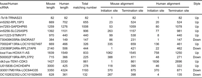
Generation of the kidney stone disease mouse model While calcium crystal deposition was undetectable in the control group (Figure 1A), several refractive irregular crystals were observed in the junction of the renal cortex and medulla in the experimental group after 5 days of glyoxylate administration (Figure 1B). Additionally, tubular epithelial cells were deformed and focally sloughed from the basement membranes. These findings indicated the successful establishment of the model.
lncRNA expression in the mouse kidneys was induced by glyoxylate administration
The hierarchical clustering showed systematic differ-ences in the expression of lncRNAs and protein-coding RNAs between the kidneys of the mice administered glyoxylate and those in the control group (Figure 2A). We identified 2165 differentially expressed genes; among these, 1421 genes were upregulated, and 744 genes were downregulated in the glyoxylate-exposed mice. A total of 376 lncRNAs were expressed in the CaOx-exposed group, of which 154 were upregulated, and 222 were downregulated compared to those in the control group. These genes were classified into different functional categories according to the GO terms for the cellular components. We found that these differentially expressed transcripts were mainly associated with the functions of the proteinaceous extracellular matrix (ECM), basement membrane, and collagen (Figure 2B). The main KEGG pathways for the differentially expressed genes involved Table 1.Real-time RT-PCR primers.
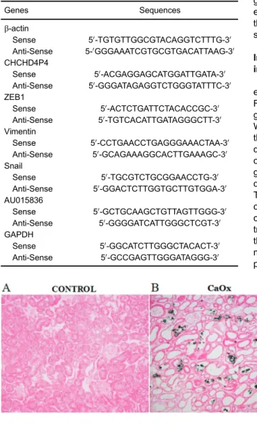
Genes Sequences
b-actin
Sense 50-TGTGTTGGCGTACAGGTCTTTG-30 Anti-Sense 5-0GGGAAATCGTGCGTGACATTAAG-30 CHCHD4P4
Sense 50-ACGAGGAGCATGGATTGATA-30 Anti-Sense 50-GGGATAGAGGTCTGGGTATTTC-30 ZEB1
Sense 50-ACTCTGATTCTACACCGC-30 Anti-Sense 50-TGTCACATTGATAGGGCTT-30 Vimentin
Sense 50-CCTGAACCTGAGGGAAACTAA-30 Anti-Sense 50-GCAGAAAGGCACTTGAAAGC-30 Snail
Sense 50-TGCGTCTGCGGAACCTG-30 Anti-Sense 50-GGACTCTTGGTGCTTGTGGA-30 AU015836
Sense 50-GCTGCAAGCTGTTAGTTGGG-30 Anti-Sense 50-GGGGATCATTGGGCTCGT-30 GAPDH
Sense 50-GGCATCTTGGGCTACACT-30 Anti-Sense 50-GCCGAGTTGGGATAGGG-30
cytokine-cytokine receptor interactions, ECM-receptor inter-actions, complement and coagulation cascades, and focal adhesion (Figure 2C). Thus, both the GO and pathway analyses suggested that the majority of the differentially expressed mRNAs may be related to the extracellular matrix.
CHCHD4P4 was upregulated in HK-2 cells that were exposed to COM
To analyze the human lncRNAs that are homologs of the mouse lncRNAs, we applied the BLAST algorithm to
mice. Interestingly, the human homolog of AU015836, CHCHD4P4, was also found to be upregulatedin vitroin the human proximal tubular epithelial cells that were exposed to COM compared to that in the untreated cells (Figure 3B). CHCHD4P4 is a human lncRNA that is 425 bp in length and is located on chromosome 3. Approximately 70% of the CHCHD4P4 lncRNAs were found in the nuclei, and the remainder were located in the cytoplasm (Figure 3C).
CHCHD4P4 promoted EMT in HK-2 cells that were exposed to COM
To assess whether CHCHD4P4 regulates EMT, we
first examined the effect of CHCHD4P4 on cellular phenotypes. The overexpression of CHCHD4P4 (Figure 4A) induced mesenchymal-like morphological features in the HK-2 cells and increased the transcript levels of
ZEB1, Vimentin, and Snail (Figure 4B). The
immuno-fluorescence and western blotting indicated that the
Figure 3.lncRNA CHCHD4P4 is upregulated in the HK-2 cells that were exposed to calcium oxalate monohydrate.A, Application of the BLAST algorithm in mouse and human lncRNAs identi-fied 15 pairs of homologous lncRNAs (Table 2). B, Validation of the differential expression of CHCHD4P4 and AU015836 in the CaOx models and control groups using RT-PCR. C, Nuclear and cytoplasmic CHCHD4P4 RNAs from the HK-2 cells. Data are reported a means±SD. *Po0.05, **Po0.01 (t-test).
Table 2.Fifteen pairs of homologous long non-coding RNAs (lncRNAs).
Mouse/Human Mouse Human Total Mouse alignment Human alignment Style
lncRNA length length matching number
Initiation site Termination site Initiation site Terminaton site
n-Ts18-TRNAS23 82 82 82 1 82 1 82 Up
Gm5292-RPL15P3 669 702 655 23 524 20 524 Up
Gm7293-GAPDHP65 1259 1276 1008 53 1059 80 1078 Up
Gm5256-SLC25A5P5 1392 1101 906 263 1157 77 981 Up
Gm11223-STMN1P1 970 440 440 99 538 8 440 Up
5730408K05Rik-SNORA57 384 149 147 86 231 1 147 Down
1700024F13Rik-LOC101927497 669 466 326 335 659 136 461 Up
B230369F24Rik-RPL27AP6 2140 506 444 1 421 22 462 Down
Hoxa11as-HOXA11-AS 1839 1549 833 1062 1545 758 1250 Up
1500002F19Rik-RPL37P2 715 366 225 388 611 48 271 Down
Cdk3-ps-TEN1-CDK3 1427 3330 861 1 861 1836 2696 Up
AU015836-CHCHD4P4 3000 425 278 118 395 46 322 Up
A930017M01Rik-LOC644335 3350 4324 1193 370 875 370 871 Down
overexpression of CHCHD4P4 resulted in higher vimen-tin protein levels and lower E-cadherin protein levels compared to the levels in the controls (COM-pcDNA3.1) (Figure 4C and D). More importantly, the silencing of CHCHD4P4 (Figure 5A) produced the opposite results (Figure 5B–D).
CHCHD4P4 inhibited proliferation in HK-2 cells that were exposed to COM
was the most obvious following the COM+ pcDNA3.1-CHCHD4P4 treatment. In our attempt to further investigate this result, we found that there were more apoptotic cells in the COM+pcDNA3.1-CHCHD4P4 group (Figure 6C). In contrast, only a slight difference was observed in the cell cycle progression (Figure 6D) between the COM+ pcDNA3.1-CHCHD4P4 and COM+pcDNA3.1 samples. This result suggested that CHCHD4P4 possibly limits the HK-2 cell proliferation by promoting apoptosisin vitroand
not by altering the cell cycle. Consistent with this result, the depletion of CHCHD4P4 produced the opposite effects (Figure 7A–D).
Discussion
failure (2,11). A decline in renal function is frequently accompanied by interstitial fibrosis, which is a common pathological occurrence in chronic kidney diseases (3,4). Therefore, the development of medical treatments that disrupt the progression of renal damage is important for protecting kidney function in patients with crystal nephrop-athies. Although the generation of experimental animal models of kidney stone disease has aided the study of these conditions (12–14), few effective therapeutic targets have been validated in clinical practice. An increasing number of studies have revealed that EMT is a key feature
of kidneyfibrosis (15–18). Injury to tubule epithelial cells (TECs) leads to a loss of functional parenchyma and induces evasive survival mechanisms, such as EMT initiation mediated through the TGF-b1-induced expres-sion of Twist1 and Snail (19).
certain lncRNA. We hypothesized that the EMT-related genes are regulated by a specific lncRNA during renal damage caused by CaOx.
Through a microarray analysis, hundreds of lncRNAs were found to be differentially expressed (fold-change 41.5 and Po0.05) in the kidney tissues from mice that were exposed to glyoxylate compared with those from mice in the control group. The differentially expressed genes were classified into different functional categories according to the GO database for cellular components. The GO categories for the differentially expressed genes
comprises a critical series of events in which ECM and cell-cell interactions are altered to release epithelial cells from the surrounding tissue (24).
The genome sequences of mice and humans are highly divergent. Therefore, we applied the BLAST algorithm to mouse and human lncRNAs to identify homologous lncRNAs. Interestingly, we found that CHCHD4P4 is the human homolog of AU015836, and both genes were upregulated in the CaOx models compared to their levels in the respec-tive control groups. We constructed stable CHCHD4P4-overexpressing and CHCHD4P4-knockdown HK-2 cells to evaluate the biological role of this molecule. We found that the majority of the CHCHD4P4 transcripts were distributed in the nuclei of the cells. Moreover, the mesenchymal pheno-type was reduced, and expression of epithelial markers was promoted in the CHCHD4P4-knockdown HK-2 cells. In contrast, the exogenous expression of CHCHD4P4 promoted EMT. The EMT process is associated with a decrease in cell proliferation in various disease conditions, such as lung cancer (25), urinary obstruction (7), and colo-rectal cancer (26). Therefore, we sought to determine the functional relevance of the association between CHCHD4P4 and cell proliferation. Interestingly, we observed that CHCHD4P4 inhibited cell proliferation. Some studies have proven that apoptosis and EMT in TECs result in a cycle of damage and host response, leading to chronic fibrosis. Preventing EMT in injured TECs results in the preserva-tion of their funcpreserva-tion, decreased pathological secrepreserva-tion, and alleviation of cell cycle arrest, all of which improve organ function (27,28). In our study, TECs exposed to
COM showed EMT, which promoted apoptosis. However, the process was not associated with the cell cycle.
Furthermore, consistent with the result of a previous study (5), EMT occurred in the mice exposed to glyoxylate, which was not the case in the control mice. Our current work shows that the CHCHD4P4 lncRNA, which is the human homolog of AU01856, participated in the regulation of EMT, cell proliferation and apoptosis in COM-exposed HK-2 cells.
Ourfindings showed that CHCHD4P4 promoted EMT and inhibited cell proliferation in COM-exposed HK-2 cells, suggesting that CHCHD4P4 played a critical role in renal damage and nephrogenesis. Understanding the precise molecular mechanisms by which this lncRNA functions will be critical for exploring potential new strategies for the early diagnosis and treatment of kidney stone dis-ease. Further research is necessary to explore the mech-anism by which the EMT-related genes are regulated by CHCHD4P4.
Acknowledgments
The microarray analyses were performed and the results were analyzed by Shanghai Sencichip Infotech Co., Ltd. (Shanghai, China) and Novel Bioinformatics Company (Shanghai, China). Dr. Jun Xiong, Department of Anatomy, Second Military Medical University, provided the HK-2 cell line. This study was supported by the National Scientific Foundation of China (Grant Nos. 81270773 and 81600521).
References
1. Moe OW. Kidney stones: pathophysiology and medical management. Lancet 2006; 367: 333–344, doi: 10.1016/ S0140-6736(06)68071-9.
2. Perazella MA. Crystal-induced acute renal failure.Am J Med 1999; 106: 459–465, doi: 10.1016/S0002-9343(99)00041-8. 3. Kanlaya R, Sintiprungrat K, Thongboonkerd V. Secreted products of macrophages exposed to calcium oxalate crystals induce epithelial mesenchymal transition of renal tubular cells via RhoA-dependent TGF-b1 pathway.Cell Biochem Biophys 2013; 67: 1207–1215, doi: 10.1007/s12013-013-9639-z. 4. Liu M, Liu YZ, Feng Y, Xu YF, Che JP, Wang GC, Zheng JH.
Novel evidence demonstrates that epithelial-mesenchymal transition contributes to nephrolithiasis-inducedrenal fi bro-sis. J Surg Res 2013; 182: 146–152, doi: 10.1016/j.jss. 2012.07.066.
5. Hu H, Chen W, Ding J, Jia M, Yin J, Guo Z. Fasudil prevents calcium oxalate crystal deposit and renalfibrogenesis in glyoxylate-induced nephrolithic mice.Exp Mol Pathol2015; 98: 277–285, doi: 10.1016/j.yex-mp.2015.02.006.
6. Wu CF, Chiang WC, Lai CF, Chang FC, Chen YT, Chou YH, et al. Transforming growth factorb-1 stimulates profibrotic epithelial signaling to activate pericyte-myofibroblast transition in obstructive kidneyfibrosis.Am J Pathol2013; 182: 118– 131, doi: 10.1016/j.ajpath.2012.09.009.
7. Lovisa S, LeBleu VS, Tampe B, Sugimoto H, Vadnagara K, Carstens JL, et al. Epithelial-to-mesenchymal transition induces cell cycle arrest and parenchymal damage in renal fibrosis. Nat Med 2015; 21: 998–1009, doi: 10.1038/nm. 3902.
8. Mercer TR1, Dinger ME, Mattick JS. Long non-coding RNAs: insights into functions. Nat Rev Genet 2009; 10: 155–159, doi: 10.1038/nrg2521.
9. Zhou Q, Chung AC, Huang XR, Dong Y, Yu X, Lan HY. Identification of novel long noncoding RNAs associated with TGF-b/Smad3-mediated renal inflammation andfibrosis by RNA sequencing.Am J Pathol 2014; 184: 409–417, doi: 10.1016/j.ajpath.2013.10.007.
10. Zhou Q, Huang XR, Yu J, Yu X, Lan HY. Long noncoding RNA Arid2-IR Is a novel therapeutic target for renal infl am-mation. Mol Ther 2015; 23: 10341043, doi: 10.1038/mt. 2015.31.
11. Rule AD, Krambeck AE, Lieske JC. Chronic kidney disease in kidney stone formers.Clin J Am Soc Nephrol2011; 6: 2069–2075, doi: 10.2215/CJN.10651110.
Urol Res 2012; 40: 121–129, doi: 10.1007/s00240-011-0400-z.
13. Knauf F, Asplin JR, Granja I, Schmidt IM, Moeckel GW, David RJ, et al. NALP3-mediated inflammation is a principal cause of progressive renal failure in oxalate nephropathy. Kidney Int2013; 84: 895–901, doi: 10.1038/ki.2013.207. 14. Mulay SR, Kulkarni OP, Rupanagudi KV, Migliorini A,
Darisipudi MN, Vilaysane A, et al. Calcium oxalate crystals induce renal inflammation by NLRP3-mediated IL-1b secre-tion. J Clin Invest 2013; 123: 236-246, doi: 10.1172/ JCI63679.
15. Iwano M, Plieth D, Danoff TM, Xue C, Okada H, Neilson EG. Evidence that fibroblasts derive from epithelium during tissue fibrosis. J Clin Invest 2002; 110: 341–350, doi: 10.1172/JCI0215518.
16. LeBleu VS, Taduri G, O’Connell J, Teng Y, Cooke VG, Woda C, et al. Origin and function of myofibroblasts in kidneyfibrosis. Nat Med2013; 19: 1047–1053, doi: 10.1038/nm.3218. 17. Zeisberg M, Kalluri R. Fibroblasts emerge via
epithelial-mesenchymal transition in chronic kidney fibrosis. Front Biosci2008; 13: 6991–6998, doi: 10.2741/3204.
18. Zeisberg EM, Potenta SE, Sugimoto H, Zeisberg M, Kalluri R. Fibroblasts in kidneyfibrosis emerge via endothelial-to-mesenchymal transition.J Am Soc Nephrol2008; 19: 2282– 2287, doi: 10.1681/ASN.2008050513.
19. Wynn TA. Cellular and molecular mechanisms offibrosis. J Pathol2008; 214: 199–210, doi: 10.1002/path.2277. 20. Sanchez-Elsner T, Gou D, Kremmer E, Sauer F. Noncoding
RNAs of trithorax response elements recruit Drosophila
Ash1 to Ultrabithorax.Science2006; 311: 1118–1123, doi: 10.1126/science.1117705.
21. Ponting CP, Oliver PL, Reik W. Evolution and functions of long noncoding RNAs. Cell 2009; 136: 629–641, doi: 10.1016/j.cell.2009.02.006.
22. Martens JA, Laprade L, Winston F. Intergenic transcription is required to repress the Saccharomyces cerevisiaeSER3 gene.Nature2004; 429: 571–574, doi: 10.1038/nature02538. 23. Carrieri C, Cimatti L, Biagioli M, Beugnet A, Zucchelli S, Fedele S, et al. Long non-coding antisense RNA controls Uchl1 translation through an embedded SINEB2 repeat. Nature2012; 491: 454–457, doi: 10.1038/nature11508. 24. Radisky DC. Epithelial-mesenchymal transition. J Cell Sci
2005; 118 (Part 19): 4325–4326, doi: 10.1002/jcb.21821. 25. Ploenes T, Scholtes B, Krohn A, Burger M, Passlick B,
Müller-Quernheim J, et al. CC-chemokine ligand 18 induces epithelial to mesenchymal transition in lung cancer A549 cells and elevates the invasive potential.PLoS One2013; 8: e53068, doi: 10.1371/journal.pone.0053068.
26. Wang B, Li W, Liu H, Yang L, Liao Q, Cui S, et al. miR-29b suppresses tumor growth and metastasis in colorectal cancer via downregulating Tiam1 expression and inhibiting epithelial-mesenchymal transition.Cell Death Dis2014; 5: e1335, doi: 10.1038/cddis.2014.304.
27. Canaud G, Bonventre JV. Cell cycle arrest and the evolution of chronic kidney disease from acute kidney injury.Nephrol Dial Transplant2015; 30: 575–583, doi: 10.1093/ndt/gfu230. 28. Wynn TA. Fibrosis under arrest. Nat Med2010; 16: 523–