Effects of entorhinal cortex lesions
on memory in different tasks
Centro de Memória, Departamento de Bioquímica, Instituto de Ciências Básicas da Saúde,
Universidade Federal do Rio Grande do Sul, 90035-003 Porto Alegre, RS, Brasil
G.P. Gutierrez-Figueroa, C. Dalmaz and I. Izquierdo
Abstract
Lesions of the entorhinal cortex produce retrograde memory impair-ment in both animals and humans. Here we report the effects of bilateral entorhinal cortex lesions caused by the stereotaxic infusion of N-methyl-D-aspartate (NMDA) in rats at two different moments, before or after the training session, on memory of different tasks: two-way shuttle avoidance, inhibitory avoidance and habituation to an open field. Pre- or post-training entorhinal cortex lesions caused an impairment of performance in the shuttle avoidance task, which agrees with the previously described role of this area in the processing of memories acquired in successive sessions. In the inhibitory avoid-ance task, only the post-training lesions had an effect (amnesia). No effect was observed on the open field task. The findings suggest that the role of the entorhinal cortex in memory processing is task-depend-ent, perhaps related to the complexity of each task.
Correspondence
C. Dalmaz Centro de Memória Departamento de Bioquímica Instituto de Ciências Básicas da Saúde, UFRGS
Rua Ramiro Barcellos, 2600 90035-003 Porto Alegre, RS Brasil
Fax: 55 (051) 316-3505
Research supported by FINEP. G.P. Gutierrez-Figueroa is a visiting fellow from the University of Quito, Ecuador.
Received December 26, 1995 Accepted March 10, 1997
Key words •Entorhinal cortex
•Memory
•NMDA lesions
Introduction
The entorhinal cortex is superficial to the amygdala, forming the anterior part of the parahippocampal gyrus (1). It processes dif-ferent types of memories, including aversive and spatial memories (2-4) both in rats (2,5,6) and primates (3,7). In humans, the most prominent lesions of Alzheimer’s disease are found in the entorhinal cortex (1,8).
The entorhinal cortex has two-way mono-and polysynaptic connections with the amygdala, the hippocampus and the medial septum (9), structures known to be involved in the processing of memories (10). Thus, the entorhinal cortex has been suggested to be an
area for the integration of information previ-ously processed by the amygdala, hippocam-pus and/or septum and for the distribution of this information to other parts of the brain (1,10). Experiments using infusion of selective neurotransmitter receptor agonists and antago-nists into the entorhinal cortex at different times after training and at the time of testing suggest that this structure has a delayed post-training role in memory (4,10) particularly in the integration or summation of memories ac-quired in consecutive trials or episodes (11).
Material and Methods
Subjects
Male adult Wistar rats (90-120 days, weighing 230-270 g) from our breeding stock were housed in groups of 4 animals per cage and kept under a normal 12-h dark/light cycle with food and water ad libitum.
Surgical procedures
The animals were anesthetized with Thionembutal (thiopental sodium, 40 mg/ kg, ip), and placed in a stereotaxic apparatus. The needle (31 gauge) connected to a Hamilton syringe (5 µl) and attached to the stereotaxic injector was gently lowered into the entorhinal cortex through a skull hole using the following coordinates (bregma sys-tem), according to the atlas of Paxinos and Watson (12): AP = -0.67 cm, LL = +0.50 cm, and DV = -0.58 cm from the dura mater, with the nosebar at -0.33 cm from the interaural line. A bilateral infusion (0.5 µl/structure) of 10 mg/ml NMDA was manually delivered for 2 min, for a total dose of 5 µg per struc-ture. The needle was withdrawn slowly after 5 min (13,14). Immediately after surgery the animals received an intramuscular injection of penicillin. The animals were allowed to recover for 1 week before being submitted to the behavioral tasks (training and testing, in the case of pretraining lesion) or to the test session (post-training lesion). Control ani-mals were injected with saline.
Behavioral procedures
Step-down inhibitory avoidance. The animals were trained and tested for the one-way step-down inhibitory task using a 50 x 25 x 25-cm plywood box with a glass-wall front and a floor consisting of 1-mm bronze bars spaced 10 mm apart (15). The left end of the grid was covered with a 5-cm high, 25-cm wide, and 7.5-25-cm long wood platform.
During the training trial, the animals were gently held by their bodies and lowered onto the platform with their noses pointing to the left corner, when a timer was activated. The timer measured the latency to step down (i.e., all four paws on the grid) when an intermittent footshock (0.5 mA, 60 Hz) was delivered until the animal climbed back onto the platform. In the test session, the animals were again placed on the platform. No footshock was given in the testing session. A 300-s ceiling was imposed on testing session latency measurements; latencies ≥300 s were counted as 300 s. Differences in testing-training session latencies were used as reten-tion scores.
Two-way active avoidance. The animals were trained and tested in a two-way active avoidance task in an automatic 50 x 25 x 25-cm opaque acrylic shuttle-box whose floor was a grid of bronze bars 1 mm in diameter spaced 10 mm apart (16). Both the training and the test sessions consisted of 30-tone footshock trials preceded by 3-min free ex-ploration of the shuttle-box, with no stimula-tion. In each session, the intertrial interval varied at random from 10 to 50 s. Each 7-s tone was immediately followed by a 0.5-mA footshock delivered until the animals crossed the midline. The shock was omitted if the animals crossed to the opposite side of the grid during the tone (avoidance response). The difference in the avoidance responses between the test and the training trials was used as the measure of retention (17,18).
used as the measure of retention.
Histological analysis
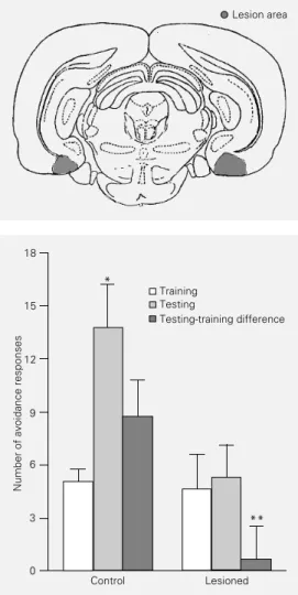
After the behavioral studies, exact place-ment of the site of injection (lesion) was determined histologically. The rats were anes-thetized with an overdose of Thionembutal and perfused with saline solution followed by 10% formaldehyde solution. Slices were stained with hematoxylin-eosin. The loca-tion of the lesion is shown in Figure 1. Behavioral data from only the animals with the lesion located in the intended site were used.
Experimental design
In each experiment the rats were ran-domly assigned to the different experimental groups.
In Experiment 1, the animals were sub-mitted to the surgical procedures. After one week for recovery, lesioned and control rats were submitted to only one behavioral task: step-down inhibitory avoidance, two-way active avoidance or habituation to the open field.
In Experiment 2, the animals were di-vided into three groups, and each group was trained in one of the behavioral tasks used: step-down inhibitory avoidance, two-way ac-tive avoidance or habituation to the open field. Twenty-four hours later, the rats were submitted to the surgical procedures, with half of the animals in each group receiving saline and the other half NMDA injections. After one week for recovery, the animals were submitted to the respective test session. In both experiments, the researcher who collected the behavioral data was blind to the condition of the animals (control or lesioned).
Statistical analysis
Parametric data are reported as means ±
SEM and were analyzed by the Student t -test. Nonparametric data are reported as median (interquartile range) and were ana-lyzed by the Mann-Whitney U-test.
Results
Experiment 1: Effect of pretraining lesion of the entorhinal cortex on memory of different tasks
In the two-way active avoidance task there was no significant difference in train-ing performance between groups (t(14) = 0.18; P>0.05, Student t-test). Control ani-mals showed better retention scores when compared to NMDA-lesioned animals (t(14) = 2.91; P<0.02, Student t-test) (Figure 2).
Figure 1 - Drawing of a repre-sentative coronal section show-ing the localization of the lesion (shaded area).
Figure 2 - Effect of a pretraining lesion of the entorhinal cortex on performance in a two-way ac-tive avoidance task. Data are re-ported as means ± SEM (N = 8 animals/group). *P<0.005 com-pared to the training session (paired Student t-test). **P<0.02 compared to the control group (unpaired Student t-test).
Number of avoidance responses
18
Training Testing
Testing-training difference
**
15
12
9
6
3
0
Control Lesioned
Lesion area
Comparison of training and testing sessions showed a significant difference in the con-trol group (t(7) = 4.27; P<0.005, Student paired t-test), while no difference was found in the NMDA-lesion group (t(7) = 0.33; P>0.05, paired Student t-test).
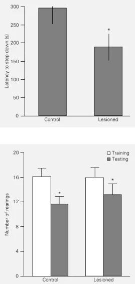
In the step-down inhibitory avoidance task there was a significant difference be-tween groups in the training session (t(14) = 2.80; P<0.02, Student t-test). The NMDA-lesioned group had a lower latency to step down (3.63 ± 0.91) compared to the saline group (8.50 ± 1.49). In the testing session, there was no difference in performance be-tween groups (U = 24.0; P>0.05, Mann-Whitney U-test) (Figure 3).
In the habituation to the open field there was no significant difference in training or testing performance between groups (P>0.05, Student t-test). Both groups showed a sig-nificant difference between training and test-ing sessions (P<0.05, paired Student t-test), which is interpreted as an adequate memory for the task (Figure 4).
Experiment 2: Effect of post-training lesion of the entorhinal cortex on memory of different tasks
In the two-way active avoidance task there was no significant difference in train-ing performance between groups (t(16) = 1.65; P>0.05, Student t-test). Control ani-mals showed better retention scores when compared to NMDA-lesioned animals (P<0.001, Student t-test) (Figure 5). Com-parison of training and testing sessions showed a significant difference in the con-trol group (t(8) = 4.5; P<0.002, paired Stu-dent t-test), while no difference was found in the NMDA-lesioned group (t(8) = 1.96; P>0.05, paired Student t-test).
In the step-down inhibitory avoidance task there was no significant difference be-tween groups in the training session (t(14) = 1.83; P>0.05, Student t-test). In the testing session there was a significant difference in
Figure 3 - Effect of a pretraining lesion of the entorhinal cortex on performance in a step-down inhibitory avoidance task. Data are reported as median (inter-quartile range) (N = 8 animals/ group). There were no signifi-cant differences between groups (P>0.05, Mann-Whitney U-test).
Latency to step down (s)
300
250
200
150
100
50
0
Control Lesioned
Figure 4 - Effect of a pretraining lesion of the entorhinal cortex on performance in an open field task. Data are reported as means ± SEM (N = 10 animals/ group). There were no significant differ-ences between groups (P>0.05, Student t-test). *P<0.05 com-pared to the training session (paired Student t-test). Number of rearings
16
12
8
4
0
Training Testing
Control Lesioned
*
Figure 5 - Effect of a post-train-ing lesion of the entorhinal cor-tex on performance in a two-way active avoidance task. Data are reported as means ± SEM (N = 9 animals/group). *P<0.002 com-pared to the training session (paired Student t-test). **P< 0.001 compared to the control group (unpaired Student t-test).
Number of avoidance responses
18
15
12
9
6
3
0
-3
-6
Control Lesioned
*
**
Training Testing Testing-training difference
performance between groups (U = 8; P<0.02, Mann-Whitney U-test). The NMDA-lesioned group had a lower latency to step down, suggesting a deficit of memory for this task (Figure 6).
In the habituation to the open field there was no significant difference in training or testing performance between groups (P>0.05, Student t-test). Both groups showed a sig-nificant difference between training and test-ing sessions (P<0.05, paired Student t-test), which is interpreted as an adequate memory for the task (Figure 7).
Discussion
The results show that bilateral NMDA lesions of the entorhinal cortex have task-specific effects on memory. In the two-way shuttle avoidance, memory impairment was observed with both pre- and post-training lesions. In the inhibitory avoidance, only post-training lesions had an amnestic effect, which was far from complete. In this task, pretraining lesions caused a significant dif-ference between groups in the training ses-sion, that may have been due to motor im-pairment, with a consequent alteration in behavior. However, this was not the case, since intertrial crossings in the two-way ac-tive avoidance, or crossings in the habitua-tion to the open field presented no differ-ence. Furthermore, in the testing session of inhibitory avoidance there was no difference in performance between groups. In the open field task neither pre- nor post-training le-sions had any effect.
The higher sensitivity of the shuttle avoid-ance task to entorhinal cortex lesions is con-sistent with the idea that this area may play an integrative role in information processing of successively acquired memories, since this task involves several consecutive trials (4,11,19-21).
The entorhinal cortex could, in principle, be involved in different aspects of memory processing: acquisition, consolidation and
retrieval. The impairment of performance in shuttle avoidance caused by pretraining le-sions may be due to an effect on any of these phases. The results with post-training le-sions suggest that its primary effect might be on consolidation and/or retrieval. Previous studies (4,10,11) have suggested a major role for this structure in late consolidation. Willner et al. (20), however, have shown that the entorhinal cortex does also participate in acquisition, although its role does not be-come manifest unless this structure is hin-dered within the late post-training period.
Another way of viewing these findings is that the effect of the NMDA lesions of the entorhinal cortex is correlated with the asso-ciative characteristics of the task. In the
Latency to step down (s)
300
250
200
150
100
50
0
Control Lesioned
*
Figure 6 - Effect of a post-train-ing lesion of the entorhinal cor-tex on performance in a step-down inhibitory avoidance task. Data are reported as median (in-terquartile range) (N = 8 animals/ group). *P<0.05 compared to the control group (Mann-Whitney U-test).
Figure 7 - Effect of a post-train-ing lesion of the entorhinal cor-tex on performance in an open field task. Data are reported as means ± SEM (N = 10 animals/ group). There were no signifi-cant differences between groups (P>0.05, Student t-test). *P<0.005 compared to the train-ing session (paired Student t -test).
Number of rearings
20
16
12
8
4
0
Control Lesioned
*
*
shuttle avoidance paradigm, indeed, the ani-mals must compare the outcome of their past behavior in each consecutive trial; this is needed just once in the inhibitory avoidance task, and is in fact irrelevant in the open field
task. Thus, the present data might be consid-ered to support previous findings that sug-gest a comparator or sorting-out role for the entorhinal cortex at the time of acquisition and retrieval (20,22).
References
1. Van Hoesen GW, Hyman BT & Damasio AR (1991). Entorhinal cortex pathology in Alzheimer´s disease. Hippocampus, 1: 1-8.
2. Thompson R (1976). Entorhinal-subicular lesions: amnestic effects on an assort-ment of learned responses in the white rat. Bulletin of the Psychological Society, 8: 433-434.
3. Zola-Morgan S, Squire LR, Amaral DG & Suzuki W (1989). Lesions of perirhinal and parahippocampal cortex that spare the amygdala and hippocampal formation pro-duce severe memory impairment. Jour-nal of Neuroscience, 9: 4355-4370. 4. Ferreira MBC, Da Silva RC, Medina JH &
Izquierdo I (1992). Late post-training memory processing by entorhinal cortex: role of NMDA and GABA-A receptors.
Pharmacology, Biochemistry and Behav-ior, 41: 767-771.
5. Glasier M, Chen X, Sutton R & Stein D (1991). Effects of unilateral entorhinal cor-tex lesion and GM1 ganglioside treatment on water maze performance. Society for Neuroscience Abstracts, 17: 132. 6. Johnson DL & Kesner RP (1991).
Differ-ential effects of entorhinal cortex and hip-pocampal lesions on performance of a spatial location recognition. Society for Neuroscience Abstracts, 17: 131. 7. Zola-Morgan S, Squire LR, Clower RP &
Rempel NL (1993). Damage to the perirhi-nal cortex exacerbates memory impair-ment following lesions to the hippocam-pal formation. Journal of Neuroscience, 13: 251-265.
8. Hyman BT, van Hoesen GW & Damasio AR (1990). Memory related neural sys-tems in Alzheimer’s disease: An anatomic study. Neurology, 40: 1721-1730.
9. Witter MP, Groenewegen HJ, Lopes da Silva FH & Lohman AHM (1989). Func-tional organization of the extrinsic and in-trinsic circuitry of the parahippocampal re-gion. Progress in Neurobiology, 33: 161-253.
10. Izquierdo I & Medina JH (1995). Correla-tion between the pharmacology of memory and the pharmacology of long-term potentiation. Neurobiology of Learn-ing and Memory, 63: 17-29.
11. Ferreira MBC, Wolfman C, Walz R, Da Silva RC, Zanatta MS, Medina JH & Izquierdo I (1992). NMDA-dependent, GABA-A-sensitive role of the entorhinal cortex in posttraining memory process-ing. Behavioral Pharmacology, 3: 387-394. 12. Paxinos G & Watson C (1986). The Rat Brain in Stereotaxic Coordinates. 2nd edn. Academic Press, Sydney.
13. Cahill L & McGaugh JL (1990). Amygda-loid complex lesions differentially affect retention of task using appetitive and aver-sive reinforcement. Behavioral Neurosci-ence, 104: 532-543.
14. Sananes BC & Davis M (1992). N-methyl-D-aspartate lesions of the lateral and basolateral nuclei of the amygdala block fear, potentiate startle and shock sensiti-zation startle. Behavioral Neuroscience, 106: 72-80.
15. Bianchin M, Walz R, Ruschel AC, Zanatta MS, Da Silva RC, Bueno e Silva M, Paczko N, Medina JH & Izquierdo I (1993). Memory expression is blocked by the in-fusion of CNQX into the hippocampus and/or amygdala several days after train-ing. Behavioral and Neural Biology, 59: 83-86.
16. Pereira ME, Rosat RM, Huang CH, Godoy MG & Izquierdo I (1989). Inhibition by di-azepam of the effect of additional training and extinction on the retention of shuttle avoidance behavior in rats. Behavioral Neuroscience, 103: 202-205.
17. Izquierdo I (1979). Effect of naloxone and morphine on various forms of memory in the rat: possible role of endogenous opi-ate mechanisms in memory consolida-tion. Psychopharmacology, 66: 199-203. 18. Netto CA & Maltchik M (1990). Distinct
mechanisms underlying memory modula-tion after the first and the second session of two avoidance tasks. Behavioral and Neural Biology, 53: 29-38.
19. Izquierdo I (1992). The neurobiology of memory consolidation. Neuroscience, 18: 1-11.
20. Willner P, Bianchin M, Walz R, Bueno e Silva M, Zanatta MS & Izquierdo I (1993). Muscimol infused into the entorhinal cor-tex prior to training blocks the involve-ment of this area in post-training memory processing. Behavioral Pharmacology, 4: 95-99.
21. Zola-Morgan S (1990). The neuropsychol-ogy of memory. Annals of the New York Academy of Sciences, 608: 434-456. 22. Jerusalinsky D, Quillfeldt JA, Walz R, Da