Involvement of the hippocampus,
amygdala, entorhinal cortex and
posterior parietal cortex in memory
consolidation
1Centro de Memória, Departamento de Bioquímica, Instituto de Biociências,
Universidade Federal do Rio Grande do Sul, 90046-900 Porto Alegre, RS, Brasil
2Laboratorio de Neurorreceptores, Instituto de Biología Celular, Facultad de Medicina,
(1121) Buenos Aires, Argentina M.S. Zanatta1,
J.H. Quillfeldt1,
E. Schaeffer1,
P.K. Schmitz1,
J. Quevedo1,
J.H. Medina2
and I. Izquierdo1
Abstract
A total of 182 young adult male Wistar rats were bilaterally implanted with cannulae into the CA1 region of the dorsal hippocampus and into the amygdaloid nucleus, the entorhinal cortex, and the posterior parietal cortex. After recovery, the animals were trained in a step-down inhibitory avoidance task. At various times after training (0, 30, 60 or 90 min) the animals received a 0.5-µl microinfusion of vehicle (saline) or 0.5 µg of muscimol dissolved in the vehicle. A retention test was carried out 24 h after training. Retention test performance was hindered by muscimol administered into both the hippocampus and amygdala at 0 but not at 30 min posttraining. The drug was amnestic when given into the entorhinal cortex 30, 60 or 90 min after training, or into the parietal cortex 60 or 90 min after training, but not before. These findings suggest a sequential entry in operation, during the posttraining period, of the hippocampus and amygdala, the entorhinal cortex, and the posterior parietal cortex in memory processing.
Correspondence
I. Izquierdo
Departamento de Bioquímica Instituto de Biociências, UFRGS 90046-900 Porto Alegre, RS Brasil
Fax: 55 (051) 227-1343 Research supported by FAPERGS, CNPq and Fundação Vitae.
Received February 15, 1996 Accepted December 4, 1996
Key words
•Memory consolidation •Hippocampus and amygdala •Entorhinal cortex
•Parietal cortex
Introduction
The hippocampus and amygdala are con-nected with each other and with the entorhinal cortex, and the latter is connected with the posterior parietal area by afferent and effer-ent pathways (1,2). Lesions and pharmaco-logical manipulations of the hippocampus, amygdala and entorhinal cortex are known to have profound effects on memory con-solidation and retrieval (1,3-9). Lesions of
the posterior parietal area in the rat have less dramatic, but also deleterious effects on spa-tial memory (10,11) and on at least some (11) but not all types of non-spatial memory (12). Single unit activity in the parietal cor-tex changes during different forms of learn-ing in cats (13).
hippocampus or amygdala (5,14) and 1-3 h after training into the entorhinal cortex (3,4,7). This observation suggests a role for these structures in different phases of memory consolidation (3-8).
The objective of the present study was to determine the effect of muscimol infused bilaterally into different brain areas at differ-ent times after training on retdiffer-ention of inhib-itory avoidance. The findings suggest a role of the hippocampus and amygdala imme-diately after training (which is in agreement with previous results reported by Izquierdo et al. (6)), a role of the entorhinal cortex starting 30 min later, and a role of the parietal cortex starting 60 min after acquisition.
Material and Methods
A total of 182 male Wistar rats (age, 3-4 months; weight, 220-350 g) were used. The animals were implanted under thionembutal anesthesia (30 mg/kg, ip) with 27-g guide cannulae aimed 1.0 mm above the following structures: the CA1 region of the dorsal hip-pocampus (A -4.3, L ±4.0, V 3.4) and the junction between the central and the lateral nuclei of the amygdala (A -2.3, L ±4.5, V 8.4) (N = 40), the surface of the entorhinal cortex (A -7.0, L 5.0, V 8.4) (N = 70), and the surface of the junction between the posterior parietal I and II regions (A -0.3, L ±6.9, V 4.5) (N = 72). The coordinates correspond to Figures 35, 27, 46 and 19, respectively, of the atlas of Paxinos and Watson (15).
Once recovered from surgery, the ani-mals were trained in a step-down inhibitory avoidance task (3-5,8,9,16). The rats were placed on a 2.5 cm high, 7.0 cm x 25.0 cm platform facing a 42.0 x 25.0 cm grid of parallel 0.1-cm caliber stainless steel bars spaced 1.0 cm apart, and their latency to step down placing their four paws on the grid was measured. During the training sessions, im-mediately after stepping down, the animals received a 0.3-mA, 2.0-s scrambled footshock. During the test sessions no
footshock was given. The training-test inter-val was 24 h. Test minus training session step-down latency was taken as a measure of retention.
At the time of infusion, 30-g cannulae were fitted into the guide cannula. The infu-sion cannula was placed immediately (0 min) or 30 min after avoidance training into the hippocampus, amygdala, or amygdala and hippocampus, and 0, 30, 60 and 90 min after training into the entorhinal or parietal cor-tex. The tip of the infusion cannula pro-truded 1.0 mm beyond that of the guide cannula and was therefore aimed at a) CA1 in the dorsal hippocampus, and the junction between the central and the basolateral amygdaloid nucleus, b) the surface of the entorhinal cortex, and c) the surface of the posterior parietal cortex. The animals re-ceived bilateral 0.5-µl infusions of saline (0.9% NaCl) or muscimol (0.5 µg/side) dis-solved in saline. The pH of the solutions was adjusted to 7.4 with 0.1 M sodium phosphate buffer. Infusions were manual and were car-ried out over a period of 45 s each, and the infusion cannula was left in place for an additional 15 s. Infusions were carried out first on the left side and then on the right side. Thus, the entire infusion procedure took about 4 min for the hippocampus and amygdala groups, and 2 min for the entorhinal and parietal cortex groups.
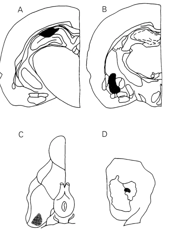
Two to 24 h after the end of the behavior-al experiments behavior-all animbehavior-als received a 0.5-µl infusion of 4% methylene blue through the infusion cannulae. Post-mortem verification of the cannula placements showed that in 36 of 40 animals implanted into the hippocam-pus and amygdala, in 68 of 70 animals im-planted into the entorhinal cortex, and in 71 of 72 animals implanted into the parietal cortex, the infusion cannula tips reached the desired structures and were within a 0.5-mm radius of the aimed locations. The correct placements of the infusion cannula tips are shown in Figure 1.
Figure 1 - Schematic drawing of rat brain stions at planes A -4.3 and -2.3, respectively, of the atlas of Paxinos and Watson (15), showing the areas reached by the microinfusion cannula tips aimed at the CA1 region of the dorsal hippocampus (A) and between the central and the basolateral nuclei of the amygdala (B) (stippled). C, Schematic drawing of the lower aspect of the rat brain, showing the area reached by the microinfusion cannula in the entorhinal cortex (stippled). D, Schematic drawing of the lateral aspect of a rat brain hemisphere illustrating the subdivisions of the parietal cortex and the area reached by the microinfusions given into this area (stippled). Only placements considered to be correct are shown here (see Material and Methods).
A
B
was carried out only for the animals with correct cannula locations. Training step-down latency differences between groups were evaluated by Kruskal-Wallis analysis of vari-ance for nonparametric data. Test minus train-ing session latency scores of different groups were compared by the individual Mann-Whitney U-test (two-tailed).
Results
Training session step-down latency dif-ferences between groups were not signifi-cant (median 5.9 s, range 1.1 to 21.9 s, H [19,155] = 1.76, P>0.1).
The results of the retention test perfor-mance are shown in Figure 2. Muscimol caused full retrograde amnesia when infused
into the hippocampus and the amygdala im-mediately (0 min), but not 30 min after train-ing. Muscimol was also amnestic when given into the entorhinal cortex 30, 60 or 90 min (but not 0 min) after training, or when given 60 or 90 min (but not 0 or 30 min) posttraining into the posterior parietal cortex.
Discussion
The present data show that, during the posttraining period, the hippocampus, amygdala, entorhinal and posterior parietal cortex are necessary for memory processing, and suggest that these structures enter into operation sequentially in the following or-der: first, immediately after training, the hip-pocampus and amygdala; 30 min later, the entorhinal cortex; 60 min after training, the parietal cortex. In the case of the entorhinal cortex, this is earlier than suggested by pre-vious experiments in which posttraining in-tervals between 0 and 90 min were not stud-ied (3,4,8), and in agreement with a study on the amnestic effect of protein kinase C in-hibitors infused into this region 30 min after training (9).
The findings on immediate posttraining muscimol administration into both hippo-campus and amygdala confirm those ob-tained previously when the drug was infused into each structure separately (5) or simulta-neously into the two structures (8). Clearly, the present data do not discriminate between these two structures in terms of the amnestic effect of muscimol. The distinction, how-ever, has been clearly made elsewhere (5,6, see 17). The hippocampus and amygdala enter into play at the time of training and in the period immediately after training, par-ticularly in this task. The hippocampus is believed to be in charge of spatial, contex-tual and other cognitive aspects of memory of the training experience and the amygdala is believed to be in charge of the emotional aspects, particularly those of an aversive nature (6,17,18).
Retention test performance (s)
60
50
40
10
0 30
20
PARIE
a b
0 30 60 90
Time (min) 70
60 50 40
10 0 -10 30 20
ENTO
a a a
0 30 60 90
50
40
30
20
10
0
HIPP + AMY
0 30
a
Figure 2 - Effect of muscimol in-fused bilaterally into both hippo-campus and amygdala (HIPP + AMY), entorhinal cortex (ENTO) or posterior parietal cortex (PARIE) on retention test perfor-mance. Data are reported on the ordinates as median (interquar-tile range) test minus training session step-down latency, in seconds. The drug (0.5 µg in 0.5 µl per side) was amnestic when infused 0 min after training but not later in HIPP + AMY; 30-90 min after training, but not be-fore, when given into ENTO, and 60-90 min after training, but not before, when given into PARIE.
aP<0.002, U values 0 to 10; bP<0.02, U values 11 to 19
The findings about muscimol given into the entorhinal cortex 90 or 180 min after training confirm those of Ferreira et al. (3,4). The sequential involvement of the different brain regions in posttraining memory pro-cessing may be mediated by the anatomical connections among these brain regions (2). The reason for the 30-min delay between the interventions of the different areas is not known. Previous findings have suggested that post-acquisition activity in the hippo-campus and amygdala, presumably long-term potentiation (LTP), must build up before it is able to trigger similar activity in the entorhinal cortex (3,4,7). Perhaps a similar explanation may account for the 30-min delay between the participation of the entorhinal cortex and that of the parietal cortex in memory pro-cessing. Indeed, a study similar to the pres-ent one was carried out using AP5 (D-2-amino-5-phosphonopentanoate), an antago-nist of glutamatergic N-methyl-D-aspartate (NMDA) receptors, and the results were very similar to those reported here for muscimol: AP5 was amnestic when given early into both hippocampus and amygdala, and after 30 and 60 min when given into the entorhinal and parietal cortex, respectively (16). For evidence on the possible role of LTP in the hippocampus, amygdala and entorhinal cor-tex in memory consolidation, see Refs. 6 and 7. The present findings illustrate the advan-tage of circumscribed drug infusion proce-dures over lesion proceproce-dures for the study of the role and timing of brain structures in
learning and memory. During a few days, extensive synaptic rearrangements occur in areas (e.g., the hippocampus) to which lesioned fibers project (e.g., those emerging from the entorhinal cortex), and vicarious structures and systems enter into play (19). This fact precludes detailed interpretations of lesion studies in terms of the functions of either the injured region or its projection sites (7). On the other hand, localized microinfusions of drugs acting at specific receptor sites, such as muscimol, exert pre-cise effects limited both in time (60 min) and space (0.5-mm radius) (20).
Previous lesion studies have failed to detect dramatic effects of posterior parietal cortex lesions on memory (e.g., 11,12), which was surprising in view of the multiple con-nections of this region with the entorhinal cortex (1,2). Lesion studies do not permit an accurate investigation of time-dependent pro-cesses. In contrast, the present experiments using the time-honored procedure of inject-ing drugs at different times after traininject-ing (21) clearly point to an important role of the posterior parietal cortex in memory consoli-dation processes, starting 60 min after train-ing.
Acknowledgments
We are thankful to M. Bianchin, R.C. Da-Silva, J.B. dos-Santos and S.G. Petry for their collaboration in some of the experi-ments.
References
1. Hyman BT, van Hoesen GT & Damasio AR (1990). Memory-related neural sys-tems in Alzheimers disease: an anatomic study. Neurology, 40: 1721-1730. 2. Witter MP, Groenewegen HJ, Lopes da
Silva FH & Lohman AHM (1989). Func-tional organization of the extrinsic and in-trinsic circuitry of the parahippocampal re-gion. Progress in Neurobiology, 33: 161-253.
3. Ferreira MBC, Da-Silva RC, Medina JH & Izquierdo I (1992). Late post-training memory processing by the entorhinal cor-tex: role of NMDA and GABA-A recep-tors. Pharmacology, Biochemistry and Be-havior, 41: 767-771.
4. Ferreira MBC, Wolfman C, Walz R, Da-Silva RC, Medina JH & Izquierdo I (1992). NMDA-dependent, GABA-A-sensitive role of the entorhinal cortex in post-training memory processing. Behavioural Pharma-cology, 3: 387-394.
5. Izquierdo I, Da Cunha C, Rosat R, Jerusalinsky D, Ferreira MBC & Medina JH (1992). Neurotransmitter receptors in-volved in post-training memory process-ing by the amygdala, medial septum and hippocampus of the rat. Behavioral and Neural Biology, 58: 16-26.
6. Izquierdo I, Medina JH, Bianchin M, Walz R, Zanatta MS, Da-Silva RC, Bueno-e-Silva M, Ruschel AC & Paczko N (1993). Memory processing by limbic system: role of specific neurotransmitter systems.
7. Izquierdo I & Medina JH (1995). Correla-tion between the pharmacology of memory and the pharmacology of long-term potentiation. Neurobiology of Learn-ing and Memory, 63: 19-32.
8. Jerusalinsky D, Quillfeldt JA, Walz R, Da-Silva RC, Bueno-e-Da-Silva M, Bianchin M, Zanatta MS, Ruschel AC, Schmitz PK, Paczko N, Medina JH & Izquierdo I (1994). Effect of the infusion of the GABA-A re-ceptor agonist, muscimol, on the role of the entorhinal cortex, amygdala and hip-pocampus in memory processes. Behav-ioral and Neural Biology, 61: 132-138. 9. Jerusalinsky D, Quillfeldt JA, Walz R,
Da-Silva RC, Medina JH & Izquierdo I (1994). Infusion of a protein kinase C inhibitor into the amygdala or entorhinal cortex causes retrograde amnesia in rats.
Comunicaciones Biológicas, 11: 179-187. 10. McDaniel WF, Davall EJ & Walker PE (1989). ACTH 4-9 analog can retard spatial alternation learning in brain damaged and normal rats. Behavioral and Neural Biol-ogy, 52: 271-278.
11. Thomas GJ & Gash DM (1990). Move-ment-associated neural excitation as a fac-tor in spatial representational memory in rats. Behavioral Neuroscience, 104: 552-563.
12. Compton DM, McDaniel WF & Dietrich KL (1994). Non-spatial learning following posterior parietal or hippocampal lesions.
NeuroReport, 5: 2189-2192.
13. Markowitsch HJ & Pritzel M (1987). Single unit activity in cat prefrontal and parietal cortex during performance of a symmetri-cally reinforced go-no go task. Inter-national Journal of Neuroscience, 32: 719-746.
14. Brioni JD, Nagahara A & McGaugh JL (1989). Involvement of the amygdala GABAergic system in the modulation of memory storage. Brain Research, 47: 105-112.
15. Paxinos G & Watson C (1986). The Rat Brain in Stereotaxic Coordinates. 2nd edn.
Academic Press, San Diego.
16. Zanatta MS, Schaeffer E, Schmitz PK, Medina JH, Quevedo J, Quillfeldt JA & Izquierdo I (1996). Sequential involvement of NMDA-dependent mechanisms in hip-pocampus, amygdala, entorhinal cortex and parietal cortex in memory process-ing. Behavioural Pharmacology, 6: 341-346.
17. Cahill L & McGaugh JL (1996). Modula-tion of memory storage. Current Opinion in Neurobiology, 6: 237-242.
18. Bechara A, Tranel D, Damasio H, Adolphs R, Rockland C & Damasio AR (1995). Double dissociation of conditioning and declarative knowledge relative to the amygdala and hippocampus in humans.
Science, 269: 1115-1118.
19. Reeves TM & Smith RC (1987). Reinner-vation of the dentate gyrus and recovery of alternation behavior following entorhi-nal lesions. Behavioral Neuroscience, 101: 179-186.
20. Martin JH (1991). Autoradiographic esti-mation of the extent of reversible inacti-vation produced by microinjection of lidocaine and muscimol in the rat. Neuro-science Letters, 127: 161-164.