The calcium -m o dulate d pro te ins,
S1 0 0 A1 and S1 0 0 B, as po te ntial
re gulato rs o f the dynam ics o f
type III inte rm e diate filam e nts
Section of Anatomy, Department of Experimental Medicine and Biochemical Sciences, University of Perugia, Perugia, Italy M. Garbuglia, M. Verzini,
G. Sorci, R. Bianchi, I. Giambanco, A.L. Agneletti and R. Donato
Abstract
The Ca2+-modulated, dimeric proteins of the EF-hand (helix-loop-helix) type, S100A1 and S100B, that have been shown to inhibit microtubule (MT) protein assembly and to promote MT disassembly, interact with the type III intermediate filament (IF) subunits, desmin and glial fibrillary acidic protein (GFAP), with a stoichiometry of 2 mol of IF subunit/mol of S100A1 or S100B dimer and an affinity of 0.5-1.0 µM in the presence of a few micromolar concentrations of Ca2+. Binding of S100A1 and S100B results in inhibition of desmin and GFAP assemblies into IFs and stimulation of the disassembly of preformed desmin and GFAP IFs. S100A1 and S100B interact with a stretch of residues in the N-terminal (head) domain of desmin and GFAP, thereby blocking the head-to-tail process of IF elongation. The C-terminal extension of S100A1 (and, likely, S100B) represents a critical part of the site that recognizes desmin and GFAP. S100B is localized to IFs within cells, suggesting that it might have a role in remodeling IFs upon elevation of cytosolic Ca2+ concentration by avoiding excess IF assembly and/or promoting IF disassembly in vivo. S100A1, that is not localized to IFs, might also play a role in the regulation of IF dynamics by binding to and sequestering unassembled IF subunits. Together, these observations suggest that S100A1 and S100B may be regarded as Ca2+-dependent regulators of the state of assembly of two important elements of the cytoskeleton, IFs and MTs, and, potentially, of MT- and IF-based activities.
Co rre spo nde nce
R. Donato Section of Anatomy
Department of Experimental Medicine and Biochemical Sciences
University of Perugia
Via del Giochetto, C.P. 81 Succ. 3 06122 Perugia
Italy
Fax: + 39-075-585-3451 E-mail: donato@ unipg.it Presented at the XXVIII Annual Meeting of the Brazilian Society of Biochemistry and Molecular Biology, Caxambu, MG, Brasil, May 22-25, 1999.
Research supported by Telethon-Italy (Project No. 922) and a European Community grant (Contract No. BIO 4CT960083) to R. Donato
Received June 30, 1999 Accepted July 12, 1999
Ke y wo rds
·S100A1 ·S100B
·Intermediate filaments ·Calcium
·Regulatory effects ·Assembly-disassembly
Intro ductio n
Intermediate filaments (IFs) constitute a major cytoskeleton component of most, if not all, eukaryotic cells (1-5). IFs, which have been subdivided into several major types depending on their subunit composition and
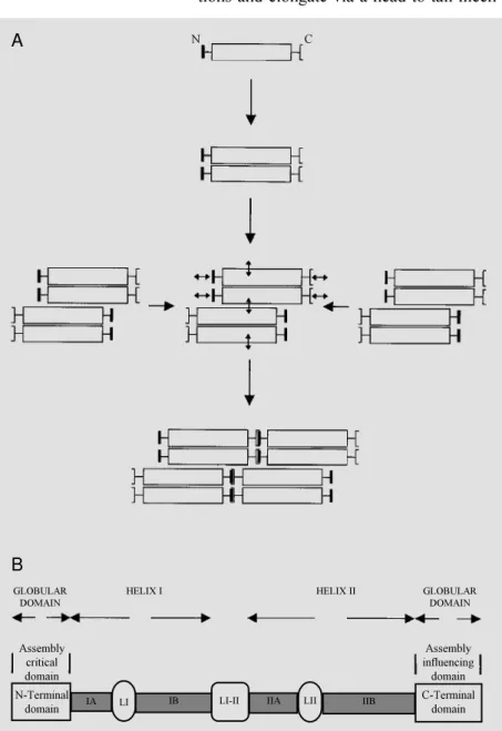
obligatory heteropolymers) in which dimers made of parallel and in register monomers associate to form tetramers in which couples of dimers are arranged in a staggered and antiparallel manner (Figure 1A). Tetramers then form octamers through lateral associa-tions and elongate via a head-to-tail
mech-anism (Figure 1A). Typically, an IF subunit consists of a conserved central core (rod domain) composed of two helical regions periodically interrupted by short, non-heli-cal stretches, that is flanked by an N-termi-nal (head) domain and a C-termiN-termi-nal (tail) domain (Figure 1B). The N-terminal domain is critical for filament elongation, whereas the C-terminal domain appears less critical. Rather, a stretch of residues found in the C-terminus of the rod domain appears impor-tant for IF formation. The rod domain is responsible for lateral association of sub-units into dimers/tetramers/octamers (1-5).
IFs have been long considered as static cytoskeleton elements owing to their resis-tance to extraction with non-ionic detergents in the presence of high concentrations of KCl and their solubility in high concentra-tions of urea (1-5). In recent years, however,
in vitro and in vivo evidence has been
pro-vided showing that IFs coexist with a pool of unassembled subunits and that polymerized subunits exchange with their unpolymerized counterparts (6-11), i.e., IFs undergo assem-bly-disassembly depending on cell needs and/ or functional states. Phosphorylation is a-mong the factors that play an important role in remodeling IFs. Phosphorylation of pre-formed IFs results in disassembly and phos-phorylation of unassembled subunits ren-ders them assembly-incompetent (12-17). Several kinases can act to regulate the IF dynamics, and at the onset of mitosis a spe-cific kinase (cdc2 kinase) is expressed that phosphorylates preformed IFs (12-17). This process is functional to the shape changes that occur at the onset of mitosis when IFs disassemble and reassemble to form a cage-like structure around chromosomes and the mitotic spindle of daughter cells (12-17).
However, it is reasonable to assume that IF remodeling also occurs in non-dividing cells during locomotion and/or at cell sites where the cell needs to be less rigid in order to accomplish specific functions. In this re-gard, factors other than kinases have been
N-Terminal
domain LI
LI-II
N C
GLOBULAR DOMAIN
HELIX I HELIX II GLOBULAR
DOMAIN
Assembly critical domain
Assembly influencing
domain B
A
IA IB IIA LII IIB C-Terminal
domain
postulated to exist that contribute to modify-ing the topographical distribution and the tridimensional organization of IFs via disas-sembly and reasdisas-sembly (3).
S100 pro te ins
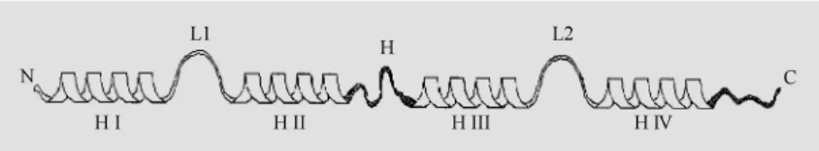
S100A1 and S100B are members of a multigenic family of Ca2+-modulated pro-teins of the EF-hand (helix-loop-helix) type that comprises 19 members showing 25 to 65% sequence identity. S100 proteins have been implicated in the regulation of several activities such as protein phosphorylation, the cell cycle, Ca2+ homeostasis, the dynam-ics of cytoskeleton elements, and enzyme activities (18-22). With the exception of calbindin D9k, which is a Ca2+ modulator protein implicated in buffering cytosolic Ca2+, all other S100 members are considered Ca2+ sensor proteins implicated in Ca2+ signal transduction via their interaction with defi-nite target proteins. Each S100A1 or S100B polypeptide chain (Mr »10 kDa) (like any other S100 member) contains two Ca2+ -bind-ing loops, one in the N-terminal half and the other one in the C-terminal half, each of which is flanked by a-helices for a total of four helices (helix I to helix IV)/molecule (23,24) (Figure 2). The Ca2+-binding loop in the N-terminal half of the chain is non-con-ventional (longer and rearranged) and binds Ca2+ with low affinity, whereas the Ca2+ -binding loop in the C-terminal half is ca-nonical and binds Ca2+ with a relatively high affinity. The two EF-hands are connected by a linker (the hinge region), and helix IV is followed by a C-terminal extension (Figure 2). The two proteins display 56% sequence identity, with the highest amount of identity in the EF-hands and the least amount in the hinge region and the C-terminal extension. It is suggested that differences in sequence and length of these two regions specify the bio-logical activities of S100A1 and S100B (and other S100 members) (18-22).
S100A1 is most abundant in slow-twitch
skeletal muscle cells, some epithelial cells, and some neuronal populations, whereas S100B is most abundant in glial cells, mel-anocytes, chondrocytes, adipocytes, and neu-ronal subpopulations (18-22). By immuno-cytochemistry at both the light and the elec-tron microscope levels, S100B has been lo-calized to IFs, axonemal microtubules (MTs), centrioles, the centrosomes, the midbody, some cytoplasmic MTs, and intracellular membranes in glial cells and several cell lines (25-28), whereas S100A1 has been localized to intracellular membranes, includ-ing the triads, in skeletal muscle cells, and close to Z-discs in sarcomeres (29,30).
Within cells, S100A1 and S100B exist as homodimers (with a small fraction of S100A1/S100B heterodimers) in which the two monomers are held together by non-covalent bonds between helices IV and IV and helices I and I, with some contribution by C-terminal extensions, and are related by a two-fold axis of rotation (31-34). A similar configuration has been detected in other S100 members (35,36), suggesting that the S100 proteins that dimerize share a common struc-tural motif. However, given the differences in sequence and length of the hinge region and the C-terminal extension and the orien-tation of helices in individual S100 proteins, it is conceivable that each of these might be implicated in the regulation of specific ac-tivities by interacting with definite target proteins (18-22).
Upon Ca2+ binding, the most dramatic change in the S100B structure is a tion of helix III with a consequent
reorienta-Figure 2 - Schematic representation of the secondary structure of an S100 protein. Each Ca2+-binding loop (L1 and L2, in the N- and the C-terminal half, respectively) is flanked by a
-helices (-helices I and II, and -helices III and IV for L1 and L2, respectively). A linker region (hinge region, H) connects helix II to helix III. Helix IV is follow ed by a C-terminal extension. The hinge region and the C-terminal extension (in black) display the least amount of sequence homology among the S100 members (see Refs. 18-24).
N
H I
C L1
H II
H L2
tion of the hinge region (33,37,38). As a result, a wide cleft forms that is defined by residues in the hinge region, helix III, helix IV and the C-terminal extension, which form an ample surface believed to mediate the interaction with target proteins. It is sug-gested that analogous changes take place in all other S100 members that dimerize and act via an interaction with target proteins in a Ca2+-dependent manner. Since in each S100 dimer the above binding surfaces are found on opposite sides, an S100 dimer might func-tionally crossbridge two homologous or het-erologous target proteins. Clearly, this mode of interaction is different from that of other EF-hand proteins such as calmodulin and troponin C; the two halves (lobes) of cal-modulin or troponin C wrap around one molecule of a definite target protein, whereas S100 dimers open up and bind couples of target proteins on opposite sides (39-41).
S100B and S100A1 re gulate the dynam ics o f m icro tubule s
Several S100 proteins have been shown to be localized to cytoskeleton constituents, to translocate to cytoskeleton elements upon elevation of cytosolic Ca2+, and to interact with cytoskeletal proteins, suggesting that some S100 members might have a role in the regulation of cell shape changes, locomo-tion, and the tridimensional organization of the cytoskeleton (18-22). Previous studies have shown that S100B and S100A1 inhibit the assembly of MT protein and greatly in-crease the Ca2+-sensitivity of preformed MTs,
in vitro (42-47). Specifically, S100B and S100A1 were shown to bind to and to se-quester unassembled tubulin, thereby inhib-iting MT formation, and to interact with the MT wall, thereby causing a rapid disassem-bly, in a Ca2+- and pH-dependent manner. However, whereas S100B is found associ-ated with MTs in vivo (25-28), S100A1 is
not (28,48), pointing to differences in the mechanism of action of individual proteins
on MT dynamics. S100B and S100A1 might have a role in avoiding excess MT formation and/or participate in MT disassembly at the onset of mitosis or at discrete sites in non-dividing cells. Also, since S100B and S100A1 are also found associated with internal mem-branes, including Golgi membranes (25-28,48), they might have a role in MT-mem-brane interactions that are relevant in the regulation of MT-endosome interactions, which are considered to be important in the mechanism of fusion of endosomes (49), transport from early endosomes to late endosomes (50), and apical recycling (51).
S100B and S100A1 re gulate the dynamics o f inte rme diate filame nts
when added to growing IFs suggest that both S100B and S100A1 also interfere with the IF elongation process (53-56).
By chemical crosslinking and fluores-cent spectroscopy S100B and S100A1 were shown to bind reversibly to unassembled GFAP and desmin with a stoichiometry of 2 mol of IF subunit/mol of S100 dimer with an affinity of 0.5-1.0 µM, in agreement with the notion that an S100 dimer has two binding surfaces on opposite sides (53,56).
Several lines of evidence suggest that S100B and S100A1 interact with the N-terminal head of both GFAP and desmin, thereby sequestering unassembled subunits and blocking the head-to-tail association of subunits into IF polymers (53,56-58) (Figure 3). 1) Neither headless GFAP or desmin nor headless/tailless GFAP or desmin (i.e., their rod domain) as obtained by enzymatic cleav-age was able to interact with either S100 protein. 2) By chemical crosslinking, heterocomplexes made of two copies of GFAP or desmin plus two copies of S100B or S100A1, in addition to complexes made of 1 mol of S100 monomer plus 1 mol of GFAP or desmin, could be resolved by West-ern blotting using a polyclonal anti-S100B/ A1 antiserum. 3) The synthetic peptide, TRTK-12 (TRTKIDWNKILS), correspond-ing to a consensus sequence found in an-other S100B- and S100A1-target protein, i.e., the a-subunit of the actin capping pro-tein, CapZ, and able to compete with CapZa for binding to either S100 protein (59,60), displayed a high sequence identity with a stretch of residues found in the N-terminal head of GFAP containing the so-called RP-box motif shown to be critical for GFAP (as well as desmin and vimentin) assembly. 4) TRTK-12 competed with GFAP or desmin for binding to S100B or S100A1 and blocked the ability of either S100 member to inhibit the GFAP and desmin assemblies and to stimulate GFAP and desmin IF disassembly, in a dose- and Ca2+-dependent manner. Inci-dentally, these observations strongly suggest
that those proteins that contain the CapZa consensus sequence (K/R)(L/I)XWXXIL might be S100B- and S100A1-binding pro-teins; in fact, other S100B and S100A1 tar-get proteins (i.e., neurogranin, neuromodulin/ GAP-43, p53, caldesmon) were shown to contain the above consensus sequence (22). Also, these observations strongly suggest that S100B and S100A1 use the same site for recognizing those target proteins that share the above consensus sequence.
C C N N
C C
C C
N N
N N
C C
N N
N
C
S100A12 or S100B2
GFAP or Desmin
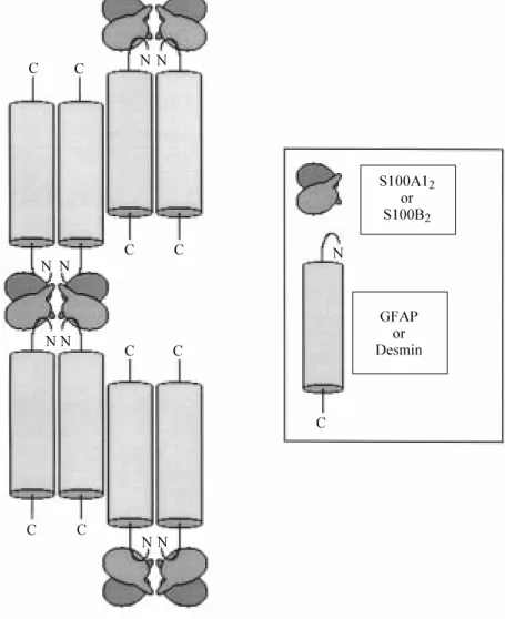
Figure 3 - Schematic representation of the Ca2+-dependent interaction betw een S100A12
or S100B2 dimers and desmin or glial fibrillary acidic protein (GFAP) tetramers. A pair of
S1002 dimers crossbridges tw o desmin or GFAP dimers by interacting w ith the desmin or
Annexin VI and annexin V, two mem-bers of a multigenic family of Ca2+ -depend-ent phospholipid-, membrane- and cytoskel-eton-binding proteins (61,62), bind to S100A1 and S100B, but only annexin VI blocks the ability of the two S100 proteins to inhibit GFAP and desmin assemblies, in a Ca2+- and dose-dependent manner (63). In those cells that co-express S100A1 or S100B, and annexin VI, the latter might act as a regulator of S100A1/S100B effects.
Recent evidence suggests that the C-ter-minal extension of S100A1 (and, by anal-ogy, S100B) is critically implicated in the recognition of GFAP (as well as CapZa, desmin, tubulin, p53) (64). In fact, a mutant S100A1 lacking the C-terminal extension (S100A1D88-93), that was shown to form homodimers, to undergo Ca2+-dependent con-formational changes, and to bind to phenyl-Sepharose in a Ca2+-dependent manner (64,65), proved unable to interact with GFAP, tubulin, p53 and TRTK-12, and to inhibit GFAP assembly.
On the basis of the above data, we sug-gest that couples of S100B or S100A1 ho-modimers (and, eventually, S100B/S100A1 heterodimers) functionally crossbridge cou-ples of GFAP or desmin dimers on opposite sides via interaction of their C-terminal ex-tensions with the GFAP or desmin N-termi-nal domain, thereby blocking the head-to-tail mechanism of IF subunit elongation, as mentioned above (Figure 3). At present we do not know whether or not other regions of the binding surface identified in Ca2+-loaded S100B or S100A1 take part in the recogni-tion of type III IF subunits.
D o e s S100B o r S100A1 have a ro le in type III IF dynamics in vivo?
S100B, but not S100A1 was localized to GFAP, vimentin and desmin IFs in several cell lines as well as to GFAP IFs in Müller cells in bovine retina, and was observed to follow the destiny of these IFs after cell
treatment with colchicine, taxol, cold, or inhibitors of phosphatases (all conditions that cause a dramatic rearrangement of IFs or, in the case of phosphatase inhibitors, IF disassembly and condensation of residual IFs in a paranuclear structure also containing MTs) (27,28,52). Also, at several cell sites MTs with their attached S100B appear sur-rounded and/or flanked by IFs with their attached S100B (32).
Thus, S100B seems strategically posi-tioned along IFs and MTs possibly to contri-bute to crossbridging of IFs to MTs and/or to take part in the regulation of the dynamics of these cytoskeleton constituents. Also, S100B inhibits GFAP and vimentin phosphoryla-tion (66), which might be a reflecphosphoryla-tion of S100B ability to interact with the N-terminal domain of type III IFs and/or represent a means of regulating their assembly-disas-sembly. Lastly, astrocytes in the brain of mutant mice expressing a much reduced amount of S100B exhibit larger amounts of GFAP IFs (67), a finding that has been re-lated (67) to the ability of S100B to inhibit GFAP IF assembly.
Together with the in vitro data mentioned
the MT wall, thereby increasing the Ca2+ -sensitivity of MTs and both proteins use their C-terminal extension for binding to tubulin; ii) no obvious IF disassembly can be observed under these conditions, further sup-porting the conclusion that S100B and S100A1 cause IF disassembly by sequester-ing unassembled IF subunits, and iii) some bundling of IFs can be observed under these
conditions due to disruption of the MT net-work (Sorci G, Agneletti AL and Donato R, unpublished data).
It would be of interest to analyze the effects of overexpression or inhibition of S100B expression in terms of number, distri-bution and total mass of MTs and IFs in resting, activated and dividing cells.
Re fe re nce s
1. Albers K & Fuchs E (1992). The molecular biology of intermediate filaments. Inter-national Review of Cytology, 134: 243-279.
2. Parry DAD & Steinert PM (1992). Interme-diate filament structure. Current Opinion in Cell Biology, 4: 94-98.
3. Georgatos SD (1993). Dynamics of inter-mediate filaments. Recent progress and unansw ered questions. FEBS Letters, 318: 101-107.
4. Fuchs E & Weber K (1994). Intermediate filaments: structure, function, and dis-ease. Annual Review of Biochemistry, 63: 345-382.
5. Hermann H & Aebi U (1998). Intermediate filam ent assem bly: fibrillogenesis is driven by decisive dimer-dimer interac-tions. Current Opinion in Structural Biol-ogy, 8: 177-185.
6. Angelides KJ, Smith KE & Tokeda M (1989.) Assembly and exchange of inter-mediate filament proteins in neurons: neurofilaments are dynamic structures. Journal of Cell Biology, 108: 1495-1506. 7. Vikstrom KL, Borisy GG & Goldman RD
(1989). Dynamic aspects of intermediate filament netw orks in BHK-21 cells. Pro-ceedings of the National Academy of Sci-ences, USA, 86: 549-553.
8. M iller RA, Vikstrom K & Goldman RD (1991). Keratin incorporation into interme-diate filament netw orks is a rapid pro-cess. Journal of Cell Biology, 113: 843-855.
9. Nakamura Y, Takeda M , Angelides KJ, Tada K, Hariguchi S & Nishimura T (1991). Assembly, disassembly, and subunit ex-change of glial fibrillary acidic protein. Glia, 4: 101-110.
10. Vikstrom KL, Lim S-S, Goldman RD & Borisy GG (1992). Steady state dynamics of intermediate filament netw orks. Jour-nal of Cell Biology, 118: 121-129.
11. Hatzfeld M & Weber K (1992). A synthetic peptide representing the consensus se-quence motif at the carboxy-terminal end of the rod domain inhibits intermediate filament assembly and disassembles pre-formed filaments. Journal of Cell Biology, 116: 157-166.
12. Evans RM & Fink LM (1982). An alteration in the phosphorylation of vimentin-type intermediate filaments is associated w ith mitosis in cultured mammalian cells. Cell, 29: 43-52.
13. Celis JE, Larsen PM , Fey SJ & Celis A (1983). Phosphorylation of keratin and vi-mentin polypeptides in normal and trans-formed mitotic human epithelial amnion cells: behavior of keratin and vimentin fila-ments during mitosis. Journal of Cell Biol-ogy, 97: 1429-1434.
14. Ottaviano Y & Gerace L (1985). Phospho-rylation of the nuclear lamins during inter-phase and mitosis. Journal of Biological Chemistry, 260: 624-632.
15. Chou Y-H, Rosevear E & Goldman R (1989). Phosphorylation and disassembly of intermediate filaments in mitotic cells. Proceedings of the National Academy of Sciences, USA, 86: 1885-1889.
16. Inagaki M , Gonda Y, Nishizaw a K, Kitamura S, Sato C, Ando S, Tanabe K, Kikuchi K, Tsuiki S & Nishi Y (1990). Phos-phorylation sites linked to glial filaments disassembly in vitro is located in a non-a -helical head domain. Journal of Biological Chemistry, 265: 4722-4729.
17. Chou Y-H, Bischoff JR, Beach D & Goldman RD (1990). Intermediate fila-ment reorganization during mitosis is me-diated by p34cdc2 phosphorylation of
vimentin. Cell, 62: 1063-1071.
18. Donato R (1991). Perspectives in S-100 protein biology. Cell Calcium, 12: 713-726. 19. Zimmer DB, Cornw all EH, Landar A & Song W (1995). The S100 protein family:
history, function, and expression. Brain Research Bulletin, 37: 417-429. 20. Schäfer BW & Heizmann CW (1996). The
S100 family of EF-hand calcium-binding proteins: functions and pathology. Trends in Biochemical Sciences, 21: 134-140. 21. Heizmann CW & Cox JA (1998). New
per-spectives on S100 proteins: a multi-func-tional Ca2+-, Zn2+- and Cu2+-binding
pro-tein family. Biometals, 11: 383-397. 22. Donato R (1999). Functional roles of S100
proteins, calcium-binding proteins of the EF-hand type. Biochimica et Biophysica Acta, 1450: 191-231.
23. Isobe T & Okuyama T (1978). The amino acid sequence of the S100 protein (PAP I-b protein) and its relation to the calcium binding proteins. European Journal of Bio-chemistry, 89: 379-388.
24. Isobe T & Okuyama T (1981). The amino acid sequence of the a-subunit in bovine brain S100a protein. European Journal of Biochemistry, 116: 79-86.
25. Rambotti M G, Saccardi C, Spreca A, Aisa M C, Giambanco I & Donato R (1989). Im-munocytochemical localization of S100b protein in olfactory and supporting cells of lamb olfactory epithelium. Journal of His-tochemistry and CyHis-tochemistry, 37: 1825-1889.
26. Rambotti M G, Spreca A, Leoncini P, Est enoz M , Cost ant ino-Ceccarini E, Giambanco I & Donato R (1990). Detec-tion of S100b protein in triton-cytoskel-etons: an immunocytochemical study on cultured Schw ann cells. Journal of His-tochemistry and CyHis-tochemistry, 38: 1583-1589.
27. Sorci G, Agneletti AL, Bianchi R & Donato R (1998). Association of S100B w ith inter-mediate filaments and microtubules in glial cells. Biochimica et Biophysica Acta, 1448: 277-289.
M G & Donato R (1999). Replicating myo-blasts and fused myotubes express the calcium-modulated proteins S100A1 and S100B. Cell Calcium, 25: 93-106. 29. Haimoto H & Kato K (1988). S100ao (aa)
protein in cardiac tissue. Isolation from human cardiac muscle and ultrastructural localization. European Journal of Bio-chemistry, 171: 409-415.
30. Donato R, Giambanco I, Aisa M C, Di Geronimo G, Ceccarelli P, Rambotti M G & Spreca A (1989). Cardiac S100ao protein:
purification by a simple procedure and re-lated immunocytochemical and immu-nochemical studies. Cell Calcium, 10: 81-92.
31. Drohat AC, Amburgey JC, Abildgaard F, Starich M R, Baldisseri D & Weber D (1996). Solution structure of rat apo-S100B (ßß) as determined by NM R spec-troscopy. Biochemistry, 35: 11577-11588. 32. Kilby PM , Van Eldik LJ & Roberts GCK (1996). The solution structure of the bo-vine S100B protein dimer in the calcium free state. Structure, 4: 1041-1052. 33. M atsumura H, Shiba T, Inoue T, Harada S
& Kai Y (1998). A novel mode of target recognition suggested by the 2.0 Å struc-ture of holo S100B from bovine brain. Structure, 6: 233-241.
34. Smith SP & Shaw GS (1997). Assignment and secondary structure of calcium-bound human S100B. Journal of Biomolecular NM R,10: 77-88.
35. Potts BCM , Smith J, Akke M , M acke TJ, Okazaki K, Hidaka H, Case DA & Chazin WJ (1995). The structure of calcyclin re-veals a novel homodimeric fold for S100 Ca2+-binding proteins. Nature Structural
Biology, 2: 790-796.
36. Brodersen DE, Etzerodt M , M adsen P, Celis JE, Thøgersen HC, Nyborg J & Kjildgaard M (1998). EF-hands at atomic resolution: the structure of human psoria-sin (S100A7) solved by M AD phapsoria-sing. Structure, 6: 477-489.
37. Drohat AC, Baldisseri DM , Rustandi RR & Weber DJ (1998). Solution structure of calcium-bound rat S100B (ßß) as deter-mined by NM R spectroscopy. Biochemis-try, 37: 2729-2740.
38. Smith SP & Shaw G (1998). A novel cal-cium-sensitive sw itch revealed by the structure of human S100B in the calcium-bound form. Structure, 6: 211-222. 39. Kuboniw a H, Thrando N, Grzesiek S, Ren
H, Klee CB & Bax A (1995). Solution struc-ture of calcium-free calmodulin. Nature Structural Biology, 9: 768-776.
40. M eador WE, M eans AR & Quiocho FA (1992). Recognition by calmodulin: 2.4 Å
structure of a calmodulin peptide com-plex. Science, 257: 1251-1255.
41. Ikura M , Chou GM , Gronengorg AM , Zhu G, Klee CB & Bax A (1992). Solution struc-ture of a calmodulin-target peptide com-plex by multidimensional NM R. Science, 256: 632-638.
42. Endo T & Hidaka H (1983). Effect of S100 protein on microtubule assembly-disas-sembly. FEBS Letters, 161: 235-238. 43. Donato R (1983). Effect of S100 protein
on assembly of brain microtubule proteins in vitro. FEBS Letters, 162: 310-313. 44. Donato R (1985). Calcium-sensitivity of
brain microtubule proteins in the pres-ence of S100 proteins. Cell Calcium, 6: 343-361.
45. Donato R (1988). Calcium-independent, pH-regulated effects of S100 proteins on assembly-disassembly of brain microtu-bule protein in vitro. Journal of Biological Chemistry, 263: 106-110.
46. Baudier J, Briving C, Deinum J, Kaglid K, Sorskog L & Wallin M (1982). Effect of S100 proteins and calmodulin on Ca2+
-induced disassembly of brain microtubule proteins in vitro. FEBS Letters, 147: 165-167.
47. Donato R, Giambanco I & Aisa M C (1989). M olecular interaction of S100 proteins w ith microtubule proteins in vitro. Journal of Neurochemistry, 53: 566-571. 48. Zimmer DB & Landar A (1995). Analysis
of S100A1 expression during skeletal muscle and neuronal cell differentiation. Journal of Neurochemistry, 64: 2727-2736.
49. Bomsel M , Parton R, Kuznetsov SA, Schroer TA & Gruenberg J (1990). M icro-tubule- and motor-dependent fusion in vi-tro betw een apical and basolateral en-docytic vesicles from M DCK cells. Cell, 62: 719-731.
50. Gruenberg J, Griffiths G & How ell KE (1989). Characterization of early endo-somes and putative endocytic carrier vesicles in vivo and w ith an assay of vesicle fusion in vitro. Journal of Cell Biol-ogy, 108: 1301-1316.
51. Breitfeld PP, M cKinnon WC & M ostov KE (1990). Effect of nocodazole on vesicular traffic to the apical and basolateral sur-faces of polarized M DCK cells. Journal of Cell Biology, 111: 2365-2373.
52. Rambotti M G, Giambanco I, Spreca A & Donato R (1999). S100B and S100A1 pro-teins in bovine retina: their calcium-de-pendent stimulation of a membrane-bound guanylate cyclase activity as inves-tigated by ultracytochemistry. Neurosci-ence, 92: 1089-1101.
53. Bianchi R, Giambanco I & Donato R (1993). S100 protein, but not calmodulin, binds to the glial fibrillary acidic protein and inhibits its polymerization in a Ca2+
-dependent manner. Journal of Biological Chemistry, 268: 12669-12674.
54. Bianchi R, Verzini M , Garbuglia M , Giambanco I & Donato R (1994). M ech-anism of S100 protein-dependent inhibi-tion of glial fibrillary acidic protein (GFAP) polymerization. Biochimica et Biophysica Acta,1223: 354-360.
55. Bianchi R, Garbuglia M , Verzini M , Giambanco I, Spreca A & Donato R (1995). S100 protein and annexin II2-p112
(calpac-tin I) act in concert to regulate the state of assembly of GFAP intermediate filaments in vitro. Biochemical and Biophysical Re-search Communications,208: 910-918. 56. Garbuglia M , Verzini M , Giambanco I,
Spreca A & Donato R (1996). Effects of calcium-binding proteins (S100ao, S100a,
S100b) on desmin assembly in vitro. FASEB Journal, 10: 317-324.
57. Bianchi R, Garbuglia M , Verzini M , Giambanco I, Ivanenkov VV, Dimlich RVW, Jamieson Jr GA & Donato R (1996). S100 (a and ß)-binding peptide (TRTK-12) blocks S100/GFAP interaction: identification of a putative S100 target epitope w ithin the head domain of GFAP. Biochimica et Bio-physica Acta,1313: 258-267.
58. Garbuglia M , Verzini M , Dimlich RVW, Jamieson Jr GA & Donato R (1996). Char-acterization of type III intermediate fila-ment regulatory protein target epitopes: S100 (ß and/or a) binds the N-terminal head domain; annexin II2-p112 binds the
rod domain. Biochimica et Biophysica Acta,1313: 268-276.
59. Ivanenkov VV, Jam ieson Jr GA, Gruenstein E & Dimlich RVW (1995). Characterization of S100b binding epi-topes: identification of a novel target, the actin capping protein, Cap Z. Journal of Biological Chemistry, 270: 14651-14658. 60. Ivanenkov VV, Dimlich RVW & Jamieson
Jr GA (1996). Interaction of S100ao
pro-tein w ith the actin capping propro-tein, CapZ: characterization of a putative S100ao
bind-ing site in CapZa-subunit. Biochemical and Biophysical Research Communica-tions,221: 45-50.
61. Raynal P & Pollard HB (1994). Annexins: the problem of assessing the biological role for a gene family of multifunctional calcium- and phospholipid-binding pro-teins. Biochimica et Biophysica Acta, 1197: 63-93.
Bio-physica Acta,1357: 129-154.
63. Garbuglia M , Verzini M & Donato R (1998). Annexin VI binds to S100A1 and S100B and blocks the ability of S100A1 and S100B to inhibit desmin and GFAP as-semblies into intermediate filaments. Cell Calcium, 24: 177-191.
64. Garbuglia M , Verzini M , Rustandi RR, Osterloh D, Weber DJ, Gerke V & Donato R (1999). Role of the C-terminal extension in the interaction of S100A1 w ith GFAP, tubulin, the S100A1- and S100B-inhibito-ry peptide, TRTK-12, and a peptide
de-rived from p53, and the S100A1 inhibitory effect on GFAP polymerization. Biochemi-cal and BiophysiBiochemi-cal Research Communi-cations,254: 36-41.
65. Osterloh D, Ivanenkov VV & Gerke V (1998). Hydrophobic residues in the C-terminal extension of S100A1 are essen-tial for target protein binding not for dimer-ization. Cell Calcium, 24: 137-151. 66. Ziegler DR, Innocente CE, Leal RB,
Rodnight R & Gonçalves CA (1998). The S100B protein inhibits phosphorylation of GFAP and vimentin in a cytoskeletal
frac-tion from immature rat hippocampus. Neurochemical Research, 23: 1259-1263. 67. Ueda S, Gu XF, Whitaker-Azmitia PM , Naruse I & Azmitia EC (1994). Neuro-glial neurothrophic interaction in the S-100ß retarded mutant mouse (Polydactyly Nagoya). I. Immunocytochemical and neu-rochemical studies. Brain Research, 633: 275-283.