L-carnitine as an ergogenic aid for
patients with chronic obstructive
pulmonary disease submitted to
whole-body and respiratory
muscle training programs
1Laboratório de Fisioterapia Cardiovascular, Núcleo de Pesquisas em Exercício Físico, 2Laboratório de Fisiologia do Exercício, Departamento de Ciências Fisiológicas, 3Laboratório de Eletromiografia e Espirometria, Universidade Federal de São Carlos,
São Carlos, SP, Brasil
4Departamento de Alimentos e Nutrição, Universidade Estadual Paulista Júlio de
Mesquita Filho, Araraquara, SP, Brasil
5Departamento de Clínica Médica, Faculdade de Medicina de Ribeirão Preto,
Universidade de São Paulo, Ribeirão Preto, SP, Brasil
6Departamento de Fisioterapia, Faculdade de Ciências da Saúde,
Universidade Metodista de Piracicaba, Piracicaba, SP, Brasil A. Borghi-Silva1,
V. Baldissera2,
L.M.M. Sampaio3,
V.A. Pires-DiLorenzo3,
M. Jamami3, A. Demonte4,
J.S. Marchini5 and D. Costa3,6
Abstract
The effects of adding L-carnitine to a whole-body and respiratory training program were determined in moderate-to-severe chronic obstructive pulmonary disease (COPD) patients. Sixteen COPD pa-tients (66 ± 7 years) were randomly assigned to L-carnitine (CG) or placebo group (PG) that received either L-carnitine or saline solution (2 g/day, orally) for 6 weeks (forced expiratory volume on first second was 38 ± 16 and 36 ± 12%, respectively). Both groups participated in three weekly 30-min treadmill and threshold inspiratory muscle train-ing sessions, with 3 sets of 10 loaded inspirations (40%) at maximal inspiratory pressure. Nutritional status, exercise tolerance on a tread-mill and six-minute walking test, blood lactate, heart rate, blood pressure, and respiratory muscle strength were determined as baseline and on day 42. Maximal capacity in the incremental exercise test was significantly improved in both groups (P < 0.05). Blood lactate, blood pressure, oxygen saturation, and heart rate at identical exercise levels were lower in CG after training (P < 0.05). Inspiratory muscle strength and walking test tolerance were significantly improved in both groups, but the gains of CG were significantly higher than those of PG (40 ± 14 vs 14 ± 5 cmH2O, and 87 ± 30 vs 34 ± 29 m, respectively; P < 0.05).
Blood lactate concentration was significantly lower in CG than in PG (1.6 ± 0.7 vs 2.3 ± 0.7 mM, P < 0.05). The present data suggest that carnitine can improve exercise tolerance and inspiratory muscle strength in COPD patients, as well as reduce lactate production.
Correspondence
A. Borghi-Silva Laboratório de Fisioterapia Cardiovascular
Núcleo de Pesquisas em Exercício Físico, UFSCar
Rod. Washington Luis, km 235 13565-905 São Carlos, SP Brasil
Fax: +55-16-3351-2081 E-mail: [email protected]
Research supported by FAPESP (No. 00/00311-6) and CNPq.
Received April 12, 2005 Accepted November 29, 2005
Key words
•Chronic obstructive
pulmonary disease
•L-carnitine •Exercise training
Introduction
Patients with chronic obstructive pulmo-nary disease (COPD) are intolerant to exer-cise mainly due to limited ventilatory capac-ity (1). This leads to deconditioning, early lactic acidosis, and reduced capillarityand muscle strength (2). An exercise training (ET) program is the main component of pulmonary rehabilitation for COPD patients (3), increasing exercise capacity and reduc-ing dyspnea. Therefore, ET can result in beneficial changes in exercise capacity and has been shown to consistently improve the quality of life of COPD patients (4).
Although training has been shown to be an essential component of the rehabilitation program, patients do not benefit from it to the same extent (5). Indeed, conflictive re-sults concerning lactate reduction and im-proved physical exercise capacity or muscle strength after training have been reported (6). Patients with reduced exercise capacity who experience less ventilatory limitation for exercise and more reduced respiratory and peripheral muscle strength are more likely to improve their exercise tolerance with a physical training program (5).
In addition to exercise, nutritional fac-tors such as L-carnitine supplementation (7) have been used to improve physical perfor-mance. L-carnitine is a quaternary amine whose function has been related to lipid metabolism, sparing muscle glycogen, im-proving tolerance to physical activity, and reducing muscle fatigue (7). It has also been demonstrated in skeletal and cardiac muscle, in plasma, kidney, liver, and brain.
The importance of the biochemical func-tions of carnitine in the altered muscle phys-iology associated with clinical carnitine de-ficiency supports the critical role of car-nitine in muscle bioenergetics (8). In an experimental study, L-carnitine directly im-proved the fatigue characteristics of muscles enriched in type I fibers when evaluated by the kinetics of contraction and relaxation
during a stimulation protocol (9).
In healthy persons, the beneficial effects of long-term treatment with L-carnitine on physical performance cannot be explained by an increase in muscle carnitine stores (10). The effects of supplementation on pathologic conditions that affect exercise performance are less clear. Some studies have shown improvement of exercise per-formance in patients with cardiovascular (11) and peripheral vascular disease (12). How-ever, to our knowledge, the present study is the first to have been conducted on COPD patients. Thus, the purpose of the present study was to determine whether carnitine supplementation could improve the exercise tolerance, pulmonary function, nutrition, dyspnea, and respiratory muscle strength of COPD patients submitted to a whole-body and respiratory training program.
Patients and Methods
Subjects
drugs with no dosage changes. The study protocol was approved by the Ethics Com-mittee for Human Research of the Federal University of São Carlos and all patients gave institutionally reviewed written in-formed consent to participate in the study.
Experimental design
Patients were randomly allocated to two groups by drawing lots. The L-carnitine group (N = 8) received a 2 g/day oral L-carnitine supplementation in two daily doses for 6 weeks, dispensed in 10-mL glass bottles con-taining 1 g each, and the control group (N = 8) received placebo (saline solution, similar in presentation to the L-carnitine prepara-tion) for the same period of time. The sup-plementation and placebo were similar in color, shape and taste. The patients in each group did not know if they were receiving carnitine supplementation or placebo (single-blind controlled trial). Both groups took part in an exercise and respiratory training pro-gram which consisted of medical follow-up by a physician and individual training super-vised by physical therapists. L-carnitine was obtained from Sintofarma Laboratory (São Paulo, SP, Brazil). The purity of L-carnitine was evaluated by enzymatic method, and was found to be consistent with the concen-trations stated on the labeling (87-90%).
Lung function tests. All patients under-went spirometry with the determination of FEV1, forced vital capacity and maximal voluntary ventilation according to Ameri-can Thoracic Society recommendations (13). The values obtained were compared to the predicted normal values of Knudson et al. (14). Spirometry was performed using a Vitalograph® Hand-Held 2120 instrument (Ennis, Ireland), which was calibrated be-fore each test according to manufacturer recommendations using a 1-L syringe.
The maximal respiratory pressures were assessed by maximal inspiratory (PImax) and expiratory pressure (PEmax) at residual
vol-ume and at total lung capacity, respectively, with an analog pressure gauge according to the method of Black and Hyatt (15). Patients were asked to make maximal inspiratory and expiratory efforts against an obstructed mouth-piece with a small leak to prevent patients from closing their glottis during the maneu-ver. Patients sustained their maximal effort for one second and the best of three consecu-tive attempts was used. The predicted values for the Brazilian population reported by Neder et al. (16) were compared with those obtained for patients with COPD.
Body composition. After the patient ar-rived at the laboratory at 8 am before break-fast, weight and height were measured using a mechanical scale (Welmy, São Paulo, SP, Brazil)and body mass index was calculated. Three consecutive measurements of triceps skinfold (TSF) thickness were used as an indirect estimate of body fat (17). Three measurements of midarm circumference (MAC) were performed in the nondominant arm, positioned parallel to the trunk. A non-distensible tape was placed around the mid-point of the arm without compressing the arm tissue, halfway between the tip of the shoulder (acromial process) and the tip of the elbow (olecranon process). Arm muscle circumference (AMC) was derived from the following formula(18): AMC (cm) = MAC -π x TSF (cm).
intake were estimated using the Nutri® Pro-gram (Federal University of São Paulo, UNIFESP-EPM, São Paulo, SP, Brazil) to determine if both groups presented a similar pattern of calorie intake. All patients were instructed to maintain their usual diet during the study.
Exercise capacity. The maximal incre-mental exercise test was performed on a treadmill (Imbrasport, Milennium® - ATL, Porto Alegre, RS, Brazil). During the test, arterial oxygen saturation was measured by pulse oximetry (SpO2; Nonim 8500A®, Ply-mouth, MN, USA), and arterial blood pres-sure and heart rate (HR) were meapres-sured at 2-min intervals. HR was measured from the R-to-R interval on the electrocardiogram (TC 500, ECAFIX, São Paulo, SP, Brazil), and arterial blood pressure was measured with a manual sphygmomanometer. At the begin-ning and at the end of the test the patients were evaluated for possible breathing diffi-culty (dyspnea) using Borg’s CR10 scale (19).
Patients first performed an incremental exercise test that included a 2-min rest pe-riod in the sitting position. They began walk-ing at 2.0 km/h with a constant 3% grade, followed by an increase in speed rate of 0.5 km/h every 2 min until exhaustion. The test could be interrupted either by the patients, because of dyspnea, leg fatigue or disabling symptoms, or by the investigator, for safety reasons. If SpO2 dropped below 80% or if threatening signs and symptoms occurred such as electrocardiography alterations and if HR reached the age-predicted maximum, the test could be interrupted.HR reserve was calculated (20) as 100% - (100 x peak HR)/ (220 - age). The metabolic equivalent (MET) was estimated at peak oxygen consumption (VO2) from the following formula (21): VO2 (mL kg-1 min-1) = (S x 0.1) + (S x G x 1.8) + 3.5, within a range of intensities from 3.2 to 6.4 km/h, where: S = speed in m/min, G = grade in % (3% = 0.03) and 1 MET = 3.5 mLO2 kg-1 min-1.
Blood lactate concentration. Blood sam-ples were drawn from each ear lobe. The first drop of blood was discarded to avoid contamination with lactate eliminated through sweat. Blood lactate concentration was measured at rest, every 2 min during the test and 2 min after the end of the maximal exercise test. Each blood sample was col-lected with a capillary tube previously cali-brated with 25 µL heparin. After collection, the blood samples were transferred to 2-mL tubes containing 50 µL 1% sodium fluoride in order to prevent glycolysis. Blood was stored at -10ºC for later analysis. Blood lac-tate concentration was determined by an electroenzymatic method (YSI 1500® - Sport Lactate Analyzer, Yellow Springs, OH, USA).
Six-minute walking test (SMWT). Func-tional exercise performance was measured by the SMWT. This test was performed in a 30-m corridor, and encouragement was stan-dardized (22). To avoid learning effects, the better of two reproducible walks was used on the occasion of the first visit. SpO2, HR and Borg’s scale were also measured during the test. The test could be interrupted by the patient because of dyspnea and/or leg fa-tigue. On the occasion of the second visit, the SMWT was performed under the same conditions as used for the pretest. The pre-dicted values of distances walked were com-pared with those obtained by the patients, according to the 2002 ATS Statement (22).
Exercise training program
Blood pressure was measured before and after each training session. The IMT was performed with a pressure threshold resis-tive training device. During training, an at-tempt was made to apply a load correspond-ing to 40% of the patient’s baseline PImax (23). Nose clips were used to occlude nasal airflow. During each session, the patients breathed through an IMT mouthpiece de-vice, with 3 sets of 10 loaded inspirations.
Statistical analysis
Values are reported as means ± SD. Due to non-Gaussian distribution and/or inho-mogeneity of variance of variable values, nonparametric tests were selected for statis-tical analysis (24). Thus, the Mann-Whitney and Wilcoxon nonparametric tests were used for intergroup and intragroup comparisons, respectively, with the level of significance set at P < 0.05. The gain obtained by the groups was derived from absolute delta com-parisons (post-treatment minus pretreatment values).
Results
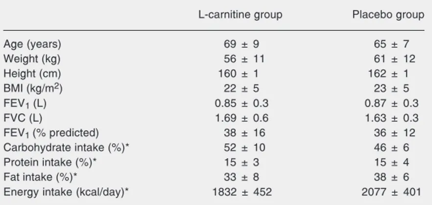
Baseline patient characteristics are pre-sented in Table 1. There were no significant differences in age, anthropometry, lung func-tion, protein, fat, or total energy intake be-tween groups. The results of the pulmonary function tests were characterized, on aver-age, by moderate-to-severe airflow obstruc-tion (FEV1 around 40% of the predicted value in both groups). The various proce-dures were well tolerated by all patients, and no untoward effects of carnitine were re-ported.
Effects on body composition and dietary intake
No significant changes were observed in body composition after the exercise training program in either group, as shown in Table
2. There were no significant differences in energy intake of carbohydrates, lipids, pro-tein, or total energy in either group.
Physiological parameters and performance during the incremental test
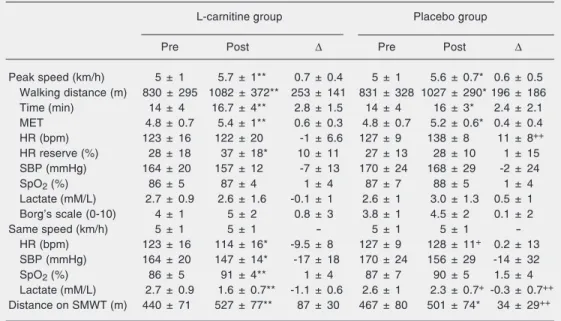
Table 3 shows the distance walked in the incremental test on a treadmill, as well as the time and the maximal speed reached. Peak metabolic equivalent obtained before the physical and respiratory program was around 5 METs for both groups. Six weeks of train-ing produced a significant increase in time and speed compared to pretreatment in both groups, and the HR reserve significantly increased (P < 0.05) only in the carnitine group. No significant differences in systolic blood pressure, SpO2, Borg scale, or blood lactate were observed between the two groups when compared at peak speed. A significant increase in HR peak was shown only in the placebo groupfrom absolute delta compari-son (P < 0.05). In the incremental exercise test, comparison of responses to identical exercise levels showed that HR, systolic blood pressure and blood lactate concentra-tion were significantly lower, and that SpO2 significantly increased only in the
L-car-Table 1. Characteristics of the patients in the L-carnitine and placebo groups.
L-carnitine group Placebo group
Age (years) 69 ± 9 65 ± 7
Weight (kg) 56 ± 11 61 ± 12
Height (cm) 160 ± 1 162 ± 1
BMI (kg/m2) 22 ± 5 23 ± 5
FEV1 (L) 0.85 ± 0.3 0.87 ± 0.3
FVC (L) 1.69 ± 0.6 1.63 ± 0.3
FEV1 (% predicted) 38 ± 16 36 ± 12
Carbohydrate intake (%)* 52 ± 10 46 ± 6
Protein intake (%)* 15 ± 3 15 ± 4
Fat intake (%)* 33 ± 8 38 ± 6
Energy intake (kcal/day)* 1832 ± 452 2077 ± 401
Data are reported as mean ± SD for 8 patients in each group. There were no significant differences between the L-carnitine and placebo groups. BMI = body mass index; FEV1 = forced expiratory volume in 1 s; FVC = forced vital capacity. *Estimated from
nitine group (Table 3). In addition, the re-duction in blood lactate concentration was
significantly greater for the L-carnitine group (P < 0.05).
Table 3. Effect of L-carnitine supplementation associated with exercise training on peak exercise and physiological responses at identical levels of exercise.
L-carnitine group Placebo group
Pre Post ∆ Pre Post ∆
Peak speed (km/h) 5 ± 1 5.7 ± 1** 0.7 ± 0.4 5 ± 1 5.6 ± 0.7* 0.6 ± 0.5
Walking distance (m) 830 ± 295 1082 ± 372** 253 ± 141 831 ± 328 1027 ± 290* 196 ± 186
Time (min) 14 ± 4 16.7 ± 4** 2.8 ± 1.5 14 ± 4 16 ± 3* 2.4 ± 2.1
MET 4.8 ± 0.7 5.4 ± 1** 0.6 ± 0.3 4.8 ± 0.7 5.2 ± 0.6* 0.4 ± 0.4
HR (bpm) 123 ± 16 122 ± 20 -1 ± 6.6 127 ± 9 138 ± 8 11 ± 8++
HR reserve (%) 28 ± 18 37 ± 18* 10 ± 11 27 ± 13 28 ± 10 1 ± 15
SBP (mmHg) 164 ± 20 157 ± 12 -7 ± 13 170 ± 24 168 ± 29 -2 ± 24
SpO2 (%) 86 ± 5 87 ± 4 1 ± 4 87 ± 7 88 ± 5 1 ± 4
Lactate (mM/L) 2.7 ± 0.9 2.6 ± 1.6 -0.1 ± 1 2.6 ± 1 3.0 ± 1.3 0.5 ± 1
Borg’s scale (0-10) 4 ± 1 5 ± 2 0.8 ± 3 3.8 ± 1 4.5 ± 2 0.1 ± 2
Same speed (km/h) 5 ± 1 5 ± 1 - 5 ± 1 5 ± 1
-HR (bpm) 123 ± 16 114 ± 16* -9.5 ± 8 127 ± 9 128 ± 11+ 0.2 ± 13
SBP (mmHg) 164 ± 20 147 ± 14* -17 ± 18 170 ± 24 156 ± 29 -14 ± 32
SpO2 (%) 86 ± 5 91 ± 4** 1 ± 4 87 ± 7 90 ± 5 1.5 ± 4
Lactate (mM/L) 2.7 ± 0.9 1.6 ± 0.7** -1.1 ± 0.6 2.6 ± 1 2.3 ± 0.7+ -0.3 ± 0.7++
Distance on SMWT (m) 440 ± 71 527 ± 77** 87 ± 30 467 ± 80 501 ± 74* 34 ± 29++
Data are reported as means ± SD for 8 patients in each group. Physiological values were compared at peak speed and at identical levels of exercise. MET = metabolic equivalent (3.5 mL O2 kg-1 min-1); HR = heart rate;
SBP = systolic blood pressure; SpO2 = arterial oxygen saturation estimated by pulse oximetry; SMWT =
six-minute walking test; ∆ = absolute delta comparison (post-treatment minus pretreatment values). There were
no significant pre-training differences between groups.
*P < 0.05 compared to pre-training response in placebo and L-carnitine groups (Wilcoxon test). **P < 0.01 compared to pre-training response in L-carnitine group (Wilcoxon test). +Significantly different from
post-training response in L-carnitine group (Mann-Whitney test). ++Significantly different from absolute delta in
L-carnitine group (Mann-Whitney test).
Table 2. Effects of L-carnitine supplementation associated with exercise training on body composition and dietary intake.
L-carnitine group Placebo group
Pre Post ∆ Pre Post ∆
BMI (kg/m2) 22 ± 5 21 ± 4 -1 ± 1 23 ± 5 23 ± 5 0 ± 1
TSF (mm) 13 ± 9 12 ± 6 -1 ± 3 13 ± 7 13 ± 7 1 ± 2
MAC (cm) 25 ± 4 26 ± 4 0 ± 1 27 ± 4 28 ± 4 0 ± 1
AMC (cm) 21 ± 2 22 ± 3 1 ± 1 24 ± 2 24 ± 2 1 ± 4
Carbohydrate intake (%)* 52 ± 10 52 ± 7 1 ± 10 46 ± 6 48 ± 6 2 ± 5
Protein intake (%)* 15 ± 3 16 ± 6 1 ± 6 15 ± 4 16 ± 2 1 ± 3
Fat intake (%)* 33 ± 8 32 ± 7 -1 ± 6 38 ± 6 36 ± 5 -3 ± 5
Energy intake (kcal/day)* 1832 ± 452 1885 ± 421 88 ± 317 2077 ± 401 2066 ± 337 -29 ± 168
Data are reported as mean ± SD for 8 patients in each group. There were no significant differences between the L-carnitine and placebo groups. BMI = body mass index; TSF = triceps skinfold; MAC = midarm circumference; AMC = arm muscle circumference; ∆ = absolute delta comparison (post-treatment minus
Performance in the six-minute walking test
No significant differences were observed between groups before training. The L-car-nitine group showed a significant improve-ment in walking distance performance after treatment (P < 0.01), as did the placebo group (P < 0.05). However, the absolute delta in walking distance obtained by the L-carnitine group was significantly greater (P < 0.05), as illustrated in Table 3.
When the predicted values for distance walked were compared with those obtained by our patients, significant reductions (P < 0.05) were observed (497 ± 53 vs 453 ± 54 m, respectively).
Maximal respiratory pressure
No significant differences in maximal respiratory pressure were observed between groups before training. After training, PImax increased significantly not only in the L-carnitine group (from 49 ± 12 to 89 ± 22 cmH2O) but also in the placebo group (from 54 ± 19 to 68 ± 21 cmH2O) with P < 0.01. Similar results were obtained for PEmax in the L-carnitine group (from 66 ± 18 to 90 ± 32 cmH2O, P < 0.01) and the placebo group (from 69 ± 26 to 81 ± 25 cmH2O, P < 0.05). However, the increase in inspiratory muscle strength (PImax) was significantly greater for the L-carnitine group than the placebo group (40 ± 14 vs 14 ± 5 cmH2O, respectively, P < 0.05).
When the predicted values were com-pared to the PImax and PEmax values obtained by patients with COPD (92 ± 12 vs 51 ± 16 cmH2O for PImax, and 97 ± 18 vs 68 ± 22 cmH2O for PEmax, respectively), we observed that both values were lower in comparison to healthy subjects of the Brazilian population.
Discussion
The main results of the present study show the beneficial effects of L-carnitine
supplementation in enhancing physiological responses at identical levels of exercise, re-ducing lactate concentration, improving ex-ercise tolerance and inspiratory muscle strength in COPD.
On the other hand, the supplementation was not associated with modification of the degree of airflow obstruction, improvement of nutritional status or increase in muscle mass. Although TSF and upper arm circum-ference measurements are extensively used to evaluate body composition, they are not sensitive, especially over short time periods, to detect changes in muscle mass after thera-peutic interventions.
In the present study, both groups in-creased their maximal exercise tolerance. Therefore, the physical training program was efficient in improving the performance of these patients. Our results agree with those obtained by other investigators who reported the importance of physical training in COPD patients (25-27). Previous studies have ob-served positive effects on maximal exercise capacity with L-carnitine supplementation alone in patients with cardiac insufficiency (11)and peripheral arterial disease (12).
However, patients receiving L-carnitine supplementation demonstrated a greater in-crement of lactate removal rate. Casaburi et al. (25) showed that ET (without a nutrient supplement) results in significant increases of peak blood lactate concentration and no significant responses to identical exercise levels in the incremental test in COPD pa-tients. Our results showed an expressive re-duction in blood lactate concentration at iden-tical exercise levels only in the L-carnitine-supplemented patients.
modifications, which contribute to improv-ing exercise performance. L-carnitine sup-plementation reduced left ventricular size and pulmonary arterial pressure in patients with congestive heart failure (29) and sys-tolic and diassys-tolic blood pressure and ST changes in patients with severe ischemia-induced cardiac insufficiency (11).
In the present study, the L-carnitine-supplemented group showed a significant reduction in HR and systolic blood pressure at submaximal intensities, as well as an in-crease in HR reserve. These changes are probably explained by the reduction of mus-cular lactate values (not measured, but lac-tate was only evaluated in plasma), which permitted a greater utilization of muscle oxy-gen during physical exercise, delaying the metaboreflex and cardiovascular adjust-ments. Premature lactic acidosis has been associated with reduced oxidative enzyme concentrations in lower limb muscles, and utilization of the bioactive nutrients involved in muscle energy and substrate metabolism can be of therapeutic importance.
Furthermore, Gosker et al. (30)observed that COPD and chronic heart failure can lead to wasting and weakness of skeletal muscle. These authors showed that patients with se-vere COPD or chronic heart failure were physically inactive, a fact assumed to have a negative or “detraining” effect on exercise capacity. In our study, we observed lower blood lactate levels, reduced maximal exer-cise capacity and high HR reserve compared to baseline values in patients with COPD, similar to other authors (25,30). Moreover, the higher HR reserve values demonstrated that patients with COPD were primarily lim-ited by their ventilatory limitation and not by cardiac dysfunction, showing that a possible cardiac impairment was subordinate to pul-monary impairment in the present study.
The subjective sensation of dyspnea was not modified in our patients by L-carnitine supplementation and/or physical training. Similar results have been reported in the
literature (31), although some investigators have observed a reduction in dyspnea (32). In our study, the Borg scale was applied only at the beginning and at the high point of exercise both before and after treatment, although it was not applied to the same workload.
Both groups significantly increased their walking distance in the SMWT, indicating increased tolerance to exercise after the pro-gram. In a previous study (33), we observed a significant increase of exercise tolerance in the SMWT in patients with mild to moder-ate COPD receiving L-carnitine supplemen-tation associated with physical exercise. The increased performance of these patients was not associated with an increase of plasma free L-carnitine levels.
Redelmeier et al. (34) reported that an increase of 54 m in the walking distance in SMWT was thought to be clinically rel-evant. Thus, we may suggest that physical exercise associated with L-carnitine supple-mentation was more effective, considering that a mean of 87 m was obtained compared to physical exercise alone, corresponding to a 34 m increase. According to some investi-gators, L-carnitine supplementation (with-out physical training) can improve the exer-cise tolerance of patients with peripheral vascular (12) and cardiovascular (11) dis-eases and of patients submitted to hemodi-alysis (35).
studies have observed the effects of L-car-nitine supplementation on muscle perfor-mance, delayed muscle fatigue in vitro (9), increased workload during an incremental test in healthy subjects (39) and athletes (40), reduced muscle fatigue in hemodialy-sis patients (35), or improved myocardial performance in congestive heart failure pa-tients (29).
The main limitation of our study was the small number of participants, a fact possibly due to the strict criteria for patient inclusion and the severity of the disease of the patients evaluated. Thus, a study with a larger num-ber of patients would be useful to confirm the present findings. Another limitation of this study was the impossibility of obtaining ventilatory and metabolic measurements due to the high cost of the methodology. More-over, this study was of the single-blind type,
with only the patients being unaware of which medication they were taking since the same researcher controlled the treatment plan, training and administration of L-carnitine or placebo.
The present study demonstrated that L-carnitine administration adds benefit to ex-ercise and respiratory strength training in outpatients with stable, moderate-to-severe COPD. Larger double-blind and controlled trials are now warranted to confirm these preliminary findings.
Acknowledgments
L-carnitine was partly supplied by Sinto-farma (São Paulo, SP, Brazil) as a donation and additional Sintofarma L-carnitine sup-plies were funded by FAPESP (No. 00/ 00311-6).
References
1. Casaburi R (2001). Skeletal muscle dysfunction in chronic obstruc-tive pulmonary disease. Medicine and Science in Sports and Exer-cise, 33 (Suppl 17): 662-670.
2. Maltais F, LeBlanc P, Whittom F et al. (2000). Oxidative enzyme activities of the vastus lateralis muscle and the functional status in patients with chronic obstructive pulmonary disease. Thorax, 55: 848-853.
3. Cooper CB (2001). Exercise in chronic obstructive pulmonary dis-ease: limitation and rehabilitation. Medicine and Science in Sports and Exercise, 33 (Suppl 7): 643-646.
4. Boueri FM, Bucher-Bartelson BL, Glenn KA et al. (2001). Quality of life with a generic instrument (short form -36) improves following pulmonary rehabilitation in patients with chronic obstructive pulmo-nary disease. Chest, 119: 77-84.
5. Troosters T, Gosselink R & Decramer M (2001). Exercise training in chronic obstructive pulmonary disease: how to distinguish respond-ers from nonrespondrespond-ers. Journal of Cardiopulmonary Rehabilita-tion, 21: 10-17.
6. American College of Chest Physicians and American Association of Cardiovascular and Pulmonary Rehabilitation (1997). Pulmonary rehabilitation joint ACCP/ACVPR evidence-based guidelines. Chest, 112: 1363-1396.
7. Brass EP (2000). Supplemental carnitine and exercise. American Journal of Clinical Nutrition, 72: 618-623.
8. Brass EP & Hiatt WR (1998). The role of carnitine and carnitine supplementation during exercise in man and in individuals with special needs. Journal of the American College of Nutrition, 17: 207-215.
9. Brass EP, Scarrow AM, Ruff LJ et al. (1993). Carnitine delays rat
skeletal muscle fatigue in vitro. Journal of Applied Physiology, 75: 1595-1600.
10. Wachter S, Vogt M, Kreis R et al. (2002). Long-term administration of L-carnitine to humans: effect on skeletal muscle carnitine content and physical performance. Clinica Chimica Acta, 318: 51-61. 11. Loster H, Miehe K, Punzel M et al. (2001). Prolonged oral L-carnitine
substitution increases bicycle ergometer performance in patients with severe, ischemically induced cardiac insufficiency. Cardiovas-cular Drugs and Therapy, 13: 537-546.
12. Barker GA, Green S, Askew CD et al. (2001). Effect of propionyl-L-carnitine on exercise performance in peripheral arterial disease. Medicine and Science in Sports and Exercise, 33: 1415-1422. 13. American Thoracic Society (1991). Lung function testing: selection
of reference values and interpretative strategies. American Review of Respiratory Disease, 144: 1202-1218.
14. Knudson RJ, Leibowitz MD & Holberg CJ (1983). Changes in the normal maximal expiratory flow-volume curve with growth and ag-ing. American Review of Respiratory Disease, 127: 725-734. 15. Black LF & Hyatt RE (1969). Maximal respiratory pressures: normal
values and relationship to age and sex. American Review of Respi-ratory Disease, 99: 696-702.
16. Neder JA, Andreoni S, Lerario MC et al. (1999). Reference values for lung function tests II. Maximal respiratory pressures and volun-tary ventilation. Brazilian Journal of Medical and Biological Re-search, 32: 719-727.
18. Blackburn GL, Bistrian BR, Mairi MS et al. (1977). Nutritional as-sessment of the hospitalized patient. Journal of Parenteral and Enteral Nutrition, 1: 1-77.
19. Borg GA (1982). Psychophysical bases of perceived exertion. Medi-cine and Science in Sports and Exercise, 14: 377-381.
20. Wasserman K, Hansen JE, Sue DY et al. (1994). Principles of Exercise Testing and Interpretation. 2nd edn. Lea & Febiger, Phila-delphia, PA, USA.
21. American College of Sports Medicine (1995). Guidelines for Exer-cise Testing and Prescription. 5th edn. Lea & Febiger, Philadelphia, PA, USA.
22. ATS Statement (2002). Guidelines for the Six-Minute Walk Test. American Journal of Respiratory and Critical Care Medicine, 166: 111-117.
23. Nield MA (1999). Inspiratory muscle training protocol using a pres-sure threshold device: effect on dyspnea in chronic obstructive pulmonary disease. Archives of Physical Medicine and Rehabilita-tion, 80: 100-102.
24. Zar JH (1984). Biostatistical Analysis. Prentice-Hall, Englewood Cliffs, NJ, USA.
25. Casaburi R, Porszasz J, Burns MR et al. (1997). Physiologic ben-efits of exercise training in rehabilitation of patients with severe chronic obstructive pulmonary disease. American Journal of Respi-ratory and Critical Care Medicine, 155: 1541-1551.
26. Maltais F, LeBlanc P, Simard C et al. (1996). Skeletal muscle adaptation to endurance training in patients with COPD. American Journal of Respiratory and Critical Care Medicine, 154: 442-447. 27. Vogiatzis I, Williamson AF, Miles J et al. (1999). Physiological
response to moderate exercise workloads in a pulmonary rehabilita-tion program in patients with varying degrees of airflow obstrucrehabilita-tion. Chest, 116: 200-207.
28. Sietsema K (2001). Cardiovascular limitations in chronic pulmonary disease. Medicine and Science in Sports and Exercise, 33 (Suppl 7): 656-661.
29. Anand I, Chandrashekhan Y, De Giuli F et al. (1998). Acute and chronic effects of propionyl-L-carnitine on the hemodynamics, exer-cise capacity, and hormones in patients with congestive heart fail-ure. Cardiovascular Drugs and Therapy, 12: 291-299.
30. Gosker HR, Lencer NHMK, Franssen FME et al. (2003). Striking similarities in systemic factors contributing to decreased exercise
capacity in patients with severe chronic heart failure or chronic obstructive pulmonary disease. Chest, 123: 1416-1424.
31. Berry MJ, Adair NE, Sevensky KS et al. (1996). Inspiratory muscle training and whole body reconditioning in chronic obstructive pulmo-nary disease. American Journal of Respiratory and Critical Care Medicine, 153: 1812-1816.
32. Reardon J, Awad E, Normandin E et al. (1994). The effect of com-prehensive outpatient pulmonary rehabilitation on dyspnea. Chest, 105: 1046-1052.
33. Silva AB, Di Lorenzo VAP, Jamami M et al. (2003). Efeitos da suplementação oral de L-carnitina associada ao treinamento físico na tolerância ao exercício de pacientes com doença pulmonar obstrutiva crônica. Jornal de Pneumologia, 29: 379-385.
34. Redelmeier DA, Bayoumi AM, Goldstein RS et al. (1997). Interpret-ing small differences in functional status: the six-minute walk test in chronic lung disease patients. American Journal of Respiratory and Critical Care Medicine, 155: 1278-1282.
35. Brass EP, Adler S, Sietsema KE et al. (2001). Intravenous L-car-nitine increases plasma carL-car-nitine, reduces fatigue, and may pre-serve exercise capacity in hemodialysis patients. American Journal of Kidney Diseases, 37: 1018-1028.
36. Riera HS, Montemayor Rubio TM, Ruiz FO et al. (2001). Inspiratory muscle training in patients with chronic obstructive pulmonary dis-ease: effect on dyspnea, exercise performance, and quality of life. Chest, 120: 748-756.
37. Larson JL, Kim MJ, Sharp JT et al. (1988). Inspiratory muscle training with a pressure threshold breathing device in patients with chronic obstructive pulmonary disease. American Review of Respi-ratory Disease, 138: 689-696.
38. Preusser BA, Winningham ML & Clanton T (1994). High- vs low-intensity inspiratory muscle interval training in patients with chronic obstructive pulmonary disease. Chest, 106: 110-117.
39. Vecchiet L, Di Lisa F, Pieralisi G et al. (1985). Influence of L-carnitine administration on maximal physical exercise. European Journal of Applied Physiology and Occupational Physiology, 54: 131-135.