O
RIGINALA
RTICLE Revista Brasileira de FisioterapiaEffects of noninvasive ventilation on dynamic
hiperinflation of patients with COPD during
activities of daily living with upper limbs
Efeitos da ventilação não-invasiva sobre a hiperinsuflação dinâmica de pacientes
com DPOC durante atividade da vida diária com os membros superiores
Isabela M. B. Sclauser Pessoa1,2, Dirceu Costa3, Marcelo Velloso4, Eliane Mancuzo5, Marco A. S. Reis6, Verônica F. Parreira4
Abstract
Background: Patients with chronic obstructive pulmonary disease (COPD) complain of dyspnea on activities of daily living (ADLs) with the upper limbs. Dynamic hyperinflation (DH) is one of the ventilatory mechanisms that may contribute towards dyspnea. To minimize the DH it is indicated the use of noninvasive ventilation (NIV). Objectives: To verify whether there is DH and dyspnea during the performance of ADL with the upper limbs with and without the use of NIV. Methods: 32 patients with moderate-to-severe COPD, aged 54 to 87 years (mean 69.4, SD 7.4) were evaluated. The subjects lift up containers weighing between 0.5 and 5.0 kg over a five-minute period, starting from the waist level and putting them onto a shelf located above head height, with and without the use of NIV (BiPAP®; IPAP 10cmH
2O; EPAP 4 cmH2O). The inspiratory capacity (IC) and dyspnea (Borg scale) were evaluated on all the patients.
The IC was measured before and after simulation of the ADL. In order to analyze the data, Student’s t test for dependent samples and the Wilcoxon test were used. Results: There were statistically significant reductions in IC after the ADL with and without NIV (p=0.01). The dyspnea increased after the ADL with and without the NIV, however between both interventional procedures protocols no between-group difference was observed. Conclusions: The simulation of an ADL with the upper limbs resulted in an increase in DH and dyspnea. The NIV supplied with pre-established pressure was not enough to prevent the DH and dyspnea.
Keywords: physicaltherapy; dynamic hyperinflation; noninvasive ventilation; chronic obstructive pulmonary disease; activities of daily living.
Resumo
Contextualização: Pacientes com doença pulmonar obstrutiva crônica (DPOC) queixam-se de dispneia nas atividades de vida diária (AVD) com os membros superiores (MMSS). A hiperinsuflação dinâmica (HD) é um dos mecanismos ventilatórios que contribuem para a dispneia. Para minimizar a HD, propõe-se a utilização de sistemas de ventilação não-invasiva (VNI). Objetivos: Verificar se existe HD e dispneia durante a realização de uma AVD com os MMSS com e sem o uso da VNI. Métodos: Participaram 32 pacientes com DPOC de moderada a muito grave, com idades entre 54 a 87 anos (69,4±7,4). Os pacientes elevaram potes com pesos de 0,5 a 5 kg durante 5 minutos, iniciando a elevação a partir da cintura pélvica em direção a uma prateleira localizada acima da cabeça, com e sem o uso da VNI (BiPAP®; IPAP 10 cmH
2O; EPAP 4 cmH2O). Foram avaliadas a capacidade inspiratória (CI) e a dispneia (Escala de Borg). A CI foi
mensurada antes e após a simulação da AVD. Na análise dos dados foram utilizados o teste t de Student para amostras dependentes e o teste de Wilcoxon. Resultados: Houve redução significativa da CI após a AVD com e sem VNI (p=0,01). A dispneia aumentou após a AVD com e sem a VNI, mas entre ambos os protocolos não houve diferença. Conclusões: A simulação da AVD com os MMSS resultou em aumento da HD e dispneia. A VNI ofertada com pressões preestabelecidas não foi suficiente para evitar a HD e a dispneia.
Palavras-chave: fisioterapia; hiperinsuflação dinâmica; ventilação não-invasiva; doença pulmonar obstrutiva crônica; AVD.
Received: 07/14/2011 – Revised: 09/07/2011 – Accepted: 10/04/2011
1 Physical Therapy Department, Universidade Católica de Minas Gerais (PUC/Minas), Belo Horizonte, MG, Brazil
2 Program of Post Graduation in Rehabilitation Sciences, Universidade Federal de Minas Gerais (UFMG), Belo Horizonte, MG, Brazil 3 Program of Post Graduation in Rehabilitation Sciences, Universidade Nove de Julho (UNINOVE), São Paulo, SP, Brazil
4 Department of Physical Therapy, UFMG, Belo Horizonte, MG, Brazil
5 Department of Medical Clinic, Faculdade de Medicina, UFMG, Belo Horizonte, MG, Brazil 6 Faculdade de Ciências Médicas de Minas Gerais (FCMMG), Belo Horizonte, MG, Brazil
Correspondence to: Verônica Franco Parreira, Departamento de Fisioterapia – EEFFTO, Universidade Federal de Minas Gerais, Av. Presidente Antônio Carlos, 6627, Pampulha, Belo Horizonte, MG, Brasil, e-mail: [email protected]
Introduction
Chronic obstructive pulmonary disease (COPD) is charac-terized by a progressive airlow obstruction and dyspnea and is the main complaint reported by patients1. Dyspnea is a
com-mon complaint in patients with chronic lung disease during physical activities, which lead to a chronic physical inactivity and sedentary lifestyle. Paradoxically, this lifestyle induces in-creased ventilatory demand for the same activity, feeding the dyspnea-inactivity-dyspnea cycle1.
he upper limbs (ULs) are used extensively in daily life for carrying out activities of daily living (ADLs). As the disease pro-gresses, patients have thoracoabdominal asynchrony, increase the oxygen consumption and increase the minute ventilation (VE) during eforts using of the ULs without support2,3.
Ac-cording to Tangri and Woolf4, patients with COPD present an
irregular, supericial and rapid breathing pattern followed by dyspnea during activities such as tying their shoes.
According to McKeough, Alison and Bye5 there are changes
in static lung volumes of patients with moderate-to-severe COPD when the ULs are raised without support above 90° of lexion. here is an increase in functional residual capacity (FRC) and a reduction in inspiratory capacity (IC), which char-acterizes the dynamic pulmonary hyperinlation (DH)6.
In the DH occurs an increase in ventilatory demand con-tributing to the severe dyspnea reported by the patients7.
Ac-cording to Porto et al.8 exercises performed with the ULs lead to
a greater DH compared with exercise performed with the lower limbs (LL). A therapeutic approach aimed to reduce DH during exercises and to interrupt the dyspnea-inactivity-dyspnea cycle is the use of noninvasive ventilation (NIV)9,10. he NIV has been
used more recently associated with the training of LL in order to increase the exercise tolerance10,11. his therapy provides a
reduction of dyspnea by decreasing the burden on the respira-tory muscles as a result of DH; resulting therefore, in a greater amount of blood low to peripheral muscles, which allows the patient to achieve higher intensities during the exercise12.
herefore, the objective of this study was to determine whether there are dynamic pulmonary hyperinlation and dyspnea during the performance of an activity with the upper limbs, simulating activities of daily living, with and without the use of noninvasive ventilation in COPD patients.
Methods
Sample
This study was conducted in a Pulmonary Function Laboratory, where 32 patients with COPD were evaluated.
The participants were recruited from a school clinic of an academic institution and from the local community. The sample size calculation was based on the inspiratory ca-pacity variable analyzed in the study of Marin et al.7, which
had an effect of moderate magnitude (d=0.71), a statistical power of 0.80 and significance level α=0.05. Considering an inferential analysis with non-directional test, significance level α=0.05 and the sample size needed to detect the de-sired effect was 22 patients.
The following inclusion criteria were established: pa-tients with COPD stages II to IV according to the criteria of the Global Initiative for Chronic Obstructive Pulmonary Disease1, presenting lung hyperinflation with values of total
lung capacity (TLC) >120% of predicted13 and/or the ratio
between the forced expiratory volume in one second and the forced vital capacity (FEV1/FVC) <70% and FEV1 <80%; air trapping represented by the increase in residual volume (RV) and the ratio RV/TLC >140% and >40% of predicted13;
in stable clinical conditions, without pulmonary infection four weeks prior the tests and reporting dyspnea during ADL the with the ULs.
Patients presenting asthma, heart failure, orthopedic impairments of the shoulder girdle, recent surgery, history of thoracic fractures, pneumothorax, and claustrophobia were excluded from the study.
his study was approved by the Ethics Committee of the Universidade Metodista de Piracicaba (UNIMEP), Piracicaba, SP, Brazil, number 75/05. All participants signed a consent form.
Instruments and measures
Static and dynamic spirometry analysis
To perform spirometry and to measure the static lung volumes, a whole-body plethysmograph (Vmax22 Autobox, Sensormedics Corporation, Yorba Linda, CA)14 was used. he
evaluations of the TLC, IC, FRC and RV were performed with the patient sitting in the plethysmograph chamber with con-stant volume and variable pressure. he pulmonary function tests were performed according to the recommendations of the American horacic Society15. In order to evaluate the
pul-monary function under optimized ventilatory conditions, the patients performed the pulmonary function tests 20 minutes after the inhalation of 400 mg of salbutamol via nebulizer7, in
the same day of the procedures described below.
Inspiratory capacity (IC) analysis
To measure the IC, patients breathed through a mouthpiece wearing a nose-clip. Each respiratory cycle was measured using an automated metabolic and ventilatory measurement system (Vmax229d Cardiopulmonary Exercise Testing Instrument
SensorMedics, Yorba Linda, CA). he equipment was calibrated prior to the beginning of the data collection. he evaluation of this variable is based on the analysis of the inspiratory and expiratory low-volume loops. he DH is demonstrated by the shift of the inspiratory and expiratory loops to the left, as a re-sult of the reduction in IC and the increase of FRC. As the TLC does not change during exercise in patients with COPD, the change in the capacities mentioned above demonstrated the occurrence of the DH16,17.
Firstly, the patient performed the spirometric FVC maneu-vers. his maneuver was chosen based on its acceptability and reprodutibility14. After four to six respiratory cycles, in the FRC
level, patients were instructed to breathe in until the TLC, with the command “take a deep breath” and at the end they returned to basal breathing in order to establish the basal IC. he greater maneuver from two reproducible IC maneuvers was selected for analysis before the beginning of the ADL simulation7. After
the intervention, the patients were asked immediately to per-form two IC maneuvers following the same verbal command. Due to the proximity of the end of the procedures the irst ma-neuver was chosen for analysis.
Analysis of respiratory distress
In order to describe the sample, the modiied Medical Research Council scale18 (MRC) was used, which includes
ive situations of physical activity that lead to dyspnea. his scale ranges from 0 to 4, where 0 = no dyspnea, except during strenuous exercise and 4 = dyspnea prevents you from leaving the house or dyspnea occurs while you dress or undress your clothes. To evaluate the efect of the intervention on dyspnea, the modiied Borg scale19 was used, where 0 represents the
absence of respiratory fatigue and 10 the greatest feeling of respiratory fatigue, on a scale raging from 0 to 10.
Intervention
he patients underwent into two diferent interventional procedures, named Protocol 1 and 2: Protocol 1: ADL - simula-tion of an ADL that involves elevasimula-tion of the upper limbs; Proto-col 2: ADL + NIV - simulation of an ADL that involves elevation of the upper limbs associated to NIV, performed with positive airway pressure ventilator (BiPAP®, Respironics® - Auto Trak Sensitivity, Murrysville, Pennsylvania, USA). he interventional procedures order was randomized by drawing lots.
Protocol 1 – ADL
he following ADL were simulated by the participants: lift containers with weights of 0.5, 1, 2, 3, 4 and 5 kg, during a pe-riod of ive minutes, with both arms extended, picking up the containers from a waist high surface and positioning them on
a shelf located above the head3. he activity started by lifting
the lightest container and continued progressively with the heaviest ones, until the completion the entire series. he series were repeated as often as necessary, within the total time of 5 minutes, and the number of completed series was not taken into consideration.
Protocol 2 - ADL ± NIV
he interventional procedure was the same that the one performed in the protocol 1, however the performance of the activity was associated with the NIV, using the BiPAP®. he inspiratory pressure (IPAP) was set at 10 cmH2O and the ex-piratory pressure (EPAP), at 4 cmH2O. All patients used a face mask (Respironics Cpap Mask Comfortfull 2w Premium, Sao Paulo). Prior to the performance of the activity associated with NIV, the patients underwent to a brief period of adaptation and orientation, which consisted in increasing the pressure gradu-ally until the preset pressure level that was established during the dyspnea analysis and the changes in vital signs: respiratory rate (RR), heart rate (HR - Polar®) and peripheral saturation of hemoglobin (SpO2 - Nonim®, USA).
Whenever it was observed a decrease in SpO2 below 90% during the performance of both interventional procedures protocols, the activities were stopped and oxygen was given by nasal cannula (protocol 1) and by face mask (protocol 2) in order to maintain SpO2 above 90%.
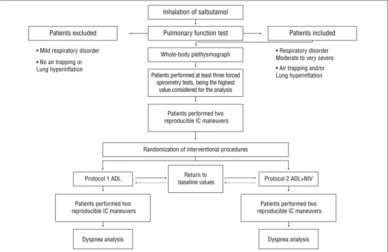
Study design and procedures
Figure 1 presents the study design and the procedures per-formed during this study. At the end of the irst interventional protocol, the patient rested long enough for the SpO2, HR and RR returned to baseline values and then they performed of the second interventional protocol. hese variables were measured 15 minutes after the performance of the irst protocol. If the return to baseline value was not reached, a new evaluation was performed every 15 minutes, establishing a maximum period of 60 minutes to return to baseline values.
Statistical analysis
Data are shown as mean ± standard deviation. he distri-bution of variables was tested using the Shapiro-Wilk tests. All variables were normally distributed with the exception of the fatigue sensation variable. Statistical analysis was performed using the Student t test for paired samples. he analysis of the fatigue sensation variable, the Wilcoxon test was used. he signiicance level of 5% was established. he analyses were performed by using the statistical package SAS® - Statistical Analysis System, 12.0, Carey, NC, USA.
Results
Initially, 33 patients were evaluated, of whom 1 participant had an obstruction stage II, however without pulmonary hy-perinlation and air-trapping, being excluded from the study. herefore, 32 patients underwent both interventional proto-cols on the same day, as the variables IC, SpO2 and HR returned to baseline values within 45 minutes.
Table 1 shows the characteristics of patients according to gender, age, body mass index (BMI), years/pack of cigarettes and pulmonary function test. he patients presented FEV1 between 22% and 64% of predicted. All participants presented air-trapping (n=32) being only 12 of them with pulmonary hyperinlation. hir-teen patients had a score of 4 on MRC scale; 15 had a score of 3 and four patients had a score of 2. All patients reported dyspnea while performing weight-bearing ADLs with the ULs elevated.
Table 2 shows the results for the IC before and after the completion of the two interventional protocols. here was a statistically signiicant decrease in IC after the completion of both interventional protocols.
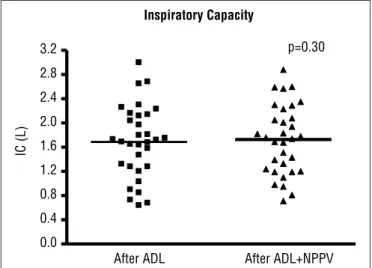
Figure 2 shows the data related to the IC after the comple-tion of the two intervencomple-tional protocols. here was no statisti-cally signiicant diference between the IC maneuvers after the completion of ADL with and without the association of NIV.
t.JMESFTQJSBUPSZEJTPSEFS t/PBJSUSBQQJOHPS -VOHIZQFSJOGMBUJPO
t3FTQJSBUPSZEJTPSEFS .PEFSBUFUPWFSZTFWFSF t"JSUSBQQJOHBOEPS -VOHIZQFSJOGMBUJPO 1BUJFOUTFYDMVEFE
1SPUPDPM"%- 1SPUPDPM"%-/*7
%ZTQOFBBOBMZTJT %ZTQOFBBOBMZTJT
1VMNPOBSZGVODUJPOUFTU
8IPMFCPEZQMFUIZTNPHSBQI
1BUJFOUTQFSGPSNFEBUMFBTUUISFFGPSDFE TQJSPNFUSZUFTUTCFJOHUIFIJHIFTU
WBMVFDPOTJEFSFEGPSUIFBOBMZTJT
1BUJFOUTQFSGPSNFEUXP SFQSPEVDJCMF*$NBOFVWFST
1BUJFOUTQFSGPSNFEUXP
SFQSPEVDJCMF*$NBOFVWFST SFQSPEVDJCMF*$NBOFVWFST1BUJFOUTQFSGPSNFEUXP 1BUJFOUTJODMVEFE *OIBMBUJPOPGTBMCVUBNPM
3BOEPNJ[BUJPOPGJOUFSWFOUJPOBMQSPDFEVSFT
3FUVSOUP CBTFMJOFWBMVFT
Figure 1. Study design and procedures.
Figure 3 shows the data related to the evaluation of dyspnea after the completion of both interventional protocols. It was observed that the performance of the interventional procedures promoted a signiicant increase in dyspnea after the completion of both protocols, with no diference between them.
Characteristics Patients with COPD (mean±SD) (n=32)
Gender: M/F 24/8
Age (yr) 69.38±7.36
Body Mass (kg) 65.04±13.76
Height (cm) 147.53±50.08
Pack-years (smoke) 78.03±42.51
FEV1-L (% predicted) 1.03±0.36 (42±13)
FVC-L (% predicted) 2.73±0.84 (89±23)
FEV1/FVC % 42±12
TLC-L (% predicted) 7.03±1.46 (120±31)
FRC-L (% predicted) 5.19±1.37 (160±52)
VR-L (% predicted) 4.27±1.35 (228±124)
RV/TLC% 61±10
IC % predicted 67±15
IC/TLC % 26±7
Table 1. Characteristics of patients with COPD.
Data are presented as mean±Standard Deviation. COPD=chronic obstructive pulmonary dise-ase; M=Male; F=Female; FEV1=forced expiratory volume in one s; FVC=forced vital capacity;
FEV1/FVC=Tiffeneau; TLC=total lung capacity; FRC=functional residual capacity; RV=residual volume; IC=inspiratory capacity and IC/TLC=inspiratory fraction.
Discussion
he main results observed in this study were: 1) there was a statistically signiicant decrease in IC with the simulation of an ADL, associated or not with noninvasive ventilation, demonstrat-ing DH; 2) a statistically signiicant diference was not observed in the comparison of the IC after the completion of both proto-cols and 3) there was a signiicant increase in dyspnea after the completion of both protocols, with no diference between them.
he DH observed in patients with COPD leads to dyspnea and limits the capacity to perform physical exercises2. he
hy-pothesis of this study was that NIV performed with two levels of positive pressure (IPAP of 10 cmH2O and EPAP of 4 cmH2O) would decrease dyspnea and pulmonary intrinsic pressure (PEEPi), and consequently would decrease the DH.
he ventilatory pattern during activities with the elevation of the ULs inluences the onset of DH in patients with COPD who have diiculty to optimize the VE by the increase in the Tidal Volume (Vt)5,20-22. In a recently study, Colucci et al.23
veri-ied the association between diferent intensities of exercises with the ULs (50, 65 and 80% of maximum load during a er-gometer cycle exercises) and the DH in patients with severe COPD. hese authors observed that higher loads of exercise were associated with DH and a lower tolerance to exercise and 80% of the maximum load represented lower work eiciency (measured by the ratio VO2 (ml/kg)/exercise duration).
he ventilatory strategy of these patients to increase the VE is to increase the RR, since the TLC does not change with exercise16,17. he premature end of exhalation increases even
more the air-trapping, decreasing therefore the IC6. In the
pres-ent study, the use of NIV with pre-determined and not indi-vidualized values (IPAP 10 cmH2O and EPAP 4 cmH2O) aimed to standardizing the intervention, and the choice of the used pressures was based on several studies,9,24,26-28 which have
dem-onstrated the beneits of using these pressures during exercises with the LLs. To our knowledge there are no studies that have associated the simulation of an ADL with NIV.
Nava et al.24 found a reduction in the activity of the
dia-phragm muscle measured by the decrease in transdiaphrag-matic pressure as well as a signiicant decrease in RR and a parallel increase in the Vt when an IPAP of 10 cmH2O or 20 cmH2O, associated with an EPAP of 5 cm H2O, was applied in stable and hypercapnic patients with severe COPD during basal respiration.
he artiicial ventilation support may improve exercise tolerance and may help patients with severe COPD to achieve higher level of training25-27. Van’t Hul et al.26 evaluated the
ef-fects of exercise training with only pressure support ventilation (PSV of 10 or 5 cmH2O) in patients with moderate to very se-vere COPD. hese authors found that in the assistance level of 10 cmH2O, there was a reduction in the VE by the reduction in the RR. he authors suggested that the changes in the respira-tory pattern may have reduced the DH, which justiied the in-creased tolerance to high-intensity training in these patients.
Maltais, Reissmann and Gottfried9 evaluated the
respira-tory pattern, dyspnea and inspirarespira-tory efort in patients with moderate-to-severe COPD, performing a constant load test on the ergometer cycle, associated with the use of SP of 10 cmH2O. hese authors found an increase in the VE by the increase in
After ADL After ADL+NPPV 0.0
0.4 0.8 1.2 1.6 2.0 2.4 2.8
3.2 p=0.30
Inspiratory Capacity
IC (L)
IC=inspiratory capacity; ADL=activity of daily living; NIV=noninvasive ventilation.
Figure 2. Data are presented as absolute values and means.
Figure 3. Data are presented as median.
Borg Scale
0.0 0.5 1.0 1.5 2.0 2.5 3.0 3.5
Before After
ADL ADL+NPPV
0.27
2.34*
*
0.25
2.67
Borg (scored)
ADL=activity of daily living; NIV=noninvasive positive pressure ventilation; *p<0.05. Iinterventional
Procedures Protocol Before After p Value
ADL 2.06±0.60 L 1.69±0.60 L 0.0001*
ADL+ NIV 2.05±0.62 L 1.73±0.57 L 0.0001*
Data are presented as mean±Standard Deviation; IC=inspiratory capacity; COPD=chronic obstructive pulmonary disease; ADL=activity of daily living; NIV=noninvasive positive pressure ventilation and *p<0.05.
Table 2. Inspiratory capacity in patients with COPD (n=32) before and after ADL with or without NIV.
References
1. Rabe KF, Hurd S, Anzueto A, Barnes PJ, Buist SA, Calverley P, et al. Global strategy for the diagnosis, management, and prevention of chronic obstructive pulmonary disease: GOLD executive summary. Am J Respir Crit Care Med. 2007;176(6):532-55.
2. O’Donnell DE. Hyperinflation, dyspnea, and exercise intolerance in chronic obstructive pulmonary disease. Proc Am Thorac Soc. 2006;3(2):180-4.
3. Velloso M, Stella SG, Cendon S, Silva AC, Jardim JR. Metabolic and ventilatory parameters of four activities of daily living accomplished with arms in COPD patients. Chest. 2003;123(4):1047-53.
4. Tangri S, Woolf CR. The breathing pattern in chronic obstructive lung disease during the performance of some common daily activities. Chest. 1973;63(1):126-7.
5. McKeough ZJ, Alison JA, Bye PT. Arm positioning alters lung volumes in subjects with COPD and healthy subjects. Aust J Physiother. 2003;49(2):133-7.
6. Dolmage TE, Goldstein RS. Repeatability of inspiratory capacity during incremental exercise in patients with severe COPD. Chest. 2002;121(3):708-14.
7. Marin JM, Carrizo SJ, Gascon M, Sanchez A, Gallego B, Celli BR. Inspiratory capacity, dynamic hyperinflation, breathlessness, and exercise performance during the 6-minute walk test in Chronic Obstructive Pulmonary Disease. Am J Respir Crit Care Med. 2001;163(6):1395-9.
8. Porto EF, Castro AA, Velloso M, Nascimento O, Dal Maso F, Jardim JR. Exercises using the upper limbs hyperinflate COPD patients more than exercises using the lower limbs at the same metabolic demand. Monaldi Arch Chest Dis. 2009;71(1):21-6.
RR and Vt, however with a signiicant decrease in the inspira-tory efort and dyspnea.
Poggi et al.27 investigated the efects of two types of artiicial
ventilation (PAV and PSV - both with IPAP, established accord-ing to patients comfort, and EPAP of 4 cmH2O) during eleva-tion of the ULs in patients with severe COPD. In both modes of ventilation, there was a reduction of the PEEPi greater than 30% compared with the control group, however this diference was not statistically signiicant. It should be noted, however, that the group that used the PAV method reported lower in-spiratory efort during the elevation of the ULs.
O’Donnell, Sanii and Younes28, in an innovative study,
evalu-ated the impact of the use of CPAP of 4 to 5 cmH2O in patients with moderate-to-severe COPD and observed a reduction of dyspnea during the performance of submaximal exercise on the treadmill, suggesting that positive pressure promoted the reduction of DH.
With regards to the IPAP established in order to pro-vide support to the inspiratory muscles, especially to the diaphragm muscle, it is possible that the selected pressure of 10 cmH2O was not adequate to achieve this goal when the simulation of a ADLs was performed. here was a signiicant increase in dyspnea, which indicates indirectly an overload of the inspiratory muscles. According to the indings of the present study, it appears that the EPAP of 4 cmH2O was insuf-icient to counterbalance the PEEPi and, therefore, to reduce the overload of the inspiratory muscles in these patients. Studies show that patients with COPD usually complain of a more intense dyspnea when performing prolonged tasks involving the upper limbs in elevated positions and without support compared to tasks with LLs3,4,8,29. his fact
demon-strates the possibility of ULs exercises to generate greater impact on DH compared with LLs exercises.
his study has some limitations. Firstly the RR, the trans-diaphragmatic pressure and the PEEPi, which could explain the absence of a reduction in the elastic and resistive work of the respiratory muscles, were not measured after the
interventional procedures protocols. herefore, the pre-established pressures may have been inadequate for some patients. Secondly, studies show that other forms of artiicial ventilation, such as proportional assisted ventilation, PAV, may provide better results compared to the two ixed pres-sure levels established in this study, since the PAV suit better to the patient’s ventilatory demand30.
he main purpose of using NIV during the exercise is to reduce dyspnea by reducing the overload imposed on the respiratory muscles as a result of DH, allowing patients to achieve higher intensity during exercise30. he reduction in the
overload of respiratory muscles during exercise reduces blood low competition between these muscles and the peripheral muscles, contributing to an increased of blood supply to the muscles12. In the Velloso et al.3 study, in patients with COPD
performing four ADLs involving the ULs (sweeping the loor, clearing a whiteboard, lifting containers of diferent weights and changing light bulbs), was observed an increase in the ratio VE on the maximum voluntary ventilation, justifying the intense dyspnea reported by these patients, although the DH has not been evaluated. Our results showed that the addition of NIV was not able to signiicantly reduce dyspnea during a simulation of ADL, which may be due to the use of pressures (IPAP and EPAP) beyond the requirements of the patients.
herefore, our results suggest that the bi-level pressure modality, with IPAP and EPAP of 10 and 4 cmH2O, respectively, was not suicient to prevent the DH and dyspnea during ADL with the ULs in patients with moderate-to-severe COPD. he search for therapeutic strategies to improve the functionality of the patient with COPD during the performance of ADLs is extremely important, considering that 80% of the functionality is performed by ULs tasks8.
We suggest future studies to examine the efects of NIV during ADL with the ULs using other pressures or modalities of NIV, since the functional independence during ADL is a major goal of respiratory physical therapy in the treatment of patients with COPD.
9. Maltais F, Reissmann H, Gottfried SB. Pressure support reduces inspiratory effort and dyspnea during exercise in chronic airflow obstruction. Am J Respir Crit Care Med. 1995;151(4):1027-33.
10. Gosselink R. Breathing techniques in patients with chronic obstructive pulmonary disease (COPD). Chron Respir Dis. 2004;1(3):163-72.
11. Mehta S, Hill NS. Noninvasive ventilation. Am J Respir Crit Care Med. 2001;163(2):540-77.
12. Ambrosino N. Assisted ventilation as an aid to exercise training: a mechanical doping? Eur Respir J. 2006;27(1):3-5.
13. Albuquerque AL, Nery LE, Villaça DS, Machado TY, Oliveira CC, Paes AT, et al. Inspiratory fraction and exercise impairment in COPD patients GOLD stages II-III. Eur Respir J. 2006;28(5):939-44.
14. Diretrizes para testes de função pulmonar. Jornal de Pneumologia. 2002;28(Suppl 3):95-100.
15. Lung function testing: selection of reference values and interpretative strategies. American Thoracic Society. Am Rev Respir Dis. 1991;144(5):1202-18.
16. Stubbing DG, Pengelly LD, Morse JL, Jones NL. Pulmonary mechanics during exercise in subjects with chronic airflow obstruction. J Appl Physiol. 1980;49(3):511-5.
17. Yan S, Kaminski D, Sliwinski P. Reliability of inspiratory capacity for estimating end-expiratory lung volume changes during exercise in patients with chronic obstructive pulmonary disease. Am J Respir Crit Care Med. 1997;156(1):55-9.
18. Surveillance for respiratory hazards in the occupational setting [American Thoracic Society]. Am Rev Respir Dis. 1982;126(5):952-6.
19. Borg GA. Psychophysical bases of perceived exertion. Med Sci Sports Exerc. 1982;14(5):377-81.
20. Cerny FJ, Ucer C. Arm work interferes with normal ventilation. Appl Ergon. 2004;35(5):411-5.
21. Dolmage TE, Maestro L, Avendano MA, Goldstein RS. The ventilatory response to arm elevation of patients with chronic obstructive pulmonary disease. Chest. 1993;104(4):1097-100.
22. Gigliotti F, Coli C, Bianchi R, Grazzini M, Stendardi L, Castellani C, et al. Arm exercise and hyperinflation in patients with COPD: effect of arm training. Chest. 2005;128(3):1225-32.
23. Colucci M, Cortopassi F, Porto E, Castro A, Colucci E, Iamonti VC, et al. Upper limb exercises using varied workloads and their association with dynamic hyperinflation in patients with COPD. Chest. 2010;138(1):39-46.
24. Nava S, Ambrosino N, Rubini F, Fracchia C, Rampulla C, Torri G, et al. Effect of nasal pressure support ventilation and external PEEP on diaphragmatic activity in patients with severe stable COPD. Chest. 1993;103(1):143-50.
25. Díaz O, Bégin P, Torrealba B, Jover E, Lisboa C. Effects of noninvasive ventilation of lung hyperinflation in stable hypercapnic COPD. Eur Respir J. 2002;20(6):1490-8.
26. van’t Hul A, Gosselink R, Hollander P, Postmus P, Kwakkel G. Training with inspiratory pressure support in patients with severe COPD. Eur Respir J. 2006;27(1):65-72.
27. Poggi R, Appendini L, Polese G, Colombo R, Donner CF, Rossi A. Noninvasive proportional assist ventilation and pressure support ventilation during arm elevation in patients with chronic respiratory failure. A preliminary, physiologic study. Respir Med. 2006;100(6):972-9.
28. O’Donnell DE, Sanii R, Younes M. Improvement in exercise endurance in patients with chronic airflow limitation using continuous positive airway pressure. Am Rev Respir Dis. 1988;138(6):1510-4.
29. Panka GF, Oliveira MM, França DC, Parreira VF, Britto RR, Velloso M. Ventilatory and muscular assessment in healthy subjects during ane activity of daily living with unsupported arm elevation. Rev Bras Fisioter. 2010;14(4):337-43.
30. Moreno J, Dal Corso S, Malaguti C. Análise descritiva do uso de ventilação mecânica não invasiva durante exercício em pacientes com DPOC. ConScientiae Saúde. 2007;6(2):295-303.