Comparison of Two- and Three-dimensional
Ultrasonography in the Evaluation of Lesion
Level in Fetuses with Spina Bi
fi
da
Comparação entre a ultrassonografi
a bidimensional e a
ultrassonografia tridimensional na avaliação do nível da
lesão nos fetos com espinha bífi
da
Márcio José Rosa Requeijo
1,2Victor Bunduki
2Rossana Pulcineli Vieira Francisco
2Marco Antonio Borges Lopes
2Rodrigo Ruano
2Marcelo Zugaib
21Departamento de Obstetrícia e Ginecologia, Faculdade de Medicina
de Itajubá, Itajubá, MG, Brazil
2Departamento de Obstetrícia e Ginecologia, Faculdade de Medicina,
Universidade de São Paulo, São Paulo, SP, Brazil
Rev Bras Ginecol Obstet 2016;38:120–126.
Address for correspondence Márcio José Rosa Requeijo, MD, PhD, Faculdade de Medicina, Universidade de São Paulo, Av. Dr. Enéas de Carvalho Aguiar, 255, 10° andar, Cerqueira César, São Paulo, SP, Brasil 05403-010 (e-mail: marciorequeijo3@hotmail.com).
Keywords
►
fetus
►
two-dimensional
ultrasonography
►
three-dimensional
ultrasonography
►
spina bi
fi
da
Abstract
Purpose
To evaluate the precision of both two- and three-dimensional
ultrasonogra-phy in determining vertebral lesion level (the
fi
rst open vertebra) in patients with spina
bi
fi
da.
Methods
This was a prospective longitudinal study comprising of fetuses with open
spina bi
fi
da who were treated in the fetal medicine division of the department of
obstetrics of Hospital das Clínicas of the Universidade de São Paulo between 2004 and
2013. Vertebral lesion level was established by using both two- and three-dimensional
ultrasonography in 50 fetuses (two examiners in each method). The lesion level in the
neonatal period was established by radiological assessment of the spine. All
pregnan-cies were followed in our hospital prenatally, and delivery was scheduled to allow
immediate postnatal surgical correction.
Results
Two-dimensional sonography precisely estimated the spina bi
fi
da level in 53%
of the cases. The estimate error was within one vertebra in 80% of the cases, in up to
two vertebrae in 89%, and in up to three vertebrae in 100%, showing a good
interobserver agreement. Three-dimensional ultrasonography precisely estimated
the lesion level in 50% of the cases. The estimate error was within one vertebra in
82% of the cases, in up to two vertebrae in 90%, and in up to three vertebrae in 100%,
also showing good interobserver agreement. Whenever an estimate error was
observed, both two- and three-dimensional ultrasonography scans tended to
underes-timate the true lesion level (55.3% and 62% of the cases, respectively).
Conclusions
No relevant difference in diagnostic performance was observed
be-tween the two- and three-dimensional ultrasonography. The use of three-dimensional
received October 13, 2015 accepted January 6, 2016 published online March 21, 2016
DOI http://dx.doi.org/ 10.1055/s-0036-1580711. ISSN 0100-7203.
Introduction
Spina bifida is characterized by an incomplete formation of the structures that protect the spinal cord through inappro-priate closing of the backbone, which may present as open and hidden forms, with or without neurological involve-ment. The most common types are myelomeningocele and meningocele, which are open forms of spina bifida and may result in severe neurological sequels, with an incidence of 0.5 to 0.8 per thousand live births.1
This defect occurs as a result of genetic and environmental factors. Many causes have been proposed, such as folate deficiency, maternal diabetes, zinc deficiency, ingestion of alcohol during thefirst 3 months of pregnancy, and maternal exposure to certain drugs such as carbamazepine and val-proic acid.2Spina bifida can be prevented by supplementa-tion of folic acid in the periconcepsupplementa-tional period, starting 3 months before conception and up to the third month of pregnancy.3,4
Correction of this defect may be performed during the intrauterine or postnatal period.5–7The MOMS study (Man-agement of Myelomeningocele Study) showed that prenatal fetal surgery for myelomeningocele decreased the need of
placing a ventriculoperitoneal shunt, and improved cogni-tive and motor development within 30 months of life. One of the selection criteria in this study was fetuses with open spina bifida between T1 and S1. Therefore, the correct diagnosis of lesion level is an important factor for surgical indication.8–11With regard to the diagnosis of spina bifida in the gestational period, ultrasonography is the method of choice, as it is able to detect around 80% to 100% of cases.12 In thefirst quarter, the assessment of intracranial trans-lucency between the 11th and 14th weeks is a promising technique. In fetuses with spina bifida, the disappearance of the fourth ventricle is observed in the caudal displacement of the hindbrain.13,14In the second and third quarters, both two- and three-dimensional ultrasonography are useful for directly observing the vertebral lesion. The definition of the lesion level is considered as the determining factor of fetal prognosis and the prediction of possible secondary compli-cations, which entail counseling of the pregnant woman, her family, and the professionals involved. Bruner et al,15 how-ever, stated that the method presents some limitations as to the correct diagnosis of the lesion level, demonstrating that two-dimensional ultrasonography defines exactly the
ultrasonography showed no additional bene
fi
t in diagnosing the lesion level in the
fetuses with spina bi
fi
da. Errors in both methods showed a tendency to underestimate
lesion level.
Resumo
Objetivo
Avaliar a precisão da ultrassonogra
fi
a bidimensional e tridimensional para a
determinação do nível da lesão vertebral em casos de fetos portadores de espinha
bí
fi
da.
Métodos
Estudo prospectivo longitudinal, compreendendo fetos portadores de
espinha bí
fi
da assistidos no setor de medicina fetal de hospital de ensino do Sudeste
do Brasil, entre os anos de 2004 e 2013. Foram incluídos 50 fetos portadores de espinha
bí
fi
da pela ultrassonogra
fi
a bidimensional e tridimensional (dois examinadores em
cada método) com relação ao nível da lesão. O nível exato da lesão foi veri
fi
cado usando
radiogra
fi
a após o nascimento.
Resultados
A ultrassonogra
fi
a bidimensional estimou corretamente o nível de
espi-nha bí
fi
da em 53,0% dos casos. Em 80,0% dos casos a estimativa ocorreu com erro de
uma vértebra; 89,0% em até duas vértebras e de 100,0% em até três vértebras. A
ultrassonogra
fi
a tridimensional estimou corretamente o nível de espinha bí
fi
da em
50,0% dos casos. Em 82,0% dos casos, a estimativa ocorreu com erro de uma vértebra;
90,0% em até duas vértebras e de 100,0% em até três vértebras. Nos casos em que
houve erro na estimativa do nível da lesão, tanto na avaliação bidimensional quanto na
tridimensional, observou-se tendência a subestimar o nível da lesão vertebral (55,3% na
avaliação bidimensional e 62,0% na tridimensional), ou seja, colocando o nível
ultrassonográ
fi
co mais baixo que o observado no pós-natal.
Conclusões
Não houve diferenças relevantes entre o desempenho diagnóstico da
ultrassonogra
fi
a bidimensional e tridimensional para determinação do nível da lesão
vertebral nos casos de fetos portadores de espinha bí
fi
da. Tendência a subestimação do
nível de lesão nos casos em que houve erro tanto na ultrassonogra
fi
a bidimensional
quanto na tridimensional.
Palavras-chave
►
fetos
►
ultrassom
bidimensional
►
ultrassom
tridimensional
postdelivery levels of the vertebral lesion in only 38% of the cases, increasing this setting to 96% within two levels and reaching 100% within three levels, that is, when performed in reference centers by trained professionals. The observation of the possibility of errors up to three levels undermines its use as a decisive factor in the management of this malforma-tion because errors of that magnitude result in widely varied injury prognosis regarding ambulation and motor strength, mainly of the lower limbs, in addition to control of vesical and anal sphincters.
Three-dimensional ultrasonography has been described as an important adjuvant method for assessing numerous fetal malformations, but its superiority to two-dimensional ultrasonography with regard to the diagnosis of the injury level in fetuses with spina bifida is controversial.16–20 The absence of conclusive data in relation to the comparison between two- and three-dimensional ultrasonography in the evaluation of injury level in cases of spina bifida motivated us to conduct this study.
Methods
A prospective longitudinal study was conducted with the objective of evaluating fetuses with isolated spina bifida and without chromosomopathies who were treated in the fetal medicine division of the department of obstetrics and gyne-cology of Hospital das Clínicas of the Universidade de São Paulo (HC-FM-USP) between October 2004 and March 2013. This study was approved by the ethics committee for the analysis of research projects of the HC-FM-USP. The authors did not receivefinancial support, equipment, or medicines from any other institution and had no conflicts of interest. The inclusion and exclusion criteria in this study were as follows: diagnosis of isolated open spina bifida on fetal morphologic ultrasonography; had been evaluated twice on two-dimensional ultrasonography, with assessment of lesion level, by two examiners in charge on the same day, using the same technique; two three-dimensional ultraso-nography evaluations, with evaluation of lesion level, by two examiners in charge on the same day, using the same technique; and confirmed injury level in spina bifida by using simple postnatal radiography. The exclusion criterion was birth in another service.
From October 2004 to March 2013, in the fetal medi-cine division of HC-FM-USP, 270 fetuses were diagnosed with spina bifida. Among these fetuses, 220 did not meet the inclusion criteria and thus were excluded (122 cases due to associated malformations and 98 cases for not having been postnatally evaluated). Finally, we included 50 fetuses who were considered asfit to participate in this study.
With regard to the calculation of the sample size, our hypothesis was that 50% concordance would be found be-tween the two methods, ranging from 35% and 65%. To obtain estimates with 95% confidence intervals, we needed to include at least 43 cases in the study. As the eligible number of fetuses with spina bifida was 50, the sample size was considered satisfactory.21
The pregnant women whose fetuses were diagnosis with spina bifida were interviewed by the researchers. If they agreed to participate in the study, they were asked to sign an informed consent form. These patients were forwarded to the prenatal fetal medicine group and followed up to term. As support, the pregnant women were referred to the depart-ment of psychology of the obstetric clinic of the same institution, with thefirst consultation preferably after ultra-sonography diagnosis. The parturition was programed in the service jointly with the neurosurgery department.
The information was entered in the registration database of patients through a specific software (Microsoft Access 2007), including the examination date, overall record, ad-dress, contact number, name, mother’s age in years, parity, personal and family histories of spina bifida, gestational age at diagnosis (date of last menstruation compatible with the obstetric ultrasonography performed until the 12th week, or at least two ultrasonography examinations concordant up to the 20th week).
In relation to the specific data that correlated with the location of the spina bifida, each fetus was evaluated by the two examiners responsible for each technique. Thus, the total number of two- and three-dimensional evaluations in the study was 100 each.
After birth, radiography was performed to assess lesion level (considered the gold standard for its location).16 The injury levels obtained by using two- and three-dimensional ultrasonography were compared with the postnatal radio-logical levels and classified as coincidental in relation to the findings. That is, the lesion level in the spine on simple radiography coincided with the level found on ultrasonogra-phy. Errors were classified as follows: error at one level, the lesion level ranged from less to more than one vertebra; error at two levels, the level of lesion ranged from less to more than two vertebrae; and error at three levels, the lesion level ranged from less to more than three vertebrae.
Data were analyzed by using the Statistical Package for Social Sciences (SPSS) version 20 (IBM SPSS statistics). The categorical variables were expressed as means, medians, and standard deviations, and absolute and relative fre-quencies. The Fisher exact test was used for comparison of proportions.
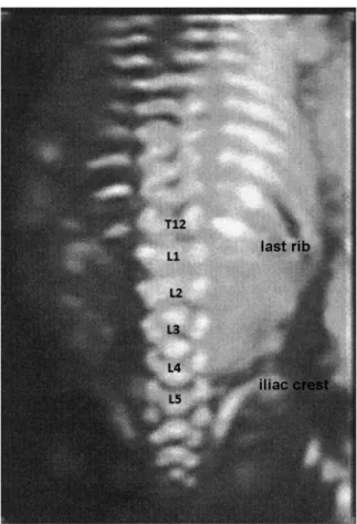
For the two-dimensional ultrasonography assessment, median sagittal, coronal, and transverse sections of the spine were obtained.15The lesion level was obtained by counting the vertebrae craniocaudally, starting from the cervical vertebrae up to thefirst abnormal opening of the side plates. That is, the vertebra was more affected cranially by the closure defect. In addition, the last ribs were used as refer-ence points, coinciding with the 12th thoracic vertebra and the iliac crest as the level of thefifth lumbar vertebra15,22 (►Fig. 1).
The three-dimensional ultrasonography assessment was performed from a sagittal section of the fetal spine. The volumetric window was adjusted to capture the whole region of the vertebral column. In fetuses with more ad-vanced gestational ages, two or more partial acquisitions of the spine were performed, which allowed the observation of spina bifida. The sweep angle was adjusted to capture only the vertebral column (15° to 30°), selecting the high-resolu-tion skeletal mode18,20(►Fig. 2).
The lesion level was determined by counting the vertebrae craniocaudally, starting from the cervical vertebrae up to the first abnormal opening of the side plates. That is, the vertebra was more cranially affected by the closing defect. In addition, the last ribs were used as reference points, coinciding with the 12th thoracic vertebra and the iliac crest as the level of thefifth lumbar vertebra.15,22
Thefirst observer performed the evaluation, noting the level of the lesion. On the same day, the second observer also performed examinations by using the same technique, with-out knowledge of the results obtained by the previous operator. The device used was the GE Voluson 730 Expert ultrasound system (Austria).
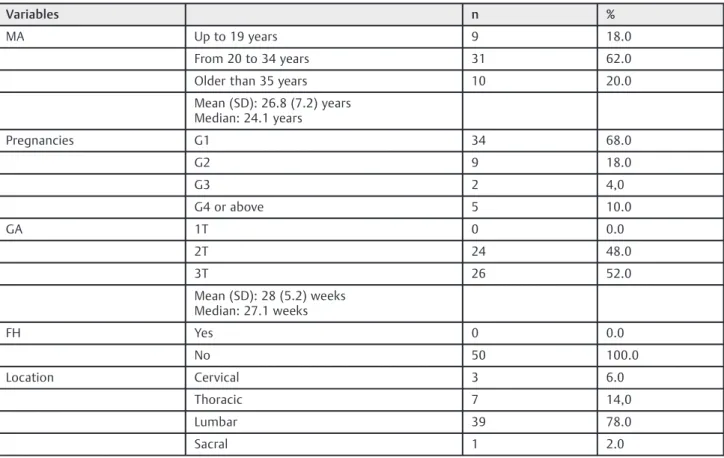
Characterization of the Studied Population
The mean maternal age in this study was 26.8 years, with a standard deviation of 7.2 years and a median of 24.1 years (range, 16.2–45.1 years). By dividing according to age group, we found an incidence of 18%, 20%, and 62% in mothers aged19 years,>35 years, and 20 to 34 years, respectively.
With regard to the number of gestations, primigravidae accounted for the highest incidence in this study, with 34 cases (68.0%), followed by second pregnancies, with 9 (18.0%); third pregnancies, with 2 (4.0%); and other parities (up to the sixth pregnancy), with 5 (10.0%). The mean (SD) gestational age when the first ultrasonography was per-formed in our institution was 28.0 (5.2) weeks, with a median of 27.1 weeks (range, 18–38 weeks). Of the cases, none was diagnosed in thefirst quarter, 48.0% (24 cases) were diagnosed in the second quarter, and 52% (26 cases) were diagnosed in the third quarter. No personal or family history of spina bifida was found in the studied population (►Table 1). The locations of the lesions in the fetuses affected were distributed as follows: cervical vertebrae, 3 cases
(6.0%); thorax, 7 cases (14.0%); lumbar spine, 39 cases (78.0%); and sacral vertebra, 1 case (2.0%).
Results
Regarding the results in the two-dimensional ultrasonogra-phy group, with a total of 100 evaluations, the level of coincidence obtained was 53% of the cases. With an error at one, two, and 3 levels, this coincidence level increased to 80%, 89%, and 100%, respectively (►Table 2).
As to the results of the three-dimensional ultrasonogra-phy group, with a total of 100 evaluations, the level of coincidence obtained was 50% of the cases. This increased to 82% with an error at one level, to 90% with an error at two Table 1 Characterization of the study population
Variables n %
MA Up to 19 years 9 18.0
From 20 to 34 years 31 62.0 Older than 35 years 10 20.0 Mean (SD): 26.8 (7.2) years
Median: 24.1 years
Pregnancies G1 34 68.0
G2 9 18.0
G3 2 4,0
G4 or above 5 10.0
GA 1T 0 0.0
2T 24 48.0
3T 26 52.0
Mean (SD): 28 (5.2) weeks Median: 27.1 weeks
FH Yes 0 0.0
No 50 100.0
Location Cervical 3 6.0
Thoracic 7 14,0
Lumbar 39 78.0
Sacral 1 2.0
Abbreviations: FH, family history; GA, gestational age at diagnosis; MA, maternal age.
Table 2 Evaluation of the combined results of the observers who performed USG-2D and USG-3D in relation to coincidence and levels of error
Coincidences n (%)
1 Level n (%)
2 Levels n (%)
3 Levels n (%)
Total Evaluations
USG-2 D 53 (53) 27 (27) Cumulative: 80%
9 (9) Cumulative: 89%
11 (11) Cumulative: 100%
100
USG-3D 50 (50) 32 (32) Cumulative: 82,0%
8 (8) Cumulative: 90%
10 (10) Cumulative: 100%
100
levels, and to 100% with an error at up to three levels. No significant difference was observed between the two-and three-dimensional ultrasonography methodology in the Fisher exact test (with a level of significance of 0.05).
In the cases in which an error occurred at one lesion level in the two-dimensional ultrasonography group, the lesion level tended to be underestimated in 55.3% of the assess-ments and overestimated at the injury level in 44.7% of the cases. In the three-dimensional ultrasonography group, the injury level tended to be underestimated in 62% of the assessments and overestimated in 38% of the cases.
Discussion
Neural tube defects cause high incidence rates of perinatal and infant mortality, and severe chronic complications in survivors. In the case of spina bifida, prenatal diagnosis enables better understanding of the problem and the associ-ated conditions that help, among other actions, in the planning of birth and interventions that may be necessary.
Individuals with spina bifida may present motor and sensory loss mainly in the lower extremities, urological complications, neurogenic bowel dysfunction, orthopedic complications, pressure ulcers, cognitive deficiency, and allergies, with varying degrees of disability and dependence for prostheses, crutches, wheelchair, or third-party aid throughout life. The severity of these complications, the point of neurological dysfunction, and the presence of asso-ciated complications, though imperfectly, may be predicted based on the lesion level in these patients. The higher the injury level, the more serious the complications.23
With regard to ultrasonography diagnosis in cases of spina bifida, the accuracy of determining the lesion level by using two-dimensional ultrasonography widely varied, with values ranging from 38% to 64%. This variation can reach up to three vertebral levels.15,22
With respect to the accuracy of three-dimensional ultra-sonography in determining lesion level, Cameron and Moran17demonstrated that three-dimensional ultrasonog-raphy can reach diagnostic accuracy within one vertebral body in80% of the patients. Buyukkurt et al16 demonstrat-ed a 79.0% level of accuracy, reaching 100% with a margin of one level.
With regard to the ultrasonography location of the lesion, our results demonstrated an accuracy of 53% for the two-dimensional ultrasonography assessment and 50.0% for the three-dimensional ultrasonography assessment. However, if we considered an error at one vertebral level, this index reached 80% and 82.0% in two- and three-dimensional ultrasonography, respectively, which are values similar to those obtained by other authors.15 However, we only reached 100% concordance, considering an error margin of three vertebral levels both in two- and three-dimensional ultrasonography.
In relation to the error in the diagnosis of the vertebral level, we observed a tendency to underestimate lesion levels. That is, we observed a tendency to place the lesion level to lower than that observed after birth, both in the two- and
three-dimensional methodology (55.3% and 62.0%, respec-tively), compared with the error by overestimation of the injury level (44.7% and 38.0%, respectively). With this obser-vation, we conclude that in case of doubt on the vertebral lesion level, we should opt for the highest lesion level to have a higher chance of concordance.
We conclude that inadequate evaluation of the anatomical reference points for the assessment of the vertebrae involved seems to be a factor that can affect results. In the evaluation of the last rib, as a point of reference for the 12ththoracic
vertebra (T12), owing to its small size, it may not be visible in cases of excessive angulation of the spine, leading to error. Excessive angulation of the spine can also make the assess-ment difficult and lead to error when using the iliac crest with reference to thefifth lumbar vertebra (L5). Therefore, the spine must be rectified if possible for the use of these points of reference.
In fetuses with gestational ages close to 20 weeks, ossifi -cation in the topography of thefifth sacral vertebra could not be observed. Whereas in fetuses in the third quarter, this assessment was possible. Taking into account this finding, the vertebrae counted toward the caudocranial direction in the second quarter could demonstrate a lower vertebral level than the actual level, which could lead to an underestimation of the lesion level. A similarfinding was reported by Kollias et al.22Therefore, the ideal technique for the evaluation of the spine should position it as rectified as possible, and the direct counting of the vertebrae should always be performed in the craniocaudal direction. Knowledge of these facts allows for more accurate diagnosis of lesion level and should not be disregarded.
Few studies have compared the accuracies of two- and three-dimensional ultrasonography for the diagnosis of spina bifida. These studies claim that three-dimensional ultrasonography is a complementing methodology and does not replace two-dimensional ultrasonography.18,19 Ourfindings are concordant with these studies. No signifi -cant differences were observed between these methodolo-gies. Thus, based on our results, we conclude that the use of three-dimensional ultrasonography had no advantages to two-dimensional ultrasonography for the diagnosis of the location of the injury level in spina bifida.
References
1 American College of Obstetricians and Gynecologists. ACOG tech-nical bulletin no. 99: prenatal detection of neural tube defects. Washington (DC): ACOG; 1986
2 Wegner C, Nau H. Alteration of embryonic folate metabolism by valproic acid during organogenesis: implications for mechanism of teratogenesis. Neurology 1992;42(4, Suppl 5): 17–24
3 McDonnell R, Johnson Z, Doyle A, Sayers G. Determinants of folic acid knowledge and use among antenatal women. J Public Health Med 1999;21(2):145–149
4 Wald NJ, Law MR, Morris JK, Wald DS. Quantifying the effect of folic acid. Lancet 2001;358(9298):2069–2073
5 Bruner JP, Tulipan N, Paschall RL, et al. Fetal surgery for myelo-meningocele and the incidence of shunt-dependent hydrocepha-lus. JAMA 1999;282(19):1819–1825
6 Hirose S, Farmer DL, Albanese CT. Fetal surgery for myelomenin-gocele. Curr Opin Obstet Gynecol 2001;13(2):215–222
7 Bruner JP, Tulipan NE, Richards WO. Endoscopic coverage of fetal open myelomeningocele in utero. Am J Obstet Gynecol 1997;176 (1 Pt 1):256–257
8 Adzick NS, Thom EA, Spong CY, et al; MOMS Investigators. A randomized trial of prenatal versus postnatal repair of myelo-meningocele. N Engl J Med 2011;364(11):993–1004
9 Meuli M, Moehrlen U. Fetal surgery for myelomeningocele is effective: a critical look at the whys. Pediatr Surg Int 2014; 30(7):689–697
10 Flake A. Percutaneous minimal-access fetoscopic surgery for myelomeningocele - not so minimal!. Ultrasound Obstet Gynecol 2014;44(5):499–500
11 Adzick NS. Fetal surgery for spina bifida: past, present, future. Semin Pediatr Surg 2013;22(1):10–17
12 Okumura M, Zugaib M. Ultra-Sonografia em obstetrícia. São Paulo: Sarvier; 2002
13 Orlandi E, Rossi C, Perino A, Cucinella G, Orlandi F. Prospective sonographic detection of spina bifida at 11-14 weeks and sys-tematic literature review. J Matern Fetal Neonatal Med 2015:1–5 [Epub ahead of print]
14 Chaoui R, Benoit B, Mitkowska-Wozniak H, Heling KS, Nicolaides KH. Assessment of intracranial translucency (IT) in the detection of spina bifida at the 11-13-week scan. Ultrasound Obstet Gynecol 2009;34(3):249–252
15 Bruner JP, Tulipan N, Dabrowiak ME, et al. Upper level of the spina bifida defect: how good are we? Ultrasound Obstet Gynecol 2004; 24(6):612–617
16 Buyukkurt S, Binokay F, Seydaoglu G, et al. Prenatal determination of the upper lesion level of spina bifida with three-dimensional ultrasound. Fetal Diagn Ther 2013;33(1):36–40
17 Cameron M, Moran P. Prenatal screening and diagnosis of neural tube defects. Prenat Diagn 2009;29(4):402–411
18 Lee W, Chaiworapongsa T, Romero R, et al. A diagnostic approach for the evaluation of spina bifida by three-dimensional ultraso-nography. J Ultrasound Med 2002;21(6):619–626
19 Carletti A, Ghi T, Ceccarini M, et al. OC160: three-dimensional ultrasonography is of limited value in diagnosing fetal spina bifida. Ultrasound Obstet Gynecol 2007;30(4):416
20 Wei Q, Cai A, Wang X, Xie L, Wang B, Wang X. Value of 3-dimensional sonography for prenatal diagnosis of vertebral formation failure. J Ultrasound Med 2013;32(4):595–607 21 Rosner BA. Fundamentals of biostatistics. 4th ed. Belmont:
Duxbury Press; 1995
22 Kollias SS, Goldstein RB, Cogen PH, Filly RA. Prenatally detected myelomeningoceles: sonographic accuracy in estimation of the spinal level. Radiology 1992;185(1):109–112