Sonographic Cervical Shortening after Labor
Induction is a Predictor of Vaginal Delivery
Ultrassonogra
fi
a para encurtamento do colo do útero
após indução de parto é um preditor de parto normal
Ugo Indraccolo
1Gennaro Scutiero
2Pantaleo Greco
21Complex Operative Unit of Obstetrics and Gynecology, Alto Tevere Hospital of Città di Castello, Città di Castello (PG), Italy
2Section of Obstetrics and Gynecology, Department of Morphology, Surgery and Experimental Medicine, University of Ferrara, Cona (FE), Italy
Rev Bras Ginecol Obstet 2016;38:585–588.
Address for correspondence Ugo Indraccolo, MD, PhD, Complex Operative Unit of Obstetrics and Gynecology, Alto Tevere Hospital of Città di Castello, Via P. Veronese 2/c, 06024 Gubbio (PG), Italy (e-mail: [email protected]).
Keywords
►
cervical shortening
►
delivery outcome
►
transvaginal
ultrasonography
►
labor induction
Abstract
Objective
Analyzing if the sonographic evaluation of the cervix (cervical shortening)
is a prognostic marker for vaginal delivery.
Methods
Women who underwent labor induction by using dinoprostone were
enrolled. Before the induction and three hours after it, the cervical length was
measured by ultrasonography to obtain the cervical shortening. The cervical
shorten-ing was introduced in logistic regression models among independent variables and for
calculating receiver operating characteristic (ROC) curves.
Results
Each centimeter in the cervical shortening increases the odds of vaginal
delivery in 24.4% within 6 hours; in 16.1% within 24 hours; and in 10.5% within 48 hours.
The best predictions for vaginal delivery are achieved for births within 6 and 24 hours,
while the cervical shortening poorly predicts vaginal delivery within 48 hours.
Conclusion
The greater the cervical shortening 3 hours after labor induction, the
higher the likelihood of vaginal delivery within 6, 24 and 48 hours.
Palavras-chave
►
encurtamento do
colo do útero
►
resultado do parto
►
ultrassonogra
fi
a
transvaginal
►
indução do parto
Resumo
Objetivo
Analisar se a avaliação ultrassonográ
fi
ca do colo do útero (encurtamento) é
um marcador prognóstico para parto normal.
Métodos
Consideramos mulheres com trabalho de parto induzido usando
dinopros-tona. Antes da indução e três horas após, a extensão cervical foi medida por
ultrassonogra
fi
a para obter o encurtamento do colo do útero. O encurtamento do
colo do útero foi aplicado em modelos de regressão dentre variáveis independentes.
Curvas de Característica de Operação do Receptor foram calculadas.
Resultados
Cada centímetro no encurtamento do colo do útero aumenta as chances
de parto normal para 24,4% dentro de 6 horas; 16,1% dentro de 24 horas; e 10,5%
dentro de 48 horas. Os melhores preditores de parto normal são alcançados para partos
dentre 6 e 24 horas, enquanto o encurtamento prevê mal o parto normal dentro de
48 horas.
received
May 16, 2016
accepted
November 8, 2016
published online
December 19, 2016
DOIhttp://dx.doi.org/ 10.1055/s-0036-1597629.
ISSN 0100-7203.
Copyright © 2016 by Thieme-Revinter Publicações Ltda, Rio de Janeiro, Brazil
THIEME
Introduction
Cervical length assessment prior to induction is a component of Bishop’s score that is already used to predict the success of the labor induction. A meta-analysis supports the ability of Bishop’s score in predicting the success of labor induction.1 The rationale of the clinical cervical length assessment has been translated in the practice to assess the cervical length by sonography. Several studies support that sonographic cervical assessments are predictors of the success of the induction,2,3but there is no consensus regarding transvagi-nal ultrasonography as a predictor of the success of the induction. Moreover, to the best of our knowledge, only the study conducted by Kang et al4has investigated the value of changes in the length of the cervix in predicting the success of the induction.
The aim of this short report is to demonstrate that the cervical shortening, as assessed by sonography, is a predictor for the success of the induction of labor. This additional evidence may be used in meta-analyses.
Methods
A sample of women undergoing labor induction for many indications by using dinoprostone (0.5 mg/3 ml– Pfizer Italia srl) intracervically were enrolled in the Obstetric and Gynecological Unit of University of Foggia (Ospedali Riunti of Foggia) between October 2006 and April 2008. The patients provided their consent to undergo trans-vaginal ultrasonography before and after labor induction for cervical assessment. Before the induction, the cervix was measured by taking a sagittal scan from the inner os to the vaginal edge. Three hours after the induction, the same assessment was repeated. The patients were enrolled during the shifts of two Authors (UI and GS), who were able to repeat the measurements in the succeeding hours after labor induction. The measurements were collected with the transvaginal probe of the sonograph (Aloka 5500, ALOKA CO. LTD, Tokyo, Japan) set at 160° and 27 Hz, with the patient in the dorsal lythotomic position. The length measured after the induction was subtracted from the value of the length measured before the induction in order to obtain the cervical shortening. This cervical shortening was introduced in the logistic regression analyses to calculate the odds ratios for vaginal delivery within 6, 12, 24 and 48 hours after the labor induction. Cervicometry before induction, multiparity, oxytocin infusion, and more administrations of prostaglandin agonists were also inde-pendent variables.
Receiver operating characteristic (ROC) curves were also calculated in order to assess the success of the prediction for the cervical shortening and the likelihood of a vaginal deliv-ery within 6, 12, 24 and 48 hours.
The Statistical Package for the Social Sciences (SPSS) software version 16.0 was used for the calculations. Alpha was set as<0.05. The study was approved by the local Ethics Committee.
Results
A sample of 63 women was enrolled (with a mean age of 28.3 years, 95% confidence interval [CI]: 26.9–29.6 years). 16 patients (25.4%) were multiparas (10 patients with only 1 previous vaginal birth; 5 patients with 2 previous vaginal births; and 1 patient with 3 previous vaginal births). The mean gestational age was 283.8 days (95% CI: 282.4–285.2 days). Inductions were indicated for patients: with over 41 weeks of gestation (20 patients–31.7%); in the 41th week of gestation (15 patients–23.8%); with premature rupture of membranes (14 patients–22.2%); with hypertensive disor-ders of the pregnancy (2 patients–3.2%); with sonographic diagnosis of intrauterine growth restriction or large for gestational age baby (5 patients–7.9%); with oligohydram-nios, or abnormal cardiotocographic patterns, or reduction of fetal movements (10 patients – 15.9%). Sometimes, the indications for the inductions overlapped.
A total of 27 patients (42.9%) underwent cesarean sections, while vacuum delivery was needed in 1 case (0.02%). The indications for cesarean section were: obstructed labor (15 cases–55.5%); abnormal cardiotocographic patterns (9 cases– 33.3%); and induction failure (4 cases–14.8%). In one case, both dystocia and abnormal fetal heart rate were found. The indication for the vacuum delivery was failure to progress.
►Table 1reports the adjusted odds ratios of the logistic
regression analyses along with the descriptive statistics. The cervical shortening after thefirst administration of intra-cervical dinoprostone is an independent predictor of deliv-ery within 6, 24 and 48 hours, while multiple administration of intracervical dinoprostone increases the odds ratios of vaginal delivery within 12 and 24 hours. Each centimeter in the cervical shortening increases in 24.4% the odds of vaginal delivery within 6 hours; in 16.1% the odds of vaginal delivery within 24 hours; and in 10.5% the odds of vaginal delivery within 48 hours.
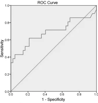
The areas under the curves (AUCs) for the cervical short-ening are: delivery within 6 hours–0.784 (standard error: 0.160,p¼0.036,►Fig. 1); delivery within 12 hours–0.621
(standard error: 0.100,p¼0.160,►Fig. 2); delivery within
24 hours–0.690 (standard error: 0.081,p¼0.014,►Fig. 3);
and delivery within 48 hours–0.630 (standard error: 0.071, p¼0.076,►Fig. 4).
Discussion
Many studies have assessed the ability of transvaginal ultra-sound examination of the cervix in predicting the successful outcome of induced labors. A wide review of these studies was recently done by Papillon-Smith and Abenhaim.5Some au-thors have assessed the cervical length parameter alone or in combination with other maternal characteristics for predict-ing the outcome of induction, while others have compared the ultrasonographic cervical length versus the digital examina-tion as a predictor of the success of the inducexamina-tion. The level of heterogeneity among the studies makes it difficult to conclude that ultrasonography cervical length is suitable for wide
Rev Bras Ginecol Obstet Vol. 38 No. 12/2016
clinical use,5and a recent meta-analysis of two randomized trials6does not prove the superiority of the ultrasonography versus Bishop’s score. Even the elastosonography of the cervix has received some criticism regarding the prediction of the outcome of the induction.7Moreover, if the induc-tion is needed, unfavorable ultrasonographicfindings and
unfavorable Bishop’s scores do not change the clinical management of the patients. Practicing obstetricians need a tool to check the effectiveness of thefirst dose of prostaglandin agonists administered for labor induction, and ultrasonograpy cervical shortening seems the easier and more precise tool.
Fig. 2 Receiver operating characteristic (ROC) curve for delivery within 12 hours.
Table 1 Descriptive statistics and results of the logistic regression analyses (adjusted odds ratio with 95% confidence intervals)
Vaginal delivery within 6 hours
Vaginal delivery within 12 hours
Vaginal delivery within 24 hours
Vaginal delivery within 48 hours
meansSD OR
95% CI of OR p
Cervical shortening 6.886.49 mm 1.244 1.030–1.503 0.024
1.060 0.947–1.187 0.308
1.161 1.029–1.310 0.016
1.105 1.010–1.209 0.030
Cervicometry before induction
26.2110.44 mm 0.823 0.673–1.007 0.058
0.940 0.862–1.024 0.156
0.933 0.859–1.014 0.103
0.953 0.888–1.022 0.180
rate (%) OR
95% CI of OR p
Multiparity 25.4% 0.490
0.008–30.962 0.736
3.320 0.527–20.919 0.201
2.791 0.556–14.002 0.212
2.298 0.624–8.465 0.211
Oxytocin infusion 65.1% 0.589 0.006–55.752 0.819
1.249 0.219–7.120 0.802
1.211 0.284–5.169 0.796
2.505 0.815–7.701 0.109
>1 dose of dinoprostone
44.5%–2 doses 11.1%–3 doses
Unreliable 0.029 0.004–0.246 0.001
0.197 0.053–0.727 0.015
1.065 0.326–3.476 0.917
Abbreviations: CI, confidence interval; OR, odds ratio; SD, standard deviation.
significant results.
Fig. 1 Receiver operating characteristic (ROC) curve for delivery within 6 hours.
This short report confirms what was already reported by Kang et al4in a wider sample of women with induced labors. Therefore, despite the biases related to the low sample size and the heterogeneity of the patients with induced labor in our study, we must reiterate the conclusion reached by Kang et al4: the presence of cervical shortening after thefirst induction is a predictor of a successful delivery if the induc-tion was repeated in the second day.
By assessing the AUCs, we found that the best predictions for vaginal delivery are achieved for births within 6 and 24 hours, while the cervical shortening poorly predicts vaginal delivery within 48 hours. Whether patients with poor cervi-cal shortening (less than 1 cm) 3 hours after induction should postpone the induction rather than receive more doses of dinoprostone should be assessed. A randomized trial should clarify this concern, in order to avoid the harms that come from the unnecessary use of prostaglandin agonists in pa-tients who are not responsive to them.
In conclusion, the greater the sonographic cervical short-ening 3 hours after labor induction, the higher the likelihood of vaginal delivery within 6, 24 and 48 hours.
This study was performed at the Institute of Obstetrics and Gynecology, Department of Surgical Sciences, University of Foggia (via Pinto–71100, Foggia, Italy) during the shifts of the Authors. The Authors do not travel currently to the University of Foggia.
References
1 Teixeira C, Lunet N, Rodrigues T, Barros H. The Bishop Score as a determinant of labour induction success: a systematic review and meta-analysis. Arch Gynecol Obstet 2012;286(3):739–753
2 Verhoeven CJ, Opmeer BC, Oei SG, Latour V, van der Post JA, Mol BW. Transvaginal sonographic assessment of cervical length and wedging for predicting outcome of labor induction at term: a systematic review and meta-analysis. Ultrasound Obstet Gynecol 2013;42(5):500–508
3 Wozniak S, Czuczwar P, Szkodziak P, Paszkowski T. Usefulness of elastography in predicting the outcome of Foley catheter labour induction. Aust N Z J Obstet Gynaecol 2015;55(3):245–250
4 Kang WS, Park KH, Kim SN, Shin DM, Hong JS, Jung HJ. Degree of cervical shortening after initial induction of labor as a predictor of subsequent successful induction. Ultrasound Obstet Gynecol 2010;36(6):749–754
5 Papillon-Smith J, Abenhaim HA. The role of sonographic cervical length in labor induction at term. J Clin Ultrasound 2015;43(1):7–16
6 Ezebialu IU, Eke AC, Eleje GU, Nwachukwu CE. Methods for assessing pre-induction cervical ripening. Cochrane Database Syst Rev 2015;(6):CD010762
7 Fruscalzo A, Mazza E, Feltovich H, Schmitz R. Cervical elastog-raphy during pregnancy: a critical review of current approaches with a focus on controversies and limitations. J Med Ultrason (2001) 2016;43(4):493–504
Fig. 3 Receiver operating characteristic (ROC) curve for delivery within 24 hours.
Fig. 4 Receiver operating characteristic (ROC) curve for delivery within 48 hours.
Rev Bras Ginecol Obstet Vol. 38 No. 12/2016