ABSTRACT
Although maxillary central incisors impaction is not a high frequent clinical condition, it is responsible for some undesirable aspects of patients smile from esthetic and functional points of view. There are some etiologic factors associated to this dental disturbance but the scientiic literature is consensual on determining the importance of the early diagnosis and proper intervention. This manuscript consists on the case report of a 10 years old boy with Class I malocclusion, who showed during the mixed dentition phase, impaction of maxillary central incisors related to deciduous teeth retention and due to the presence of 2 mesiodens. The treatment proposed involved the surgical removal of the mesiodens, bonding of orthodontic accessories to the impacted incisors crowns followed by orthodontic traction with a removable orthodontic appliance. Regardless the development stage of the dentition was not ideal for this intervention, once the diagnosis should have been done as earlier as possible, favorable esthetic and functional results were attained.
Indexing terms: Malocclusion. Orthodontics. Tooth impacted.
RESUMO
A impacção de incisivos centrais superiores é uma condição clínica que embora não seja muito frequente, é um fator desagradável do ponto de vista estético e funcional. Existem diversas causas dessa alteração e a literatura é consensual quanto à necessidade da realização do diagnóstico e da intervenção o mais precocemente possível. Este trabalho traz o relato de um caso clínico de um paciente com 10 anos de idade, na fase da dentição mista, com má oclusão de Classe I, impacção de incisivos centrais superiores associado à retenção prolongada dos decíduos e a presença de dois supra-numerários na linha média. A conduta proposta foi à remoção cirúrgica dos mesiodens, colagem de dispositivos ortodônticos nos incisivos centrais impactados e, tracionamento ortodôntico com aparelho removível. Desta forma, embora a época de tratamento não fosse a ideal, pois o diagnóstico deveria ter sido estabelecido o mais precocemente possível, alcançou-se um resultado estético e funcional favorável.
Termos de indexação: Má oclusão. Ortodontia. Dente impactado.
Orthodontic traction of impacted upper central incisors related to
mesiodens
Tracionamento ortodôntico de incisivos centrais superiores impactados associados à presença de
mesiodens
André Wilson MACHADO1
Luiz Guilherme Martins MAIA2
Alexandre Protásio VIANNA2
Luiz Gonzaga Gandini JÚNIOR2
1 Universidade Federal da Bahia, Faculdade de Odontologia, Disciplina de Ortodontia. Av. Araújo Pinho, 62, 7° Andar, 40110-040, Canela, Salvador, BA, Brasil. Correspondência para / Correspondence to: AW MACHADO. E-mail: <[email protected]>.
2 Universidade Estadual Paulista Júlio de Mesquita Filho, Faculdade de Odontologia. Departamento de Clínica Infantil. Araraquara, SP, Brasil.
occur, due to its anterior location it is a determining factor in the deterioration of esthetic appearance1.
A tooth may be regarded as impacted as a result of several etiological factors that determine a delay in the timing of eruption. Amongst the local conditions which most inluence incisor impaction are physical obstructions. These mechanical barriers are the result of various causes, such as: hyperdontia, mucosal barrier, scar tissue and tumors. Of the above, the condition that requires special
INTRODUCTION
In the intraoral evaluation, the patient was found to be in the mixed dentition phase, in the second transitional period. From the frontal evaluation, the clinical absence of the upper central incisors was conirmed, combined with the prolonged retention of the deciduous teeth (Figure 2A). From the side view, the molars and canines were in a Class I relationship (Figures 2B and 2C). Finally, the study of the occlusal photographs shows dental arches within normal criteria (Figures 2D and 2E).
CASE REPORT
Ten year-old male patient in the mixed dentition phase complained that his “front teeth were not growing”. His medical history revealed no contraindication to orthodontic treatment.
The analysis of the facial photograph of his close-up smile shows a quite unlattering appearance due to the absence of permanent central incisors and the prolonged retention of the deciduous teeth, a feature that was the reason behind seeking orthodontic treatment (Figure 1). attention is the presence of hyperdontia which, when
situated in the midline, are called mesiodens and, according to the literature, cause dental impaction in between 28% and 60% of individuals2.
According to Suri et al.2, although a variety of
terms have been used to characterize teeth with delayed or retarded eruption, this condition is characterized by two situations, as follows: a) teeth that have not erupted within the prescribed chronology based on epidemiological studies, and b) teeth that have not erupted regardless of the degree of root formation. Accordingly, impacted teeth would be those with delayed eruption related to some or other physical barrier which blocks the usual eruption route.
The literature shows various options for a clinical solution to impacted upper incisors. In short, the options range from more conservative procedures, such as the extraction of deciduous teeth, to surgical procedures followed or otherwise by orthodontic traction1. On the
other hand, it should be emphasized that before any radical intervention such as surgical exposure, it would be prudent to use instruments to open a suficient space, stimulating the natural eruption of the incisors3.
As the literature is rather sparse with regard to clinical studies with an adequately dimensioned sample, that evaluate the impact of the different approaches to treatment, the publication of clinical cases is required in order to get a better understanding of the topic.
Thus the objective of this study is to report on a clinical case, in the mixed dentition phase, with Class I malocclusion and impacted upper central incisors. The treatment consisted of the surgical removal of the root cause of the problem (mesiodens) followed by orthodontic traction of the impacted teeth.
Figure 1. Initial facial photograph of the close-up smile.
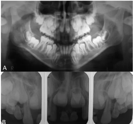
surgically remove the deciduous teeth in prolonged retention and the supernumerary teeth. In addition, at the same time as the surgery, orthodontic appliances were glued to the central incisors in order to commence the orthodontic treatment. The x-ray evaluation corroborated the clinically
obtained data and identiied two supernumerary teeth in the midline region (Figure 3).
The aim of the interceptive treatment was to
Figure 3. A) initial panoramic x-ray; B) initial periapical location.
A removable orthodontic appliance was used by the patient who was duly instructed to use it throughout the day, only removing it for meals and when engaged in sporting activity. In the anterior region of the appliance, retainers were fabricated so the patient could place 1/8’’ rubber bands of the ligature hooks fastened to the orthodontic buttons for these types of structure (Figure 4).
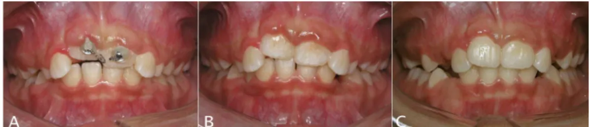
After a period of four months, the incisors had erupted in the oral cavity, at which time the traction was halted and the orthodontic buttons removed. The periodic checks of the patient began and after two months the incisors regained their normal eruptive pattern and, after ten months, they were in a quite favorable position (Figure 5). In this phase, a positive outcome could be seen of the technique carried out with adequate tooth position and good periodontal condition.
The patient’s guardian was directed on the need to sign the Free and Informed Consent Agreement for the use of the procedures and results obtained in the technical/ scientiic work, with the child’s identity being protected. The guardian provided consent and signed the form.
DISCUSSION
The frequency with which upper incisors become impacted is low, ranging from 0.06% to 0.2%4. However,
when it does occur, it is quite disturbing, from an esthetic and functional point of view.
In many situations, incisor impaction is spotted by the patients’ parents in the mixed dentition phase and, subsequently, the contact with the general or pediatric dental clinician enables early diagnosis of this alteration in eruption pattern5. However, the case in question does
not support this notion, as the central incisors should have erupted in the oral cavity at an average chronological age of 7.27 years6 and the patient was already 10 years
old. If the diagnosis had been made early, the treatment would have been made easier, evidencing the importance
of making clinicians and pediatric dentists aware of the importance of early diagnosis of malocclusion.
The clinical case presented here, in addition to the delay in incisor eruption, also presented prolonged retention of the deciduous central incisors. Moreover, the conirmation of impaction was made in the x-ray examination with the presence of two mesiodens in the midline region, acting in combination with the deciduous teeth as a physical barrier, thus preventing the eruption of the central teeth.
In this regard, Kurol7 stressed that supervision of
the development of the dentition and of early diagnosis of eruption deviations are essential for intervention at the ideal moment and for catching the problem. Batra et al.8, for example, suggest that periodic x-rays be taken
for all patients presenting with any abnormality in the chronological pattern of eruption.
As regards the treatment, Lin9 described three
options for the handling of patients with impacted incisors: a) extraction of the impacted tooth and rehabilitation with a prosthetic device or implant procedure when the growth has stopped; b) extraction of the impacted tooth and closure of the space, replacing the central incisors with the lateral ones, and c) surgical exposure followed by orthodontic traction.
On the other hand, Becker3 commented that, although
the options for treatment are many, the initial approach which is the most logical and conservative should be the orthodontic opening of space to encourage the natural eruption of the incisors. As previously mentioned, this approach would have been ideal had an early diagnosis been made.
The literature describes a number of clinical cases of spontaneous eruption of impacted incisors after the orthodontic creation of space1,10-11. This raises the question
whether, in the case in question, eruption would or would not occur spontaneously after the removal of the physical barriers. On the other hand, as the patient was subjected to surgery for the removal of supernumerary teeth, at the same time as the surgery, orthodontic devices were glued to Figure 5. Clinical outcome of the traction. A) after 4 months; B) after 6 months; C) after 16 months.
Figure 6. Final facial photograph of the close-up smile.
enable orthodontic traction to be carried out, as performed by other authors5,8-9,12-22. Thus, if spontaneous eruption of
the impacted incisors had not occurred spontaneously, further surgery would not be required.
With this approach, a modicum of care has to be taken during surgery and traction in order to ensure a inal esthetic outcome23-24. Sandler16 suggested that an excellent
option for traction would be the use of magnets and that the periodontal response would be greatly improved due to the type of force applied.
In order to carry out the traction, the impacted tooth may be supported on removable appliances or on the arch of the ixed orthodontic appliances. In these situations there are disadvantages which sometimes limit the results obtained, such as the need for patient cooperation and the presence of side effects with the orthodontic arch, respectively. On the other hand, patient cooperation with regard to the usage of the appliance and the changing of the rubber bands were satisfactory, having a positive inluence on the outcome.
The esthetic outcome found determines the success of the treatment. This inding agrees to Sarver & Ackerman25 when they stated that the upper incisors are
the key to esthetic success in orthodontic treatment. The analysis of the smile obtained demonstrates the improvement in the appearance of the smile obtained
through treatment. Moreover, as observed by Crawford14,
this type of therapeutic approach, in addition to giving back the patient a more pleasant, sociable smile, was able to restore his conidence, vanity and self-esteem, which is an important factor even at this early age.
CONCLUSION
Among the majority of upper incisor impactions, when the diagnosis is established early, the possibility of achieving more satisfactory results is increased, as well as the ease of applying treatment.
The use of the traction technique for impacted incisors, when well-planned and executed, gives acceptable success rates, signiicantly favoring esthetic appearance, which in the majority of cases contributes to improving the psychological state of the patient.
Collaborators
AW MACHADO was responsible for the diagnosis and orthodontic planning of the case as well as the composition of the article. LGM MAIA was responsible for the review of the literature and the composition of the article. AP VIANNA took part in the composition of the article. LG GANDINI JÚNIOR took part in the composition of the article.
REFERENCES
1. Machado AW, Loriato L, Souki B, Junqueira T. Erupção espontânea de incisivos centrais superiores impactados após a abertura ortodôntica de espaço. Rev Clin Ortodon Dental Press. 2007;5(6):43-52.
2. Suri L, Gagari E, Vastardis H. Delayed tooth eruption: pathogenesis, diagnosis, and treatment. A literature review. Am J Orthod Dentofacial Orthop. 2004;126(4):432-45.
3. Becker A. Early treatment for impacted maxillary incisors. Am J Orthod Dentofacial Orthop. 2002;121(6):586-7.
4. Grover PS, Lorton L. The incidence of unerupted permanent teeth and related clinical cases. Oral Surg Oral Med Oral Pathol. 1985;59(4):420-5.
5. Uematsu S, Uematsu T, Furusawa K, Degushi T, Kurihara S. Orthodontic treatment of an impacted dilacerated maxillary central incisor combined with surgical exposure and apicoectomy. Angle Orthod. 2004;74(1):132-6.
6. Burdi AR, Moyers RE. Desenvolvimento da dentição e da oclusão. In: Moyers RE. Ortodontia. 4ª ed. Rio de Janeiro: Guanabara Koogan; 1991. p. 86-126.
7. Kurol J. Early treatment of tooth-eruption disturbances. Am J Orthod Dentofacial Orthop. 2002;121(6):588-91.
8. Batra P, Duggal R, Kharbanda OP, Parkash H. Orthodontic treatment of impacted anterior teeth due to odontomas: a report of two case. J Clin Pediatr Dent. 2004;28(4):289-94. 9. Lin YJ. Treatment of an impacted dilacerated maxillary central
incisor. Am J Orthod Dentofacial Orthop. 1999;115(4):406-9. 10. Prillaman II WN, Macon R, Visser BE, Isaacson RJ. Treatment of a
class II malocclusion with impacted maxillary central incisor. Am J Orthod Dentofacial Orthop. 1997;112(4):367-71.
11. Tanaka O, Fronza F, Guariza Filho O, Ribeiro G. A disjunção palatal como auxiliar na irrupção de incisivo central impactado. J Bras Ortodon Ortop Facial. 2000;30(5):13-9.
12. Batterson KD, Curtis T, Parks C, Curtis E, Carlson C, Southard TE. Nonextraction treatment of a Class II malocclusion and impacted maxillary central incisors. Am J Orthod Dentofacial Orthop. 2004;125(1):107-14.
13. Barnett D. Treating an impacted incisor with a removable appliance. J Clin Orthod. 1978;12(5):376-7.
15. Locks A, Ritter DE, Morona AR, Haertel GB, Ribeiro GLU, Menezes LM. Tratamento ortodôntico-cirúrgico de incisivo central superior impactado com dilaceração acentuada: caso clínico. Rev Dental Press Ortodon Ortop Facial. 2000;5(5):75-9. 16. Sandler JP. An attractive solution to unerupted teeth. Am J
Orthod Dentofacial Orthop. 1990;100(6):489-93.
17. Sato K, Mitani H. Unerupted maxillary central and lateral incisors and canine with crossbite and asymetry. Am J Orthod Dentofacial Orthop. 2003;123(1):87-92.
18. Stuani AS, Souza AHF, Stuani AS, Stuani MBS. Solução alternativa para incisivo superior impactado. Rev Ibero-am Odontopediatr Odontol Bebê, 2004;38(7):335-40.
19. Suzigan LC, Stuani AS, Stuani AS, Stuani MBS. Incisivo superior impactado: técnica de erupção fechada. J Bras Ortodon Ortop Facial. 2004;50(9):156-60.
20. Swinnen K, Erum RV, Verdonck A, Carels C. An impacted central incisor with a severe root malformation. J Clin Orthod. 1999;33(9):511-5.
21. Tanaka E, Watanabe M, Nagaoka K, Yamaguchi K, Tanne K. Orthodontic traction of an impacted maxillary central incisor. J Clin Orthod. 2001;35(6):375-8.
22. Wasserstein A, Tzur B, Brezniak N. Incomplete canine transposition and maxillary central incisor impaction: a case report. Am J Orthod Dentofacial Orthop. 1997;111(6):635-9. 23. BRAND A, Akhavan M, Tong H, Kook YA, Zernik JH. Orthodontic,
genetic, and periodontal considerations in the treatment of impacted maxillary central incisors: a study of twins. Am J Orthod Dentofacial Orthop. 2000;117(1):68-74.
24. Frank CA, Long M. Periodontal concerns associated with the orthodontic treatment of impacted teeth. Am J Orthod Dentofacial Orthop. 2002;121(6):639-49.
25. Sarver DM, Ackerman JL. Orthodontics about face: the re-emergence of the esthetic paradigm. Am J Orthod Dentofacial Orthop. 2000;117(5):575-6.