ABSTRACT
The aim of the present study was to review the literature regarding the utilization of 2-octyl cyanoacrylate’s (Dermabond®, Ethicon US, USA) as a tissue adhesive in dentistry; also, to report its use in the stabilization and ixation of a free gingival graft, indicated to increase the width of the keratinized attached mucosa at the lower incisive region. Literature analysis revealed numerous indications for this tissue adhesive in the medical ield related to maxillofacial injuries. In dentistry, clinical reports, as well as controlled clinical studies conducted in humans and in animal models, using histological analysis described positive results for the use of different cyanoacrylate-based tissue adhesives. These studies reported that the use of tissue adhesives reduced the surgical procedure time period, eliminated postoperative visits as well as the discomfort of suture removal and, in addition, did not interfere with the clinical repair process. Favorable results, like the ones described in the literature, were obtained in the present case report using Dermabond®.
Indexing terms: Cyanoacrylates. Tissue adhesives. Transplants.
RESUMO
O objetivo do presente estudo foi revisar a literatura pertinente sobre a utilização do 2-octil cianoacrilato (Dermabond®, Ethicon US, USA) como adesivo tecidual na Odontologia, assim como relatar seu uso na estabilização e ixação de um enxerto gengival livre indicado para aumento da mucosa ceratinizada inserida na região dos incisivos inferiores. A análise da literatura revelou ampla aplicabilidade deste adesivo tecidual na área médica relacionada aos ferimentos maxilofaciais. Na Odontologia existem relatos clínicos e estudos controlados em humanos e em modelos animais, descrevendo resultados positivos, inclusive por meio da análise histológica, sobre a utilização de diferentes adesivos teciduais, à base do cianocrilato. Estes estudos relataram que o uso dos adesivos teciduais, reduz o tempo operatório, elimina visitas pós-operatórias, não apresenta o desconforto da remoção de suturas, além de não interferir no processo de reparo clínico. Resultados favoráveis como os descritos na literatura, foram obtidos no presente relato de caso com a utilização do Dermabond®.
Termos de indexação: Cianoacrilato. Adesivos teciduais. Transplantes.
Tissue adhesive in free gingival graft
Adesivo tecidual no enxerto gengival livre
Elton Gonçalves ZENÓBIO1
Elias Youssef Abou ABDALLAH1
Flávia Isabela BARBOSA1
Anna Cristina Petraccone CAIXETA1
Rodrigo Villamarim SOARES1
1 Pontifícia Universidade Católica de Minas Gerais, Departamento de Odontologia. Av. Dom José Gaspar, 500/ Pr. 46, Coração Eucarístico, 30535-610, Belo Horizonte, MG, Brasil. Correspondência para / Correspondence to: EG ZENÓBIO. E-mail: <[email protected]>.
The use of cyanoacrylate-based adhesives in periodontal surgery, including free gingival grafts, has demonstrated ease and eficiency, minimizing the problems generated by suturing thread, and showing minimal toxicity and low cost6-10. Thus, with the results described in the literature, it was considered appropriate to evaluate of the use of Dermabond® as a tissue adhesive for setting free gingival grafts11-12.
Binnie & Forrest13 performed periodontal laps, in the region of the second pre-molars and the incisors, on two six-month old dogs. They used sutures for coaptation on one side, and butyl-cyanoacrylate tissue adhesive on the other. They reported that the initial repair was faster
INTRODUCTION
The success of periodontal surgery depends on the appropriate coaptation of the incised edges, elimination of empty spaces and reduction of the amount of coagulation. In free gingival graft surgery, stabilization of the graft, hemostasis and cleansing of the site remain necessary1.
The following were used in the surgical procedure: rinse with a 0.12% chlorhexidine digluconate solution for one minute and anesthesia using 2% lidocaine (1:100000) with vasoconstrictor. The surgical technique was performed the study and with no signiicant changes regarding the total area of the graft. They conclude that the modality of gingival graft ixation did not present any signiicant inluence over the clinical parameters evaluated.
CASE REPORT
The patient, a 42-year-old woman with no systemic alterations, was referred to the Department of Periodontics of the authors. Clinical examination revealed the absence of inserted keratinized mucosa in the region of the lower central incisors, the presence of inlammation and marginal edema and complaint of dificulty cleaning in the region. It was decided that a free gingival graft, attached using the tissue adhesive Dermabond®, would be performed as part of the treatment in order to increase the amount of keratinized mucosa, facilitate cleaning and reestablish periodontal health. Four weeks prior to the surgical procedure, the patient underwent professional plaque control to prepare the region for the procedure.
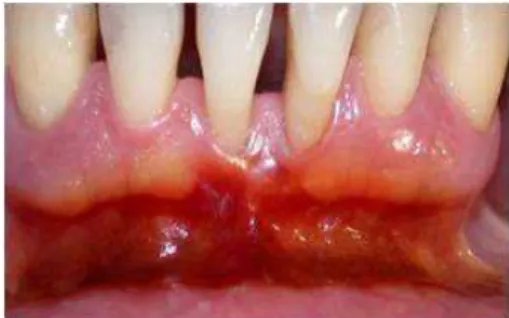
During the initial evaluation, the following periodontal parameters were obtained: gingival bleeding index, probing depth (Figure 1), gingival recession, clinical attachment level, amount of keratinized mucosa (Figure 2) and amount of inserted keratinized mucosa.
using the adhesive, that the product was easy to apply, appropriate for setting the gingival laps and that the histological exam showed no adverse effects.
Forrest14, in a longitudinal study, evaluated more than 300 patients undergoing periodontal surgery, including free gingival grafts, laterally and apically positioned laps, rhizectomies, endodontic and exodontic surgery, in which butyl-cyanoacrylate was used for the coaptation of the edges. The results were compared with edges set using sutures. The adhesive was reported to promote rapid hemostasis and clinical repair similar to the sutures, was well accepted by the patients and there were no reports of local or systemic reactions. The application of the material to the posterior region was considered dificult but, in general, the use of the adhesive was reported to reduce operating time considerably when compared with suturing techniques.
One ive-month study, comparing the tissue adhesive 2-octyl cyanoacrylate (Dermabondâ) to sutures in 136 lacerations of the face, hands and feet, was described15. The authors observed no signiicant differences between the two forms of coaptation in the repair of the wounds, and reported that Dermabond® has plasticizers in its composition that support its use in movable areas such as the face, skin over the joints, or in longer incisions. The authors also reported that the tested adhesive is effective in closing lacerations, in addition to taking less time to apply and being less painful than suturing.
Toriumi et al.16 submitted 111 patients to plastic facial procedures and compared the closing of the incisions using 2-octyl-cyanoacrylate to suturing. They observed that the time required for repair with sutures was approximately 4 times greater than with the adhesive. There was no incidence of dehiscence, hematoma or infection among the groups after one year; and, the esthetic results and patient satisfaction were superior in the 2-octyl-cyanoacryate group.
Barbosa et al.17 evaluated twenty-four subjects with gingival recession and absence of keratinized mucosa divided into two groups: free gingival grafts ixed with ethyl-cyanoacrylate (Group 1) and ixed with sutures (Group 2) to treatment. Probing depth, clinical attachment level, gingival recession, and dimensional changes of height and width were evaluated immediately post-operatively and at 15, 30, 45, and 90 days after surgery. They observed that dimensional changes related to the area of gingival graft were similar for both groups. The thickness of the gingival graft tissue inluenced the dimensional changes in the height of the grafts in the recipient bed (p < 0.047). Gingival grafts, thinner than 1 mm, showed a greater average height at the end of
Figure 1. Pre-operative case (probing depth).
according to standard form17-18, modiied for the setting of the lap using Dermabond®.
A Bard Parker no. 15C scalpel blade was used to perform the partial lap with a horizontal incision parallel to the long axis of the root, starting from the Mucogengival line to the base of the vestibule, extending to the mesial and distal, according to the width and height of the intended graft. Muscle inserts that could interfere with the stability of the graft were eliminated, maintaining a thin, smooth layer of conjunctive tissue to facilitate the correct adaptation of the graft. A perpendicular incision was made at the base of the vestibule to fenestrate the periosteum. A surgical guide having the same dimensions as the receiving area was created from sterile paper and placed in the region of the palate. Anesthesia was given by iniltration along the side of the guide, the incision was made around the guide and the graft was removed delicately. Adipose tissue and irregularities in the connective tissue were removed from the graft. The donor site was sutured using 4.0 silk threads and protected for seven days with surgical cement (Coe Packâ). The graft was placed in the recipient site and lightly pressed using sterile gauze soaked in sterile saline solution to reduce/eliminate clotting.
For setting with Dermabond®, a new, sealed tube was used which was opened at the moment of the surgery. The contents were emptied into a sterile dappen dish, were immediately collected using a clinical probe (Figure 3) and taken to the surgical site, closing the edges of the gingival graft and the recipient site. With the graft properly adapted and coopted to the recipient site, traction and tension lip movements were performed to verify if there were any interference from muscle inserts that could cause movement in the graft (Figure 4). The patient received post-operative instructions related to careful cleaning of the region for 30 days with 0.12% chlorhexidine digluconate and the use of analgesics to reduce the initial pain.
Figure 3. Tissular adhesive Dermabond®.
Figure 4. Immediate post-operative case.
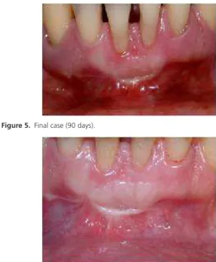
Figure 5. Final case (90 days).
Figure 6. Final case (180 days).
At seven days the graft showed normal healing and absence of movement. The patient did not report, nor was there observed, any interference caused by the method of setting. At 15 days there was no edema and integration of the gingival graft with the adjacent tissues could be observed. At 30 days, the graft appeared clinically completely healed. A signiicant increase in the keratinized mucosa and the width of the gingival margin could be seen. The clinical observations of this period were similar to those veriied at 90 and 180 days (Figures 5 and 6). All surgical procedures and the care of the patient were performed following the ethical principals contained in the Helsinki Declaration (2000), in addition to following the ANVISA standards for biosecurity and control.
DISCUSSION
been studied and proposed for this purpose. They maintain all the advantages of conventional suturing, favoring the stabilization of the blood clot and providing esthetics to the surgical site17-25.
The use of cyanoacrylate-based adhesives in periodontal surgery, including free gingival grafts, demonstrates ease and eficiency, minimizes the problems caused by suturing thread and shows minimal toxicity and low cost6-10,17.
The improvement of the periodontal clinical parameters, associated with the positive report of the patient as to the current ease of oral hygiene, reinforces the importance of the free gingival graft to increase the width of the gingival margin26 from the area of the inserted gingiva27-28 and for the maintenance of periodontal health.
CONCLUSION
In the clinical case reported, the use of Dermabond® as the tissue adhesive for the setting and stabilization of the free gingival graft reduced the operating time and post-operative discomfort, eliminated the need for suture removal, did not interfere with the clinical repair process and, as reviewed in the literature, is suggested as an alternative to suturing in mucogingival surgery.
Collaborators
EG ZENOBIO, RV SOARES, EYA ABDALLAHM and ACP CAIXETA were responsible for guiding the development of the clinical case and writing the article.
REFERÊNCES
1. Hoexter DL. The sutureless free gingival graft. J Periodontol. 1979;50(2):75-8. doi:10.1902/jop.1979.50.2.75
2. Coover HN, Joyner FB, Sheerer NH. Chemistry and performance of cyanoacrylate adhesive. Spec J Tech Papers. 1959;5(1):413-7. doi:10.1055/s-2004-831406
3. Herod EL. Cyanoacrylates in dentistry: a review of the literature. J Can Dent Assoc. 1990;56(4):331-4.
4. Doraiswamy NV, Baig H, Hammett S, Hutton M. Which tissue adhesive for wounds? Injury. 2003;35(8):636-7. doi:10.1016/ S0020-1383(02)00210-3
5. Toriumi DM, Bagal AA. Cyanoacrylate tissue adhesives for skin closure in the outpatient setting. Otolaryngol Clin North Am. 2002;35(1):103-18. doi:10.1016/S0030-6665(03)00097-5
6. Bhaskar SN, Frisch J, Margetis PM, Leonard F. Application of a new chemical adhesive in periodontic and oral surgery. Oral Surg Oral Med Oral Pathol. 1966;22:526-35.
7. Bhaskar SN, Frisch J, Cutright DE, Margetis PM. Effect of butyl cyanoacrylate on the healing of extraction wounds. Oral Surg Oral Med Oral Pathol. 1967;24(3):604-16. doi:10.1016/0030-4220(67)90200-9
8. Bhaskar SN, Beasley III JD, Cutright DE, Perez B. Free mucosal grafts in miniature swine and man. J Periodontol. 1971;42:322-30. doi:10.1902/jop.1971.42.6.322
9. Lacaz Netto R, Macedo NL. Estudo clínico da reparação do enxerto gengival livre ixado por um adesivo à base de cianoacrilato. Rev Assoc Paul Cir Dent. 1986;40(2):164-70.
10. Santos GM, Lacaz Netto R, Santos LM, Okamoto T, Rocha RF. Uso do Super-Bonder no reparo das feridas cirúrgicas. RGO - Rev Gaúch Odontol. 1990;38(6):435-9.
11. Bhaskar SN, Frisch J, Margetis PM. Tissue response of rat tongue to hexyl, heptyl and octyl cyanoacrylate. Oral Surg Oral Med Oral Pathol. 1967;24(1):137-43. doi:10.1016/0030-4220(67)90300-3
12. Bhaskar SN, Frisch J. Use of cyanoacrylate adhesives in dentistry. J Am Dent Assoc. 1968;77(4):831-7.
13. Binnie WH, Forrest JO. A study of tissue response to cyanoacrylate adhesive in periodontal surgery. J Periodontol. 1974;45(8):619-25.
14. Forrest JO. The use of cyanoacrilate in periodontal surgery. J Periodontol. 1974;45(4):225-9.
15. Quinn J, Wells G, Sutcliffe T, Jarmuske M, Maw J, Stiell I, et al. A randomized trial comparing octylcyanoacrylate tissue adhesive and sutures in the management of lacerations. JAMA. 1997;277(19):1527-30. doi:10.1001/jama.1997.03540430039030
16. Toriumi DM, O’Grady K, Desai D, Bagal A. Use of octyl-2-cyanoacrylate for skin closure in facial plastic surgery. Plast Reconstr Surg. 1998;102(6):2209-19.
17. Barbosa FI, Corrêa DS, Zenóbio EG, Costa FO, Shibli JA.Dimensional changes between free gingival grafts ixed with ethyl cyanoacrylate and silk sutures. J Int Acad Periodontol. 2009;11(2):170-6.
18. Dorfman HS, Kennedy JE, Bird WC. Longitudinal evaluation of free autogenous gingival grafts. J Clin Periodontol. 1980;7(4):316-24.
19. Miller GM, Dannenbaum R, Cohen DW. A preliminary histologic study of the wound healing of mucogingival laps when secured with the cyanoacrylate tissue adhesives. J Periodontol. 1974;45(8):608-18. Doi: 10.1902/jop.1974.45.8.2.608
20. Levin MP, Cutright DE, Bhaskar SN. Cyanoacrilates as a periodontal dressing. J Oral Med. 1975;30(2):40-3.
22. Maw JL, Quinn JV, Wells GA, Ducic Y, Odell PF, Lamothe A, et al. A prospective comparison of cyanoacrylate tissue adhesive and suture for the closure of heas and neck incision. J Otolaryngol. 1997;26(1):26-30. doi:10.1001/archderm.137.9.1177
23. Quinn J, Maw J, Ramotar K, Wenckebach G, Wells G. Octyl cyanoacrylate tissue adhesive versus suture wound repair in a contaminated wound model. Surgery. 1997;122(1):69-72. doi:10.1016/S0039-6060(97)90266-X
24. Quinn J, Wells G, Sutcliffe T, Jarmuske M, Maw J, Stiell I, et al. A randomized trial comparing octylcyanoacrylate tissue adhesive and sutures in the management of lacerations. JAMA. 1997;277(19):1527-30.
25. Bruns TB, Robinson NS, Smith RJ, Kile DL, Davis TP, Sullivan KM, et al. A new tissue adhesive for laceration repair in children. J Pediatr. 1998;132(6):1067-70. doi:10.1016/S0022-3476(98)70415-9
26. Wennström JL. Lack of association between width of attached gingival and development of soft tissue recession-A 5-year longitudinal study. J Clin Periodontol. 1987;14(3):181-4. doi:10.1111/j.1600-051X.1987.tb00964.x
27. Yared KF, Zenóbio EG, Pacheco W. Periodontal status of mandibular central incisors after orthodontic proclination in adults. Am J Orth Dentofac Orthop. 2006;130(1):1-8. doi:10.1016/j. ajodo.2006.01.015
28. Maynard JG Jr, Wilson RD. Diagnosis and management of mucogingival problems in children. Dent Clin North Am. 1980;24:683-703.