Rev Bras Anestesiol CLINICAL INFORMATION 2012; 62: 6: 878-884
878 Revista Brasileira de Anestesiologia
Vol. 62, No 6, November-December, 2012
Received from the Anesthesiology Department, Hospital Universitário de Brasília, Universidade de Brasília (UnB), Brasília, DF, Brazil.
1. Anesthesiology Department, Hospital Universitário de Brasília, Universidade de Brasília (UnB), Brazil
2. Instituto Brasília de Arritmia, Brasília, Brazil 3. PhD; Instituto Brasília de Arritmia, Brasília, Brazil 4. Anesthesiology Department, Hospital Brasília, Brasília, Brazil Submitted on October 8, 2011.
Approved on February 29, 2012. Correspondence to:
Maurício Daher, MD
Serviço de Anestesiologia, Hospital Universitário de Brasília, Universidade de Brasília
E-mail: [email protected]
CLINICAL INFORMATION
Sudden Cardiac Arrest in General Anesthesia as the First
Manifestation of Anomalous Origin of the Left Coronary Artery
Maurício Daher, TSA
1, André Rodrigues Zanatta
2, Benhur David Henz
3,
Marcelo Carneiro da Silva, TSA
4, Simone Nascimento dos Santos
2, Luiz Roberto Leite
3Summary: Daher M, Zanatta AR, Henz BD, Silva MC, Santos SN, Leite LR – Sudden Cardiac Arrest in General Anesthesia as the First
Manifesta-tion of Anomalous Origin of the Left Coronary Artery.
Background and objectives: This case report describes a rare and potentially fatal condition associated with anesthesia administration. Our
aim was to discuss the causes of sudden cardiac arrest during the perioperative period in apparently healthy patients and the pathophysiology of anomalous origin of the coronary arteries as a cause of sudden cardiac arrest.
Case Report: Female patient, 44 years old, with no previous symptoms of heart disease or arrhythmias, had a sudden cardiac arrest during
general anesthesia in two different situations. In the first episode, the patient presented signs of acute abdomen, but remained hemodynamically stable. Following induction of anesthesia, the patient exhibited bradycardia and hypotension refractory to volume replacement and vasopressors. The condition progressed to asystole. The patient was successfully resuscitated and discharged from the hospital in good condition. In the second episode, one year after the first, the patient was in good clinical condition to undergo an elective surgery. After induction of anesthesia, the patient developed ventricular tachycardia followed by asystole, which was promptly reversed. After extensive investigation, an anomalous origin of the left coronary artery was identified.
Conclusions: Our report is illustrative as it emphasizes that a thorough diagnostic investigation should be done in cases of sudden cardiac arrest during the perioperative period, even in patients that appear to be healthy.
Keywords: Anesthesia; Coronary Vessel Anomalies; Death, Sudden, Cardiac; Heart Arrest.
©2012 Elsevier Editora Ltda. All rights reserved.
INTRODUCTION
Preoperative risk stratification for non-cardiac surgery can ad-equately identify patients at high risk for cardiovascular com-plications, including sudden cardiac death 1. Some of the main risk factors are well known, such as unstable coronary artery disease, cardiac arrhythmias, heart failure, and valvular heart disease. If none of the major factors is present, perioperative morbidity and mortality risk is less than 1%, and a thorough cardiovascular diagnostic investigation would not change the perioperative management of asymptomatic patients.
Given the unpredictability of sudden cardiac death in “low-risk” patients, all efforts must be made to treat those who suffer from an unanticipated cardiac arrest during anesthesia. More
importantly, in patients presenting with perioperative cardiac events, further planned surgeries should only be scheduled after a complete diagnostic evaluation and definition of the causative event.
Second to hypertrophic cardiomyopathy, the anomalous origin of coronary arteries (AOCA) is the most common cause of sudden cardiac death in young adults in the U.S., and it is found in 13% of autopsies in these cases 2. Most cases of sud-den death secondary to AOCA are completely unexpected, as at least 50% of affected individuals have no history of angina, syncope, or palpitations 3.
We report a rare case of recurrent cardiac arrest related to an anomalous origin of the left coronary artery during general anesthesia in a patient without previous symptoms of heart disease or arrhythmias.
CASE REPORT
SUDDEN CARDIAC ARREST IN GENERAL ANESTHESIA AS THE FIRST MANIFESTATION OF ANOMALOUS ORIGIN OF THE LEFT CORONARY ARTERY
Revista Brasileira de Anestesiologia 879
Vol. 62, No 6, November-December, 2012
induction with propofol (120 mg), fentanyl (150 µg), succinyl-choline (80 mg), and cisatracurium (10 mg) was followed by persistent hypotension, refractory to repeated doses of ephed-rine and rapid infusion of crystalloids. Electrocardiogram monitoring showed sinus bradycardia followed by electrome-chanical dissociation and asystole. Cardiopulmonary resusci-tation (CPR) was immediately initiated and sinus rhythm was restablished after one hour of CPR and seven defibrillations. After surgery, the patient was taken to the intensive care unit, where she had a complicated evolution with prolonged intuba-tion, acute respiratory distress syndrome and acute kidney in-jury. Nonetheless, she was discharged with complete clinical remission and in good general condition. At the time, cardiac arrest was attributed to hypovolemia and vasodilation, likely secondary to sepsis, although her preoperative clinical status had been considered stable.
After one year, the patient was diagnosed with breast can-cer. A radical mastectomy was scheduled to be performed un-der general anesthesia. She unun-derwent preoperative clinical evaluation, which revealed no significant changes in 12-lead electrocardiogram (ECG), chest X-ray, treadmill stress test, and resting echocardiogram.
Induction with midazolam (3 mg), fentanyl (250 µg), propo-fol (180 mg), and rocuronium (30 mg) resulted in ventricular ectopic beats a brief period of ventricular tachycardia which was followed by asystole. Cardiopulmonary resuscitation was started, this time with prompt recovery of sinus rhythm. She had no neurological sequelae. Surgery was postponed, and the patient underwent a new cardiac evaluation with ECG, with and without procainamide; ambulatory ECG; echocar-diography; myocardial scintigraphy; and treadmill exercise test. All tests were normal, and she did not report any residual symptoms.
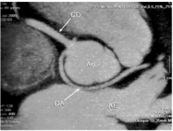
The patient was referred to our center for investigation of possible undisclosed causes of sudden cardiac arrest. Elec-trophysiological study was normal, but coronary angiography showed an anomalous origin of the left coronary artery. Multi-slice coronary computed tomography showed the anoma-lous origin and route of the left coronary artery, which was the result of a single right coronary ostium and passage of the vessel between the aorta and the left atrium alongside
the pulmonary veins (Figure 1). The anomaly determined an acute angulation of the artery at its origin (Figure 2).
The patient then underwent the breast cancer resection. Induction with etomidate (14 mg), fentanyl (250 µg), and cisa-tracurium (10 mg) and maintenance with sevoflurane and remifentanil did not result in hemodynamic complications. She had an uncomplicated postoperative evolution. After dis-charge from the hospital, she was informed about the need for surgical correction of her cardiac anomaly, but she refused to undergo a new procedure. After one year of follow-up, she reported no symptoms or showed any cardiac event.
DISCUSSION
Sudden cardiac arrest secondary to general anesthesia is an extremely rare event in individuals without apparent cardio-vascular disease. An anatomopathological study of 50 pre-viously healthy individuals, whose deaths were attributed to anesthesia, revealed an anatomical substrate in most cases for this dreadful event 4. Arrhythmogenic right ventricular car-diomyopathy, myocardial diseases, fibrosis of the Bundle of Hiss, and AOCA were the most frequently found diseases. The cases where an anomalous origin or route of the coronary artery was identified, circulatory arrest occurred during the in-duction of anesthesia 4, as in the case presented here.
Coronary artery congenital anomalies with the vessel origi-nating from the contralateral aortic cusp are one of the most important cardiovascular causes of sudden death among young adults and athletes 2,5. The pathophysiology is probably related to an acute angulation observed at the vessel’s ori-gin and an anomalous slit lumen of the artery, features that may predispose to arterial compression induced by exercise, resulting in myocardial ischemia and malignant ventricular ar-rhythmias 6. Another proposed explanation is endothelial dam-age resulting from chronic compression and turbulent flow, which could lead to vasospasm during stress conditions 7. The patient under discussion presented an unusual retroaor-tic subtype in which the left coronary artery crosses behind the aorta near the left atrium. Although most cardiac arrests secondary to AOCA are associated with peak exercise, there are reports of collapse after physical activity, in which the
isch-Figure 1 – Three-dimensional Reconstruction of CT images showing
Left Coronary Artery (DA) in Retro-aortic Course, Passing between the Aorta (Ao) and Left Atrium (LA).
DAHER, ZANATTA, HENZ ET AL.
880 Revista Brasileira de Anestesiologia
Vol. 62, No 6, November-December, 2012
emic reperfusion of the myocardial tissue could play a role in the development of malignant arrhythmias 7.
In the case here described, both events occurred during the induction of anesthesia, a period normally associated with hypotension due to lack of surgical stimuli and high plasma concentrations of anesthetic drugs. The most prominent car-diovascular effect of propofol in anesthesia is a sudden drop in blood pressure, which may decrease the systolic component in 25% to 40% compared to preanesthetic values. Diastolic component and the mean arterial pressure suffer the same effects 8, which in this case report may have been determi-nant of a sharp decrease in coronary perfusion pressure. It is noteworthy that when the hypnotic agent etomidate was used, a drug which provides greater cardiac stability, there was no occurrence of cardiovascular events. Fentanyl, despite being considered a cardiostable drug, has vagotonic effects that sometimes result in severe bradycardia and its combination with propofol is particularly hypotensive 8. Succinylcholine binds to binds to all cholinergic receptors, activating nicotinic receptors in sympathetic and parasympathetic ganglia and muscarinic receptors in the cardiac sinus node predisposes the emergence of different types of cardiac arrhythmias and may have contributed to the first episode of cardiac arrest pre-sented by the patient 8. Our hypothesis is that, because of the anomalous coronary artery anatomy, myocardial ischemia oc-curred over a period of hypotention of the aortic bulb induced by anesthetic drugs. Ventricular arrhythmias could have origi-nated as a direct consequence of ischemia or due to ischemic reperfusion of the heart tissue.
Because the presence of an AOCA is generally asymp-tomatic and general cardiac tests of most patients with this condition are normal 3, a high index of suspicion is necessary for diagnosis. Possible manifestations include syncope, palpi-tations, angina, and dyspnea disproportionate to the degree of exercise performed. Chest pain is the most likely symptom leading a coronary angiography, where the diagnosis is
typi-cally made 6. Report of any of these warning symptoms should raise suspicion of heart disease, and specific tests should be considered even in low-risk patients, especially when a major surgery is being planned. In our patient, the clinical evalua-tion and the laboratory tests done before the first surgery did not suggest any heart problems and the adverse event could not have been avoided. After the first cardiac arrest, 12-lead electrocardiogram, chest X-ray, exercise stress testing, and echocardiography were performed and revealed no cardiac abnormalities. Echocardiography can be useful for the evalu-ation of the coronary anatomy and for AOCA diagnosis, but there are many limitations, including the need of a pre-test suspicion. This case is very illustrative in showing that every effort should be made to evaluate possible cardiovascular causes of sudden cardiac arrest, especially in cases where a secondary cause is not clearly identified.
Until recently, coronary angiography was the standard technique for AOCA identification. With the technological ad-vances in magnetic resonance angiography (MRA) and multi-slice computed tomography, both techniques have surpassed the X-ray coronary angiography and are recommended for diagnosis and characterization of coronary anomalies 9. When available, MRA should be the preferred method because it offers excellent precision, without exposing the patient to ion-izing radiation or iodinated contrast media. These minimally invasive tests should be done after any case of unexpected cardiac arrest.
Revista Brasileira de Anestesiologia 883 Vol. 62, No 6, Novembro-Dezembro, 2012
PARADA CARDÍACA SÚBITA EM ANESTESIA GERAL COMO A PRIMEIRA MANIFESTAÇÃO DA ORIGEM ANÔMALA DE ARTÉRIA CORONÁRIA ESQUERDA
REFERENCES
1. Gualandro DM, Yu PC, Calderaro D et al. – II Diretriz de Avaliação Pe-rioperatória da Sociedade Brasileira de Cardiologia. Arq Bras Cardiol, 2011;96(3 supl.1):1-68.
2. Maron BJ – Sudden death in young athletes. N Engl J Med, 2003;349:1064-1075.
3. Basso C, Maron BJ, Corrado D et al. – Clinical profile of congenital coronary artery anomalies with origin from the wrong aortic sinus lead-ing to sudden death in young competitive athletes. J Am Coll Cardiol, 2000;35:1493-1501.
4. Tabib A, Loire R, Miras A et al. – Unsuspected cardiac lesions associ-ated with sudden unexpected perioperative death. Eur J Anaesthesiol, 2000;17:230-235.
5. Eckart RE, Scoville SL, Campbell CL et al. – Sudden death in young adults: a 25-year review of autopsies in military recruits. Ann Intern Med, 2004;141:829-834.
6. Angelini P – Coronary artery anomalies: an entity in search of an iden-tity. Circulation, 2007;115:1296-1305.
884 Revista Brasileira de Anestesiologia Vol. 62, No 6, Novembro-Dezembro, 2012 DAHER, ZANATTA, HENZ E COL.
8. Miller RD – Miller’s anesthesia. 7th ed. Philadelphia: Churchill Living-stone/Elsevier, 2010.
9. Bluemke DA, Achenbach S, Budoff M et al. – Noninvasive coronary artery imaging: magnetic resonance angiography and multidetector computed tomography angiography: a scientific statement from the American Heart Association Committee on Cardiovascular Imaging and Intervention of the Council on Cardiovascular Radiology and In-tervention, and the Councils on Clinical Cardiology and Cardiovascu-lar Disease in the Young. Circulation, 2008;118:586-606.