352
1. MCMSc in Clinical Surgical by UFPR; cardiovascular surgeon; manager of Surgical Center of Hospital Santa Catarina – Blumenau, SC. 2. Cardiovascular Surgeon at Hospital Santa Catarina – Blumenau, SC. 3. MCMSc in Clinical Surgical by UFPR; cirurgião cardiovascular; manager of Cardiovascular Surgery Service of Hospital Santa Catarina – Blumenau, SC.
Work done at Hospital Santa Catarina, Blumenau, SC.
Correspondence address: Décio Cavalet-Soares Abuchaim
Rua Amazonas, 301 – Blumenau, SC - CEP 89020-900. E-mail: decioabu@terra.com.br
Décio Cavalet Soares ABUCHAIM1,Martin BURGER2,Silvana Agnoletto BERWANGER2,Djalma Luis FARACO3 Rev Bras Cir Cardiovasc 2007; 22(3): 352-354 CASE REPORT
Article received in April 20th, 2007 Article accepted in July 20th, 2007
RBCCV 44205-913
Anel vascular associado a divertículo de Kommerell: relato de caso
Vascular ring related to Kommerell diverticula:
case report
Abstract
Report of a surgical treatment for vascular ring (right aortic arch and the anomalous origin of the left subclavian artery) related to Kommerell diverticulum with resection of the ligamentum arteriosum (ductus arteriosus), suture of the Kommerell diverticulum, and reimplantation of left subclavian artery in the ipsilateral carotid artery through left thoracotomy in a 13-year-old female.
Descriptors: Aorta, thoracic, abnormalities. Subclavian
artery, abnormalities. Aortic diseases, surgery.
Resumo
Relato do tratamento cirúrgico de anel vascular (arco aórtico à direita e origem anômala de artéria subclávia esquerda) relacionado a divertículo de Kommerel, com realização de secção de ligamento arterial, rafia de divertículo e reimplante de artéria subclávia esquerda em carótida ipsilateral, por toracotomia esquerda, em uma paciente de 13 anos.
Descritores: Aorta torácica, anormalidades. Artéria
353 ABUCHAIM, DCS ET AL - Vascular ring related to Kommerell
diverticula: case report
Rev Bras Cir Cardiovasc 2007; 22(3): 352-354
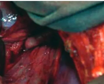
The patient underwent left thoracotomy, opening of the pleura, and exposure of the aorta through lung retraction. After the identification of the anatomical elements, the following procedures were performed: resection of the ligamentum arteriosum; suture of the Kermmell diverticulum (Figure 3), and resection and reimplant of the left subclavian artery into the left carotid artery (Figure 4). Afterwards, a procedure to free the fibrous bands adjacent (adhesive) to the esophagus and trachea was performed. The pleura was left open with the drainage tube placed in the normal position.
INTRODUCTION
Kommerell’s diverticulum, which is a rare condition, involves the left fourth aortic arch (LAA) and the anomalous origin of the right subclavian artery (RSA) (0.5%-2%), or the right aortic arch (RAA) with the anomalous origin of the left subclavian artery (LSA) (o.05%-0.1%) [1]. A cause of aberrant subclavian artery origin can be an abnormal regression of the primitive fourth aortic arch during embryogenesis. The left fourth aortic arch persists as the aortic arch, while the right fourth aortic arch remains as the right subclavian artery (RSA) and the innominate artery (obsolete term for brachiocephalic (arterial) trunk) [2].
The right aortic arch (RAA) with ligamentum arteriosum to the descending aorta is one of the two vascular rings that cause tracheoesophageal compression; usually, children with a double aortic arch had earlier onset of symptoms (stridor and dysphagia) than children with a right aortic arch and ligamentum arteriosum.
A diverticulum may become aneurysmal even after the ligamentum arteriosum resection [1], the procedure of choice usually performed.
CASE REPORT
A 13-year-old female patient with wheezing or stridor and dysphagia in the last 5 years was, at present, progressive for pasty food. She reported weight loss in the last few months, unmeasured.
It was observed, on a chest radiograph, a right aortic arch (Figure 1); on the esophagram, an extrinsic compression suggestive of vascular ring. An angiotomography showed a right aortic arch with Kommerell diverticulum at the anomalous origin of the left subclavian artery (Figure 2).
Fig. 1 – PA chest radiograph, revealing the aortic arch to the right
Fig. 2 – Chest Computerized Tomography, showing the Kommerell diverticulum
354
REFERENCES
1. Backer CL, Hillman N, Mavroudis C, Holinger LD. Resection of Kommerell’s diverticulum and left subclavian artery transfer for recurrent symptoms after vascular ring division. Eur J Cardiothorac Surg. 2002;22(1):64-9.
2. Ota T, Okada K, Takanashi S, Yamamoto S, Okita Y. Surgical treatment for Kommerell’s diverticulum. J Thorac Cardiovasc Surg. 2006;131(3):574-8.
3. Backer CL, Mavroudis C, Rigsby CK, Holinger LD. Trends in vascular ring surgery. J Thorac Cardiovasc Surg. 2005;129(6):1339-47.
4. Komiyama M, Yasui T. Left subclavian artery originating from Kommerell diverticulum in the left aortic arch. J Thorac Cardiovasc Surg. 2006;132(6):1477.
Fig. 4 – Side-to-End Anastomosis between left carotid artery and left subclavian artery
There were no further complications and the patient was transferred to the ward in the next day without the drain. The patient was discharged on postoperative day 5, eating solid food, with normal voice, and absence of differential blood pressure between upper limbs.
DISCUSSION
The Kommerell diverticulum is an important vascular structure that through dilatation can causes symptoms, such as dysphagia, dyspnea, stridor, chest pain, emphysema, and pneumonia [2]. Left subclavian artery originating from Kommerell’s diverticulum which results in absence of pulse with the arm in a supine position is very unusual [4]. By the recurrence of symptoms in patients with a Kommerell diverticulum who had simple ligamentum resection, it is recommended surgical repair and primary reimplantation of the subclavian artery [2, 3], whenever the patients present with a diameter > 50 mm [2].
The diagnosis of vascular ring is suggested by the barium esophagram showing the extrinsic compression; however, an angiotomography or a chest angioressonance should always be performed to define the anatomy. Bronchoscopy and digestive endoscopy can be necessary transoperatively to confirm the surgical outcome [3].
Left posterolateral thoracotomy gtants a better approach to the structures, allowing a complete repair, and specially the reimplantation of LSA, thus avoiding upper limb ischemia [3] and the artery subclavian steal syndrome [2]. The pleura should not be closed because its healing can cause a new esophageal compression [1]. In the presence of an aneurismal diverticulum, it is recommended to replace the descending aorta with the reconstruction in situ of the aberrant subclavian artery through the right or left thoracotomy, according to the position of aortic arch. The exposure of the posteromedial surface of the aortic arch and proximal surface of descending aorta, through median thoracotomy, is very difficult [2]. The most frequent surgical complication is the chylothorax. It is always recommended its resection close to the diverticulum [1].
CONCLUSION
The resection of the ligamentum arteriosum, the resection and reimplantation of the left subclavian artery into the left carotid artery, and the repair of Kommerell’s diverticulum, in spite of being a more complex procedure than the simple resection of the ligamentum arteriosum, the procedure of choice, has satisfactory result and can prevent the recurrence of symptoms by extrinsic compression. ABUCHAIM, DCS ET AL - Vascular ring related to Kommerell
diverticula: case report

