Ruthenium(II) complexes of 1,3-thiazolidine-2-thione: Cytotoxicity
against tumor cells and anti-
Trypanosoma cruzi
activity enhanced upon
combination with benznidazole
Rodrigo S. Corrêa
a,b,⁎
, Monize M. da Silva
a, Angelica E. Graminha
a, Cássio S. Meira
c, Jamyle A.F. dos Santos
c,
Diogo R.M. Moreira
c, Milena B.P. Soares
c,d, Gustavo Von Poelhsitz
e, Eduardo E. Castellano
f, Carlos Bloch Jr
g,
Marcia R. Cominetti
h, Alzir A. Batista
a,⁎
aDepartamento de Química, Universidade Federal de São Carlos, CP 676, CEP 13565-905 São Carlos, SP, Brazil bDepartamento de Química, ICEB, Universidade Federal de Ouro Preto, CEP 35400-000 Ouro Preto, MG, Brazil cCentro de Pesquisas Gonçalo Moniz, Fiocruz, CEP:40296-710 Salvador, BA, Brazil
dCentro de Biotecnologia e Terapia Celular, Hospital São Rafael, Avenida São Rafael, 2152, São Marcos, CEP 41253-190 Salvador, BA, Brazil eInstituto de Química, Universidade Federal de Uberlândia, CP 593, CEP 38400-902 Uberlândia, MG, Brazil
fInstituto de Física, Universidade de São Paulo, CP 369, CEP 13560-970 São Carlos, SP, Brazil
gCentro Nacional de Pesquisa de Recursos Genéticos e Biotecnologia, EMBRAPA, Estação Parque Biológico, CEP 70910-900 Brasília, DF, Brazil hDepartamento de Gerontologia, Universidade Federal de São Carlos, CP 676, CEP 13565-905 São Carlos, SP, Brazil
a b s t r a c t
a r t i c l e
i n f o
Article history:
Received 2 September 2015
Received in revised form 16 December 2015 Accepted 28 December 2015
Available online 2 January 2016
Three new mixed and mononuclear Ru(II) complexes containing 1,3-thiazolidine-2-thione (tzdtH) were synthe-sized and characterized by spectroscopic analysis, molar conductivity, cyclic voltammetry, high-resolution electrospray ionization mass spectra and X-ray diffraction. The complexes presented unique stereochemistry and the proposed formulae are: [Ru(tzdt)(bipy)(dppb)]PF6 (1), cis-[Ru(tzdt)2(PPh3)2] (2) and trans -[Ru(tzdt)(PPh3)2(bipy)]PF6(3), where dppb = 1,4-bis(diphenylphosphino)butane and bipy = 2,2′-bipyridine. These complexes demonstrated strong cytotoxicity against cancer cell lines when compared to cisplatin. Specif-ically, complex2was the most potent cytotoxic agent against MCF-7 breast cells, while complexes1and3were more active in DU-145 prostate cells. Binding of complexes to ctDNA was determined by UV–vis titration and viscosity measurements and revealed binding constant (Kb) values in range of 1.0–4.9 × 10³M−1, which are characteristic of compounds possessing weak affinity to ctDNA. In addition, these complexes presented antipar-asitic activity againstTrypanosoma cruzi. Specifically, complex3demonstrated strong potency, moderate selectivity index and acted in synergism with the approved antiparasitic drug, benznidazole. Additionally, com-plex3caused parasite cell death through a necrotic process. In conclusion, we demonstrated that Ru(II) com-plexes have powerful pharmacological activity, while the metal-free tzdtH does not provoke the same outcome. © 2015 Elsevier Inc. All rights reserved.
Keywords:
Ru(II) complexes Cytotoxicity
Trypanosoma cruzi
ctDNA-binding 1,3-Thiazolidine-2-thione
1. Introduction
Cancer is considered a group of complex and multifaceted diseases[1]. Carcinogenesis is thought to be initiated by changes to the DNA within cells and also by inhibition of growth suppressors, which, in turn, gives rise to the uncontrolled cell proliferation, invasion of surrounding and distant tissues, and ultimately leads to a risk of aggressive metastasis [2]. Prostate and breast cancers are of high incidence and mortality around the world and the development of new drugs is of interest[3]. Drugs containing transition metals hold a promising possibility for cancer
treatment. Although cisplatin has been largely employed alone or in drug combinations against prostate and breast cancers, limitations regarding resistance has been observed[4,5]. To overcome cisplatin limitation, Satraplatin, thefirst orally available Pt drug, is currently undergoing clin-ical investigation[6]. Ruthenium compounds are promising pharmaceuti-cals because of many biological features, such as reduced toxicity, suitable biodistribution, and mechanisms of action different than platinum-based compounds[7,8]. In the last years, Ru(III) complexes have entered clinical trials: NAMI-A [ImH][trans-RuCl4(DMSO)(Im)], KP1019 (indazolium trans-[tetrachloridobis(1H-indazole)ruthenate(III)]) and NKP-1339 [sodiumtrans-[tetrachloridobis(1H-indazole)ruthenate(III)][9,10]. The half-sandwich η6-arene-Ru(II) complexes are a promising class of antitumor compounds with particular emphasis on the [RuCl2(η6
-p-cymene)(PTA)], PTA = 1,3,5-triaza-7-phosphaadamantane, named as RAPTA-C[11]. This compound is highly active in vivo against metastatic
⁎ Corresponding authors.
E-mail addresses:rodrigocorrea@iceb.ufop.br(R.S. Corrêa),daab@ufscar.br (A.A. Batista).
http://dx.doi.org/10.1016/j.jinorgbio.2015.12.024 0162-0134/© 2015 Elsevier Inc. All rights reserved.
Contents lists available atScienceDirect
Journal of Inorganic Biochemistry
cells by inhibiting cathepsin B, a protease related to tumor invasion and metastasis[12,13].
Regarding parasitic infections, American trypanosomiasis (Chagas disease) is an important health problem in Latin America, affecting 8–
14 million people (14,000 deaths per year) with different forms of the pathology[14,15]. It is of great concern due to the development of chronic cardiomyopathy and other related health problems [16]. Chemotherapy is only based on nifurtimox and benznidazole (Bdz), drugs that are more than 50 years old and suffer from low efficacy and high levels of toxicity[17]. Since the discovery of an efficient Ru(II) complex containing clotrimazole againstTrypanosoma cruzi, a large number of metal complexes have been evaluated for this purpose[18]. Strategies for the development of new anti-trypanosomal metallodrugs generally involve: coordination of antiparasitic ligands to the metal, and coordination of DNA intercalators to the metal and metal compounds as direct inhibitors of parasite enzymes[19,20].
In view of this, our research group has been studying a number of pharmacological properties for Ru(II) complexes containing phosphines and diimines ligands as anticancer and anti-infectious agents[21–27].
Remarkable activities were observed for complexes with general for-mulae [Ru(pic)(dppb)(N–N)]PF6, where pic = 2-pyridinecarboxylate;
N–N = 2,2-bipyridine or 1,10′-phenanthroline. These complexes
displayed strong in vitro activity againstMycobacterium tuberculosis
and more importantly, presented activity against multi-resistant strains. In view of these results with bidentateN,O-ligand (pic), we sought to investigate Ru complexes containing bidentateN,S-ligands. In fact, heterocyclicN,S-ligands have been extensively used in the preparation of metal complexes for therapeutic application[27–29]. Among them,
an interesting ligand is 1,3-thiazolidine-2-thiol (tzdtH), which has a classical thiol/thione tautomerism as shown inFig. 1. Crystallographic studies for tzdtH structure indicate that the thione tautomer (form II; Fig. 1) is present in the solid state [30]. However, two tautomeric forms can be found in aqueous and in organic solutions (i.e., 1,4-diox-ane, CCl4, benzene, CHCl3, CH2Cl2, C2H4Cl2s, EtOH, MeOH, CH3CN, DMF
and DMSO)[31,32]. Given this promising outlook and considering the different coordination sites of the tzdtH[33–35], this work aimed to
study the reactivity of this ligand with Ru(II) phosphine precursors. To the best of our knowledge, only three ruthenium organometallic compounds with anionic tzdt−are described in the literature, however
no detailed structural as well as pharmacological evaluation were carried out[36,37]. Thus, here three new Ru(II) complexes with the formulae [Ru(tzdt)(bipy)(dppb)]PF6 (1),cis-[Ru(tzdt)2(PPh3)2] (2)
andtrans-[Ru(tzdt)(PPh3)2(bipy)]PF6(3), were obtained and evaluated
as cytotoxic and antiparasitic agents.
2. Experimental
2.1. General
Reactions and chemicals were handled under Argon atmosphere. Solvents were purified by standard methods. Chemicals used were of reagent grade or comparable purity. RuCl3·3H2O, dppb, bipy and
tzdtH were purchased from Sigma-Aldrich (St. Louis, MO) and used as supplied. The reactantscis-[RuCl2(dppb)(bipy)][38], [RuCl2(PPh3)3]
[39] and cis-[RuCl2(PPh3)2(bipy)] [40] precursors were prepared
according to literature. The IR spectra were recorded on a FT-IR Bomem-Michelson 102 spectrometer in the 4000–250 cm−1region,
using CsI pellets. Conductivity values were obtained at room tempera-ture using 10−3M solutions of the complexes in CH
2Cl2by using a
Meter Lab CDM2300 instrument.1H,31P{1H} and13C{1H} NMR were
re-corded on a Bruker DRX 400 MHz using tetramethylsilane as reference and solvent CDCl3to the complexes1and2and acetone-d6to complex 3. The31P{1H} chemical shifts are reported in relation to H
3PO4, 85%. The
UV–vis spectra of the complexes, (concentration c.a. 10−4M), were
recorded in CH2Cl2on a Hewlett Packard diode array—8452 A. Cyclic
voltammetry experiments were performed in an electrochemical analyzer BAS, model 100B and carried out at room temperature. Typical conditions were: CH2Cl2 containing 0.10 M Bu4NClO4 (TBAP) as
supporting electrolyte, and using a one-compartment cell, both working and auxiliary electrodes were stationary Pt foils, and the reference electrode was Ag/AgCl, 0.10 M TBAP in CH2Cl2. Ferrocene (Fc) was
employed for calibrating the electrochemical system and the redox potential. Under these conditions, (Fc+/Fc) couple presented 430 mV.
High resolution mass spectra of complexes were obtained by direct infusion in a MicroTof-Q II Bruker Daltonics Mass Spectrometer (Le) in the positive ion mode, employing methanol as solvent (LC/MS grade from Honeywell/B&J Brand). Elemental analyses were carried out in the Microanalytical Laboratory of Federal University of São Carlos, with an EA 1108 FISONS Instruments CHNS microanalyzer.
2.2. Synthesis and characterization
2.2.1. [Ru(tzdt)(bipy)(dppb)]PF6 (1)
In a Schlenkflask, 16 mg (0.14 mmol) of tzdtH was dissolved in 10 mL of a CH2Cl2solution containing 20μL of triethylamine. After,
100 mg (0.12 mmol) ofcis-[RuCl2(dppb)(bipy)] reactant was added
and maintained under stirring at room temperature for 3 h. Then, 30 mg (0.18 mmol) of NH4PF6was added and the volume concentrated
under reduced pressure to ca. 2 mL. Orange crystals were separated by
filtration, washed with dry diethyl ether and dried under vacuum to yield 94 mg (83%). Anal. Calc. for [RuC41H40N3P2S2]PF6: exp. (calc)
51.90 (52.01); H, 4.21 (4.26); N, 4.28 (4.44); S, 6.55 (6.77) %. Molar conductance (S cm2mol−1, CH
2Cl2) 46.5. IR (cm−1): (υC–H) 3076,
3053, 2949, 2924; (υCH2) 2854, 2679; (υCN) 1535, 1433;
(νCC(ring)+νCC(dppb)) 1483, 1309; (υC–S) 1159; (υC–P) 1094; (νring)
1043, 997; (υP–F) 843; (γCS) 764; (γring) 698; (υRu–P) 517, 507;
(υRu–S) 492; (υRu–N) 426.31P{1H} NMR (162 MHz, CDCl3, 298 K):δ
(ppm) (d, 43.2 and 44.6,2J = 35.1 Hz);1H NMR (400 MHz, CDCl 3,
298 K):δ(ppm): 8.96 (1H, d, bipy); 8.74 (1H, br. s, bipy); 8.09 (1H, m,
bipy); 8.03 (1H, m, bipy); 7.85 (1H, m, bipy); 7.73 (1H, m, bipy); 7.45 (2H, t, Hpof dppb); 7.41–6.93 (16H, m, Hoand Hmof dppb); 6.89 (2H,
t, bipy); 6.38 (2H, t, Hp of dppb); 3.40–2.30 (4H, m, CH2of tzdt);
2.30–1.03 (8H, m, CH2of dppb).13C{1H} NMR (125.74 MHz, CDCl3,
298 K):δ(ppm) 183.75 (CS); 158.09–151.45 (C-Bipy), 139.93–121.92
(C-dppb and C-Bipy); 55.26 (1C, CH2–N of tzdt) and 32.03 (1C, CH2–S
of tzdt); 31.75–21.29 (C–CH2 of dppb). UV–vis (CH2Cl2,
1.6 × 10⁻⁴M):
λ/nm (ε/M−1cm−1) 296 (15,970), 420 (2850).
2.2.2. cis-[Ru(tzdt)2(PPh3)2] (2)
In the Schlenkflask, 20 mg (0.17 mmol) of tzdtH was dissolved in 60 mL of ethanol. To this, 60 mL of CH2Cl2containing 30μL of Et3N
follow-ed by 70 mg of [RuCl2(PPh3)3] reactant were added. After stirring for
30 min, under room temperature, color mixture changed from a brownish to a yellowish suspension. Solvent was removed under reduced pressure and the yellowish solid wasfiltered and washed with ethanol and diethyl ether and then dried under vacuum to yield 50 mg (79%). Anal. Calc. for [RuC42H38N2S4P2].½H2O: exp. (calc) 57.76 (57.91); H, 4.22 (4.51); N,
3.33 (3.22); S, 15.17 (14.73) %. Molar conductance (S cm2 mol−1,
CH2Cl2) 1.8. IR (cm−1) (υC–H) 3072, 3049, 2947, 2928; (υCH2) 2849;
(υCN) 1527; 1508; (νCC(ring)+νCC(dppb)) 1479, 1385; (υC–S) 1188;
(υC–P) 1088; (νring) 1045, 993; (γCS) 750; (γring) 696; (υRu–P) 520;
(υRu–S) 497; (υRu–N) 435.31P{1H} NMR (162 MHz, CDCl3, 298 K):δ
(ppm) 54.2 (s);1H NMR (400 MHz, CDCl
3, 298 K):δ(ppm): 7.32 (12H,
m, Hoof PPh3); 7.23 (6H, t, Hpof PPh3); 7.10 (12H, t, Hmof PPh3); 3.27
(2H, ddd, CH2of tzdt); 3.20 (2H, dd, CH2of tzdt); 2.94 (2H, ddd, CH2of
tzdt); 2.65 (2H, dd, CH2of tzdt).13C{1H} NMR (125.74 MHz, CDCl3,
298 K):δ(ppm) 181.88 (CS); 137.33–127.09 (36C, C-PPh3); 56.49 (2C,
CH2-N of tzdt) and 31.72 (2C, CH2-S of tzdt). UV–vis (CH2Cl2,
4 × 10⁻⁵M):
λ/nm (ε/M−1cm−1) 310 (1993).
2.2.3. trans-[Ru(Tzdt)(Pph3)2(Bipy)]PF6(3)
In the Schlenkflask, 33 mg (0.137 mmol) of tzdtH was dissolved in a mixture of CH2Cl2:MeOH (80:20) containing 20μL Et3N. Then, 100 mg
(0.114 mmol) ofcis-[RuCl2(PPh3)2(bipy)] reactant was added and the
mixture was stirred under reflux temperature for 24 h. After this, the resulting orange solution was concentrated under reduced pressure to 2 mL and 10 mL of water was added. The resulting orange solid was
filtered, washed with warmed water, diethyl ether and then dried under vacuum to yield 113 mg (92%). Anal. Calc. for [RuC49H42N3S2P2]PF6·2H2O: exp. (calc) 54.69 (54.44); H, 4.12 (4.29);
N, 3.97 (3.89); S, 6.23 (5.93) %. Molar conductance (S cm2mol−1,
CH2Cl2) 50.2. IR (cm−1): (υC–H) 3076; 3055; 2951; 2924; (υCH2)
2852; (υCN) 1528, 1433; (νCC(ring)+νCC(dppb)) 1384; 1307; (υC–S)
1159; (υC–P) 1090; (νring) 1051, 999; (υP–F) 840; (γCS) 762; (γring)
698; (υRu–P) 519; (υRu–S) 492; (υRu–N) 438. 31P{1H} NMR
(162 MHz, CDCl3, 298 K):δ(ppm) 33.3 (s);1H NMR (400 MHz, CDCl3,
298 K):δ(ppm): 9.75 (1H, d, bipy); 9.02 (1H, d, bipy); 7.71 (2H, m, bipy); 7.65 (2H, m, bipy); 7.57 (1H, m, bipy); 7.35 (6H, m, Hp of
PPh3); 7.28 (12H, m, Hoof PPh3); 7.20 (12H, m, Hmof PPh3); 7.11 (1H,
m, bipy); 3.37 (2H, t, CH2of tzdt); 2.57 (2H, t, CH2of tzdt).13C{1H}
NMR (125.74 MHz, CDCl3, 298 K): δ (ppm) 182.52 (CS);
158.68–153.62 (C-Bipy), 136.58–123.77 (C-PPh3and C-Bipy); 57.41
(1C, CH2-N, tzdt), 31.77 (1C, CH2-S, tzdt). UV–vis (CH2Cl2,
8 × 10⁻⁵M):
λ/nm (ε/M−1cm−1) 280 (26266), 302 (17927), 348
(5488), 444 (3434).
2.3. X-ray diffraction
Single crystals of the complexes were grown from diethyl ether dif-fusion into a dichloromethane solution of complex at room temperature (293 K). X-ray diffraction experiments were carried out at room tem-perature using a suitable crystal mounted on glassfiber, and positioned on the goniometer head. Intensity data were measured on an Enraf–
Nonius Kappa-CCD diffractometer with graphite monochromated MoKαradiation (λ= 0.71073 Å). The cell refinements were performed using the software Collect[41]and Scalepack[42], and thefinal cell parameters were obtained on all reflections. The structures were solved by direct method using SHELXS-97 and refined using the software SHELXL-97. In all complexes' structures, the Gaussian method was used for the absorption corrections[43]. Non-hydrogen atoms of the complexes were unambiguously located, and a full-matrix, least-squares refinement of these atoms with anisotropic thermal parameters was carried out. In all ligands of the complexes, aromatic C–H hydrogen
atoms were positioned stereochemically and were refined withfixed individual displacement parameters [Uiso(H) = 1.2 Ueq(Csp2)] using a
riding model with aromatic, C–H bond lengths which werefixed at
0.93 Å. Methylene groups of tzdt ligand were also set as isotropic with a thermal parameter 20% greater than the equivalent isotropic displace-ment parameter of the atom to which each one was bonded and C–H
bond lengths werefixed at 0.97 Å. Tables were generated by WinGX and the structure representations by MERCURY. The CrystalExplorer 2.1 program was used to generate the Hirshfeld surfaces and thefi nger-print plot. The Hirshfeld surfaces were employed to define the intermo-lecular environment of molecules within the crystal of each complex
[44–46]. Thefingerprint plot or 2D-fingerprint graphics is constructed
by the plot ofdeversusdi(de= external distance is defined as the distance between the calculated Hirshfeld surface and the nearest atom of an adjacent molecule;di= internal distance is distance be-tween the nearest nucleus internal and the calculated Hirshfeld surface). Relationships between crystal packing pattern and molecular geometry were determined by analyzing parameters present in Hirshfeld fingerprint plots. The 2D-fingerprint also provides the percentage of each intermolecular contact occurring in the complex structure. Crystallography data were registered in the Cambridge Crystallographic Data Centre (CCDC), with the respective deposit numbers: 1037025 (1), 1037026 (2) and 1037027 (3).
2.4. DNA binding
2.4.1. Spectroscopic titration
A solution of calf thymus DNA (ctDNA, Sigma-Aldrich) was prepared in Tris–HCl buffer (5 mM Tris–HCl, pH 7.2). A solution of ctDNA in the
buffer gave a ratio of UV absorbance at 260 and 280 nm of about 1.8:1, indicating that the solution is protein-free. The concentration of ctDNA was measured from its absorption intensity at 260 nm using the molar absorption coefficient value of 6600 M−1cm−1 [47]. Solutions of
Ru(II) complexes used in the experiments were prepared in Tris–HCl
buffer containing 5% DMSO. To the ctDNA titration experiments, different concentrations of the ctDNA were used (ranging 3.8 × 10⁻⁵
to 7.6 × 10⁻⁴M), while the concentration of ruthenium complexes
were maintained at 1.6 × 10⁻⁴, 4.4 × 10⁻⁵and 8.6 × 10⁻⁵M for1,2
and3, respectively. Sample correction was done for the absorbance of DNA and the spectra were recorded after solution equilibration for 2 min. It is worth mentioning that complex2and3structures change after incubating in the buffered medium, such as observed in the31P
NMR spectrum of complexes2and3(see the Supplementary material). These chances can be attributed to exchange of the monodentate PPh3
ligand. As a result, the signal of PPh3 free is observed at around
−6.3 ppm. The intrinsic equilibrium binding constant (Kb) of the
com-plexes to ctDNA was obtained using the McGhee–von Hippel (MvH)
method[48]by using the expression of Wolfe and co-workers[49]:
ctDNA
½ =ðεa–εfÞ ¼½ctDNA=ðεb–εfÞ þ1=Kbðεb–εfÞ
in which [ctDNA] is the concentration of ctDNA in base pairs,εais the ratio of the absorbance/[Ru(II) complex],εfis the extinction coefficient of the free Ru(II) complex, andεbis the extinction coefficient of the complex in the fully bound form. The ratio of the slope to the intercept in the plot of [ctDNA]/(εa–εf) vs. [ctDNA] gives the value of Kb, which
was calculated from the metal to ligand charge transfer (MLCT) absorp-tion band (λmax). Changes in the absorption intensity increasing
concentration of ctDNA was monitored and analyzed by regression analysis. The nonlinear least-squares analysis was calculated by using OriginLab.
2.4.2. Viscosity measurements
Viscometric titrations of1–3were performed using an Ostwald viscometer in a constant temperature (37 °C). The concentration of ctDNA was 4.2 × 10⁻³
μM, and theflow times were measured with an automated timer. Each sample was measured 5 times and an average
flow time was calculated. Data were presented as (η/ηo)1/3versus
[complex]/[ctDNA], whereηis the viscosity of ctDNA in the presence of the complex andηois that of ctDNA alone. Relative viscosity for
ctDNA in either the presence or absence of complex was calculated from the relation:η= (t−to) / (to), where t is the observedflow
time of the ctDNA containing solution and tois theflow time of buffer
2.5. Cytotoxicity against cancer cells
Human tumor breast cell line MCF-7 (ATCC No. HTB-22) and human prostate tumor cell line DU-145 (ATCC: HTB-81) were cultured in RPMI-1640 medium (Sigma-Aldrich) supplemented with 20% fetal bovine serum (FBS; Cultilab, Campinas, Brazil) at 37 °C in 5% CO2. Aliquots of
200μL containing 1 × 10⁴cells were seeded in 96-well microplates
and incubated for 12 h. Drugs were dissolved in sterile DMSO (stock so-lution with maximum concentration of 20 mM) and diluted in RPMI-1640 medium from 0.05 to 200μM (final concentration of 1% DMSO per well). Negative control (without drug) and positive control (cisplat-in) were included in the plate and then incubated for 48 h at 37 °C in 5% CO2. After incubation, cells were washed twice with phosphate buffer
saline, 50μL of MTT (MTT, Life, Carlsbad, USA) at 0.5 mg mL−1was
added and incubated for 4 h, followed by adding 100μL of isopropanol. Colorimetric reading was performed in a microplate reader at 595 nm. Cell viability was measured in comparison to the negative control (cells receiving only DMSO). Inhibitory concentration to 50% (IC50)
was determined by using non-linear regression. Three independent experiments were performed.
2.6. Host cell toxicity
J774 macrophages were seeded on 96-well plates at a cell number of 5 × 10⁴cells mL−1in 200μL of RPMI medium (Sigma-Aldrich)
supplemented with 10% of FBS (Life, Carlsbad, USA) and 50μg mL−1of gentamycin (Life) and incubated for 24 h at 37 °C
and 5% CO2. After that time each compound was added at six
concen-trations (0.04 to 10μM) in triplicate and incubated for 72 h. Cell viability was determined by AlamarBlue assay (Life) according to the manufacturer's instructions. Colorimetric readings were performed after 6 h at 570 and 600 nm. Cytotoxic concentration to 50% (CC50) was calculated using data-points gathered from three
in-dependent experiments. Gentian violet (Synth, São Paulo, Brazil) was used as positive control.
2.7. Antiparasitic activity
2.7.1. Cytotoxicity for trypomastigotes
Trypomastigotes collected from the supernatants of previously infected LLC-MK2 cells were dispensed into 96-well plates at a cell number of 2 × 10⁶cells mL−1in 200
μL of RPMI medium. Compounds
were tested at concentration range of 0.02 to 10μM, in triplicate. The
plate was incubated for 24 h at 37 °C and 5% CO2. Aliquots from each
well were collected and the number of viable parasites was assessed in a Neubauer chamber and compared to untreated parasite culture. This experiment was performed three times. Benznidazole (LAFEPE, Recife, Brazil) was used as the positive control. Cytotoxic concentration to 50% (CC50) was calculated using data-points gathered from three
independent experiments.
2.7.2. Inhibition of parasite infection
Peritoneal macrophages (2 × 10⁵cells mL−1) obtained from BALB/c
mice were seeded in a 24 well-plate with rounded coverslips on the bottom in RPMI supplemented with 10% FBS and incubated for 24 h. Cells were then infected with trypomastigotes (10:1) for 2 h. Free trypomastigotes were removed by successive washes using saline solution and the cells were incubated for 24 h for internalization and differentiation of trypomastigotes into amastigotes. Following this, cultures were incubated in complete medium alone or with compounds for 72 h. Cells werefixed in absolute alcohol and the percentage of infected macrophages and the number of amastigotes/100 macro-phages was determined by manual counting after hematoxylin and eosin staining using an optical microscope (Olympus, Tokyo, Japan). The percentage of infected macrophages and the number of amastigotes per 100 macrophages was determined by counting 100 cells per slide.
The one-way ANOVA and Bonferroni for multiple comparisons were used to determine the statistical significance of the group comparisons. Benznidazole was used as the positive control.
2.8. Propidium iodide and annexin V staining
Trypomastigotes (1 × 10⁷) were incubated for 24 h at 37 °C in the
absence or presence of Ru(II) complex3(0.01, 0.015 or 0.02μM). After incubation, the parasites were labeled for propidium iodide (PI) and annexin V using the annexin V-FITC apoptosis detection kit (Sigma-Aldrich), according to the manufacturer's instructions. Acquisi-tion and analyses was performed using a FACS Caliburflow cytometer (Becton Dickinson, CA, USA), with FlowJo software (Tree Star, CA, USA). A total of 30,000 events were acquired in the region previously established as trypomastigote forms ofT. cruzi. Two independent experiments were performed.
3. Results
3.1. Synthesis, infrared spectroscopy and mass spectrometry
The chemical reactivity of tzdtH in triethylamine was studied under
the presence of metal complexes cis-[RuCl2(dppb)(bipy)],
[RuCl2(PPh3)3] andcis-[RuCl2(PPh3)2(bipy)]. This led to the formation
of complexes with formulae [Ru(tzdt)(bipy)(dppb)]PF6 (1), cis
-[Ru(tzdt)2(PPh3)2] (2) andtrans-[Ru(tzdt)(PPh3)2(bipy)]PF6(3)
con-taining the monoanionic tzdt−as chelated ligand as shown inScheme
1. Each synthetic procedure was straightforward, provided good yields and analytically pure complexes as determined by elemental analyses. Infrared spectra of complexes1–3confirmed the presence of the tzdt−ligand coordinated to the metal center. High energy region of
each spectrum exhibited bands at 2854–2849 cm−1, which were
assigned to theυCH2stretching vibration of tdzt ligand. TheυN–H
stretching vibration at 3138 cm−1in the spectrum of metal-free tzdtH
was absent in the spectra of the ruthenium complexes, suggesting that ligand is coordinated into its deprotonated form. Strong bands found in the region of 1535–1300 cm−1are characteristic ofυCN andυCC
stretching vibrations of the tdzt−, dppb and bipy ligands. In the
com-plexes, the bands related toνC…S and
δC…S absorptions of tdzt−occur
in the regions around 1188–1159 and 764–750 cm−1, respectively.
Regarding ESI-MS(+) spectra, complex 1 presented the most
intense molecular peak at 802.1179 Da, while its predicted monoisoto-pic mass is 802.1186 Da. For complexes2and3, M+peaks occurred at
862.0428 and 900.1345 Da, respectively. In both complexes2and3, peaks corresponding to [M−PPh3]+were observed.
3.2. Electrochemical study
The redox behavior of metal complexes was investigated by cyclic voltammetry (Fig. 2). Complexes1–3revealed one-electron waves for Ru(II)/Ru(III) redox process withquasi-reversible behavior at +1150, +690 and +926 mV. These values highlight the different stereochem-istry around the Ru(II) center. Complex2exhibited redox process in lower potential than complexes1and3. This can be explained because of two molecules of tzdt−and the absence of bipy, which is a
well-known π–electron acceptor ligand. In fact, the bipy-containing
complexes1and3presented Ru(II)/Ru(III) oxidation peaks around 1000 mV, which are similar values to Ru(II) complexes described in the literature[25,26]. Despite complexes1and3presenting the same ligands,3has a redox potential much lower than 1. This may be explained by the competition for electron density around the metal between the phosphorus atoms intransposition observed in complex
3[27].
This indicates that ruthenium is more easily oxidized in metal precur-sors than complexes (1–3), therefore complexes (1–3) are more stable
than their starting reactants. This stabilization is possible due to the
re-placement of two σ and π donor chlorides by a negative and
monocharged chelating tzdt−, which contains an acceptor group.
3.3. NMR spectroscopy
Resonance for complexes1and2was carried out in CDCl3, while
acetone-d6was used in complex3. In the31P{1H} NMR spectrum of
complex1, a typical AB spin system was observed with chemical shifts at 43.2(d) and 44.5(d) ppm, indicating the magnetic nonequivalence
of the two phosphorus atoms of dppb. The precursor complexcis -[RuCl2(dppb)(bipy)] shows a pair of doublets at 32.0 and 43.0 ppm
with the highfield signal corresponding to the PtransN, as previously described[40]. These assignments are based on an empirical linear cor-relation established between crystallographic determined Ru–P
dis-tances in a series of Ru–dppb complexes and the corresponding31P
chemical shift observed in solution, in which the chemical shift become more high-field with increasing Ru–P bond length[50]. In view of this
information, we suggest that in complex1the high-field doublet be-longs to the Ptransto nitrogen from bipy, because the Ru–P2 distance
of 2.3299(9) Å (transbipy) is longer than that observed for the Ru–P1 transof nitrogen of the 2-MT ligand [2.3069(9) Å].
In contrast, only one singlet is observed in the31P{1H} NMR spectra
of complexes2and3, due to the presence of two equivalent PPh3
phos-phorus atoms (Table 1). For complex2the singlet signal at 54.2 ppm is typical of PPh3transto nitrogen of N-heterocyclic ligands as observed
for similar compounds such as thecis-[Ru(pymS)2(PPh3)2], pymS =
deprotonated 2-mercaptopyrimidine[51]. For complex3the singlet is observed at 33.3 ppm, a chemical shift in lowfield is shifted when com-pared to cis-[RuCl2(PPh3)2(bipy)], where a singlet is presented at
21.5 ppm[52]. These values are typical of PPh3transto PPh3as observed
for a series of ruthenium compounds[53,54]. The1H NMR spectrum of
metal-free tzdtH in CDCl3displayed a broad singlet corresponding to
the proton of the N–H group around 7.30 ppm and a pair of triplets in
the range 3.50–4.00 ppm corresponding to the methylenic protons. Scheme 1.Routes used to prepare Ru(II)/tzdt−complexes.
Fig. 2.Cyclic voltammograms of Ru(II) complexes1,2and3. Conditions: CH2Cl2, 0.10 M Bu4NClO4as supporting electrolyte, scan rate 100 mV s−1; working and auxiliary electrodes stationary Pt foils, and Ag/AgCl as reference electrode.
Table 1
31P{1H} NMR and cyclic voltammetry data for complexes1–3. Complex δ(ppm) 2J
P–P(Hz) E½ (mV) ΔEp(mV)
1 43.2(d); 44.6(d) 35.1 1082 135
2 54.2(s) – 602 176
For complexes1–3the signal at 7.30 is absent confirming that tzdtH is coordinated to Ru(II) in a deprotonated form and the methylenic pro-tons appeared as multiplet signals in the range 2.30–3.40 ppm,
consid-erably high-field shifted when compared with free-ligand. In the aromatic region protons of dppb (complex1) and PPh3(complexes2
and3) displayed the typical pattern of multiplet signals forortho,meta
andpara hydrogens of the aromatic rings in ranges of 6.38–7.45;
7.10–7.32 and 7.20–7.35 ppm, respectively. In addition, complexes1
and3exhibited the expected deshielded doublets corresponding to the ortho hydrogens of the bipy ligands at 8.96; 8.74 and 9.75; 9.02 ppm, respectively. The 13C NMR spectra of complexes 1–3
displayed signals around 183–181 ppm, depending on the complex,
typical of the CS coordinated group. This signal is shielded compared with that observed for the free ligand which occurs at 201.7 ppm, indi-cating that sulfur is coordinated to the metal. In addition, complexes displayed signals in the range 57.4–55.3 and 32.0–31.7 ppm, typically
assigned to carbon atoms of the N-CH2 and S-CH2 groups of the
thiazolidine ring, respectively.
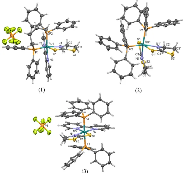
3.4. X-ray crystal structures
Suitable crystals for a single crystal X-ray structure determination were obtained by slow evaporation of a chloroform solution. The MERCURY plots inFig. 3show that these complexes possess a distorted octahedral geometry. Crystal data collections and structure refinement parameters are summarized in Table 1S. The crystallographic analysis of metal-free tzdtH described in the literature shows that C1–S1 is a
double bond, while C1–N1 single-bond[54]. When tzdtH is coordinated
to Ru, the length of these bonds significantly changes in which the C1–
S1 is longer whereas C1–N1 is shorter in all the complexes. This suggests
that the ligand adopts the canonical form (III) depicted inFig. 1. In the crystal structure of complex3, the Ru–P bond lengths are longer than
the other two complexes, possible due to the P to Ptransinfluence. In contrast, the Ru–N1 length in3is shorter than that the observed for complexes1and2, because of the P to Ntransinfluence which slightly affects the Ru–N bond length.
When we analyze the tzdt−conformation, a planar conformation in
complexes1and3is observed, while in complex2a twisted ring is ob-served in the ligand structure. Due to the intermolecular interaction and crystal packing, the free tzdtH in solid state adopts either a distorted or a planar conformation. In the crystal structure of the neutral complex2, a sulfur⋯sulfur contact is observed, which explains the tzdtH distorted conformation. The distance of S⋯S atoms in the structure of complex
2is at 3.543 Å as shown inFig. 4, being shorter than the sum of the van der Waals radius (3.60 Å).
To examine the spatial arrangement of Ru complexes, the intermo-lecular contacts of each crystal structure were determined by using the Hirshfeld surfaces and their corresponding 2D-fingerprint plots (Supplementary material). The relative contribution of the intermolec-ular contacts present in these complexes shares interesting structural features. In complex (1), the contribution is: H⋯H (55.1%), C⋯H (17.2%), F⋯H (15.9%), S⋯H (7.9%), C⋯C (1.4%), C⋯F (0.5%), S⋯F (0.4%), and N⋯H (0.4%). In comparison to complex1, the intermolecular contribution found in2is slightly different [H⋯H (63.6%), C⋯H (17.5%),
S⋯H (17.5%), C⋯C (0.8%), S⋯C (0.2%)]. For complex2,the Hirshfeld sur-face analysis highlights the intermolecular contacts between S⋯S with contribution of 0.3%, which is absent in other complexes, this kind of contact can be seen inFig. 4. The contribution to Hirshfeld surface in complex3[H⋯H (48.7%), C⋯H (19.0%), F⋯H (13.7%), S⋯H (6.3%), C⋯C (0.6%)] is similar to that observed in complex1. In all of them, the H⋯H contacts compose about 50% of the Hirshfeld surface, evidenc-ing the importance of van der Waals forces to crystal packevidenc-ing stabilization.
3.5. Pharmacological evaluation
3.5.1. Cytotoxicity in cancer cells and ctDNA binding
In vitrocytotoxicity against DU-145 prostate and MCF-7 breast can-cer cells was examined 48 h after incubation with drugs and the results were expressed by determining the IC50values. Cisplatin was the
refer-ence cytotoxic drug. For comparison reason only, metal-free ligands tzdtH, dppb, bipy and PPh3were tested as well. The results are
summa-rized inTable 2.
All the complexes displayed cytotoxicity against cancer cells, while none metal-free ligands were cytotoxic in concentrations up to 200μM. These observations strongly suggests that Ru(II) associ-ated with the ligands are responsible for the cytotoxicity in cancer
cells. Importantly, the Ru(II) complexes were more active than cisplatin. A comparison between the complexes revealed that compound1is potent against the two cancer cell lines, while2is more cytotoxic against breast than prostate cells. Complex1was particularly more potent against prostate cells, while compound3
was against breast cells. Complex2 was less active among the
complexes.
Based on the cytotoxicity of these Ru(II) complexes against can-cer cells, it was hypothesized that these complexes may interact with ctDNA. To verify this, the interaction with ctDNA was studied via spectroscopic titration (Fig. 5a). Under the presence of the Ru(II) complexes, a ctDNA hypochromism in the range of 29–35%
was observed, which indicates that metal complexes form a ternary complex with ctDNA. In addition, the binding constant (Kb) were
de-termined and the respective values found were: 1.0, 1.7 and 4.9 × 10³M−1for complexes1,2and3. These values indicate a
weak interaction with ctDNA when compared to a classical ctDNA intercalator ethidium bromide (Kb106M−1)[55]. Interestingly,
complex1was the most active anticancer drug, but it presented lower ctDNA than complex3, which was less cytotoxic. Moreover, viscosity analysis of ctDNA-binding revealed that viscosity is not modified when the concentration of a Ru(II) complex increases. This supports the idea that ruthenium complexes have a weak Fig. 4.Representation of S⋯S contact occurring in complex2.
Table 2
Cytotoxicity and antitrypanosomal activities of complexes1–3, metal-free tzdtH and reference drugs.
Compounds IC50± S.E.M. (μM) J774 macrophages, CC50± S.E.M. (μM)c SId
DU-145a MCF-7a T. cruzitrypomastigotesb
tzdtH N200 N200 N10 N10 –
1 0.3 ± 0.2 1.1 ± 0.9 0.23 ± 0.09 1.0 ± 0.16 3.7
2 4.9 ± 0.2 0.98 ± 0.2 N10 N10 –
3 0.9 ± 0.9 3.3 ± 1.3 0.010 ± 0.001 0.34 ± 0.3 34
Cisplatin 2.0 ± 0.5 8.9 ± 2.6 - – –
Benznidazole – – 10.6 ± 0.8 – –
Gentian violet – – – 0.82 ± 0.1 –
Dppb N200 N200 – – –
PPh3 180.1 ± 1.6 N200 – – –
Bipy N200 N200 – – –
IC50= inhibitory concentration to 50%; and CC50= cytotoxic concentration to 50%. SI = selectivity index. IC50and CC50values were determined from at least two independent experi-ments using concentration in triplicate.
aDetermined in cancer cells after 48 h incubation with drugs. b Determined in Y strain ofT. cruzitrypomastigotes after 24 h incubation. c Determined in J774 macrophage cell lines after 72 h incubation. d SI determined as (CC
interaction, possibly by an electrostatic mode[56]. A plausible interpretation for this observation is that the binding of Ru(II) com-plexes to ctDNA is not via intercalation, due to the absence of planar ligands.
3.5.2. Antiparasitic activity
The antiparasitic evaluation against bloodstream trypomastigotes of
T. cruziparasite revealed that metal-free tzdtH and complex2have no activity in a concentration up to 10μM. In contrast, complexes1and3 Fig. 5.(a) Electronic absorption spectra of complex1at a concentration of 1.6 × 10⁻⁴M, showing the changes when concentration of ctDNA is increased (ranging from 3.8 × 10⁻⁵to 7.6 × 10⁻⁴M). ctDNA has no absorption atλN325 nm. (b) Viscosity of ctDNA (η/ηo)1/3in the presence of complexes1–3at increasing amounts. Experiments carried out at 298 K, in a Tris–HCl buffer, pH 7.4.
exhibited strong activity (Table 2). Complex3displayed the highest an-tiparasitic activity, being more potent than benznidazole, the reference antiparasitic drug. Additionally, complex3had little effect on J774 mac-rophage viability, therefore showing that the antiparasitic activity for this complex was achieved with great selectivity index. Regarding the structure–activity relationships, active antiparasitic complexes
contain-ing a bipy ligand were observed, while complex2lacking bipy was inac-tive. Therefore, these observations suggest that the presence of bipy as well as a positive charge present in the structures of complexes1and
3contribute to antiparasitic activity.
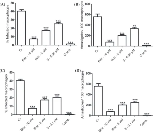
3.5.3. Evaluation in T. cruzi-infected macrophages
After observing that complex3has potent and selective activity against the extracellular parasite, its antiparasitic activity against the intracellular form ofT. cruziwas investigated (Fig. 6). In comparison to untreated infected macrophages, complex3treatment reduced the percentage of infected macrophages. Moreover, this treatment reduced the mean number of amastigotes per 100 macrophages. Importantly,
complex 3 at 0.1 μM has comparable antiparasitic activity to
benznidazole, the positive control. Therefore, these results show that this complex has antiparasitic activity against the intracellular and pro-liferative amastigote form. Since amastigote proliferation is pivotal within parasite cell cycle, it is plausible that these compounds impair the parasite cell cycle development inside host cells.
Given this strong antiparasitic activity, it was investigated whether the Ru(II) complexes have enhanced activity in drug combination
with benznidazole. As shown in Fig. 7, drug combination of
benznidazole at 5μM plus complex3at 0.05μM reduced the percentage of infected macrophages as well as the number of amastigotes more than each drug alone (Fig. 7, panels A, B). Importantly, the drug
combination displayed stronger activity than benznidazole alone at a high concentration (10μM). When the concentration of complex3
was increased at 0.1μM and added in combination to benznidazole at 5μM, in practice no intracellular parasites were observed (Fig. 7, panels
C, D). These results indicate that drug combination of benznidazole and complex3has enhanced antiparasitic activity.
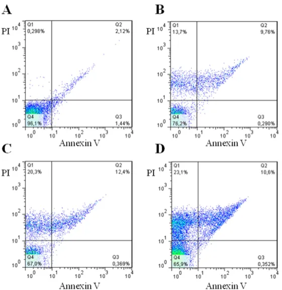
3.5.4. Parasite cell death
After ascertaining the antiparasitic activity of complex3, it was in-vestigated how this complex causes parasite cell death. In comparison to untreated trypomastigotes (Fig. 8, panel A), complex3treatment lead to single PI staining and double PI+ annexin V staining, which are characteristics of necrosis and late apoptosis, respectively. As observed by comparing panels B–D, complex3causes cell death in a concentration-dependent manner. Therefore, the Ru complex causes parasite cell death mainly by inducing necrosis.
4. Conclusions
Here we demonstrated the great chemical versatility of tzdtH, which is able to react with phosphine-, diamine- and phosphine/diamine-Ru precursors. The X-ray crystallography analyses revealed the exact structures of the complexes [Ru(tzdt)(bipy)(dppb)]PF6 (1), cis
-[Ru(tzdt)2(PPh3)2] (2) andtrans-[Ru(tzdt)(PPh3)2(bipy)]PF6(3) and
highlighted that the tzdt heterocyclic ring can assume a planar or twist-ed conformation under metal coordination. The electrochemical profile of these Ru complexes pointed out that tzdt provided resistance toward oxidation than the precursor complexes. These complexes exhibited strong anticancer and antiparasitic activity, while the metal-free tzdtH do not provoke the same outcome. Regarding the anticancer activity,
the new complexes exhibited cytotoxicity against prostate and breast cancer cells. They were more potent than cisplatin and more cytotoxic for cancer than normal cells (macrophages), indicating a degree of selectivity. Regarding the antiparasitic activity againstT. cruzi, these complexes exhibited a broad spectrum of action (extracellular, intracel-lular forms). Flow cytometry analysis revealed that complex3destroys parasite cells, indicating this is more likely a parasiticidal than a cytostatic drug. These complexes arrested the parasite cell cycle and strongly affected the intracellular development and ultimately caused irreversible parasite death through a necrotic process. An important aspect in the anticancer and antiparasitic therapy is the drug combina-tion. Here it was observed that Ru(II) complexes exhibit enhanced anti-parasitic activity when given in combination with the antianti-parasitic drug benznidazole. This points out that these complexes are suitable molecules for drug combination compositions.
Acknowledgments
We thank the Brazilian Agencies of Research: CNPq, CAPES, FAPESB and FAPESP (grant number 2012/06013-4). R.S.C. thanks FAPESP (Fundação de Amparo à Pesquisa do Estado de São Paulo) for a post-doctoral fellowship (grant numbers 2009/08131-1 and 2013/26559-4). C.S.M. is receiving a FAPESB (Fundação de Amparo à Pesquisa do Estado da Bahia) (Grant 0417/2012) scholarship. G.V.P. thanks FAPEMIG (Fundação de Amparo à Pesquisa do Estado de Minas Gerais) (Grant
APQ-04010-10). The authors acknowledge Dr. Marília I. F. Barbosa and Prof. Javier Ellena for the helpful discussions during the preparation of the manuscript.
Supplementary data
Coordinates and other crystallographic data have been deposited with the deposition codes CCDC 1037025, CCDC 1037026 and CCDC 1037027, for1,2and3, respectively. Copies of this information may be obtained from The Director, CCDC, 12 Union Road, Cambridge CB2 1EZ, UK, Fax: +44 1233 336,033, E-mail:deposit@ccdc.cam.ac.ukor www.ccdc.cam.ac.uk. Supplementary data to this article can be found online athttp://dx.doi.org/10.1016/j.jinorgbio.2015.12.024.
References
[1] B.L.D.M. Bruecher, G. Lyman, R. van Hillegersberg, R.E. Pollock, F. Lordick, H. Yang, T. Ushijima, K.G. Yeoh, T. Skricka, W. Polkowski, G. Wallner, V. Verwaal, A. Garofalo, D. D'Ugo, F. Roviello, H.U. Steinau, T.J. Wallace, M. Daumer, N. Maihle, T.J. Reid, M. Ducreux, Y. Kitagawa, A. Knuth, B. Zilberstein, S.R. Steele, I.S. Jamall, I.S. Jamall, BMC Cancer 14 (2014) 186.
[2] K.C. Oeffinger, S.S. Baxi, D.N. Friedman, C.S. Moskowitz, Semin. Oncol. 40 (2013) 676–689.
[3] J. Ferlay, E. Steliarova-Foucher, J. Lortet-Tieulent, S. Rosso, J.W.W. Coebergh, H. Comber, D. Forman, F. Bray, Eur. J. Cancer 49 (2013) 1374–1403.
[4] N.J. Wheate, S. Walker, G.E. Craig, R. Oun, Dalton Trans. 39 (2010) 8113–8127. [5] S. Dasari, P.B. Tchounwou, Eur. J. Pharmacol. 740 (2014) 364–378.
[6] J. Cetnar, G. Wilding, D. McNeel, N.K. LoConte, T.A. McFarland, J. Eickhoff, G. Liu, Urol. Oncol.-Sem. Orig. Investig. 31 (2013) 436–441.
[7] K.D. Mjos, C. Orvig, Chem. Rev. 114 (2014) 4540–4563.
[8] R.S. Correa, K.M.d. Oliveira, F.G. Delolo, A. Alvarez, R. Mocelo, A.M. Plutin, M.R. Cominetti, E.E. Castellano, A.A. Batista, J. Inorg. Biochem. 150 (2015) 63–71. [9] S. Pillozzi, L. Gasparoli, M. Stefanini, M. Ristori, M. D'Amico, E. Alessio, F. Scaletti, A.
Becchetti, A. Arcangeli, L. Messori, Dalton Trans. 43 (2014) 12150–12155. [10] R. Trondl, P. Heffeter, C.R. Kowol, M.A. Jakupec, W. Berger, B.K. Keppler, Chem. Sci. 5
(2014) 2925–2932.
[11] C. Scolaro, A. Bergamo, L. Brescacin, R. Delfino, M. Cocchietto, G. Laurenczy, T.J. Geldbach, G. Sava, P.J. Dyson, J. Med. Chem. 48 (2005) 4161–4171.
[12] C.G. Hartinger, N. Metzler-Nolte, P.J. Dyson, Organometallics 31 (2012) 5677–5685. [13] M.V. Babak, S.M. Meier, K.V.M. Huber, J. Reynisson, A.A. Legin, M.A. Jakupec, A. Roller, A. Stukalov, M. Gridling, K.L. Bennett, J. Colinge, W. Berger, P.J. Dyson, G. Superti-Furga, B.K. Keppler, C.G. Hartinger, Chem. Sci. 6 (2015) 2449–2456.
[14] P.J. Hotez, D.H. Molyneux, A. Fenwick, J. Kumaresan, S.E. Sachs, J.D. Sachs, L. Savioli, N. Engl. J. Med. 357 (2007) 1018–1027.
[15]I. Ribeiro, A.-M. Sevcsik, F. Alves, G. Diap, R. Don, M.O. Harhay, S. Chang, B. Pecoul, PLoS Negl. Trop. Dis. 3 (2009).
[16] J.A. Urbina, Acta Trop. 115 (2010) 55–68.
[17] C.A. Morillo, J.A. Marin-Neto, A. Avezum, S. Sosa-Estani, A. Rassi-Jr, F. Rosas, E. Villena, R. Quiroz, R. Bonilla, C. Britto, F. Guhl, E. Velazquez, L. Bonilla, B. Meeks, P. Rao-Melacini, J. Pogue, A. Mattos, J. Lazdins, A. Rassi, S.J. Connolly, S. Yusuf, BENEFIT Investigators, N. Engl. J. Med. 373 (2015) 1295–1306.
[18] M. Navarro, C. Gabbiani, L. Messori, D. Gambino, Drug Discov. Today 15 (2010) 1070–1078.
[19] D. Gambino, Coord. Chem. Rev. 255 (2011) 2193–2203.
[20]D.R. Moreira, A.C. Leite, R.R. dos Santos, M.B. Soares, Curr. Drug Targets 10 (2009) 212–231.
[21] F.B. Nascimento, G. Von Poelhsitz, F.R. Pavan, D.N. Sato, C.Q.F. Leite, H.S. Selistre-de-Araújo, J. Ellena, E.E. Castellano, V.M. Deflon, A.A. Batista, J. Inorg. Biochem. 102 (2008) 1783–1789.
[22] F.R. Pavan, G. Von Poelhsitz, M.I.F. Barbosa, S.R.A. Leite, A.A. Batista, J. Ellena, L.S. Sato, S.G. Franzblau, V. Moreno, D. Gambino, C.Q.F. Leite, Eur. J. Med. Chem. 46 (2011) 5099–5107.
[23]E.R. dos Santos, M.A. Mondelli, L.V. Pozzi, R.S. Correa, H.S. Salistre-de-Araujo, F.R. Pavan, C.Q.F. Leite, J. Ellena, V.R.S. Malta, S.P. Machado, A.A. Batista, Polyhedron 51 (2013) 292–297.
[24] F.R. Pavan, G. Von Poelhsitz, L.V.P. da Cunha, M.I.F. Barbosa, S.R.A. Leite, A.A. Batista, S.H. Cho, S.G. Franzblau, M.S. de Camargo, F.A. Resende, E.A. Varanda, C.Q.F. Leite, PLoS One 8 (2013).
[25] M.A. Mondelli, A.E. Graminha, R.S. Correa, M.M. da Silva, A.P. Carnizello, G. Von Poelhsitz, J. Ellena, V.M. Deflon, G.F. Caramori, M.H. Torre, D.C. Tavares, A.A. Batista, Polyhedron 68 (2014) 312–318.
[26]M.I.F. Barbosa, R.S. Corrêa, K.M.d. Oliveira, C. Rodrigues, J. Ellena, O.R. Nascimento, V.P.C. Rocha, F.R. Nonato, T.S. Macedo, J.M. Barbosa-Filho, M.B.P. Soares, A.A. Batista, J. Inorg. Biochem. 136 (2014) 33–39.
[27]M.A.P. Almeida, F.B.d. Nascimento, A.E. Graminha, A.G. Ferreira, J. Ellena, F.M.d.S. Mello, A.P.d. Lima, E.d.P. Silveira-Lacerda, A.A. Batista, Polyhedron 81 (2014) 735–742.
[28] R. Corona-Sanchez, R.A. Toscano, M. Carmen Ortega-Alfaro, C. Sandoval-Chavez, J.G. Lopez-Cortes, Dalton Trans. 42 (2013) 11992–12004.
[29] L. Li, K. Du, Y. Wang, H. Jia, X. Hou, H. Chao, L. Ji, Dalton Trans. 42 (2013) 11576–11588.
[30] E.S. Raper, R.E. Oughtred, I.W. Nowell, Inorg. Chim. Acta Lett. 77 (1983) L89–L93. [31] U.M. Rabie, M.H.M. Abou-El-Wafa, H. Nassar, Spectrochim. Acta A 78 (2011)
512–517.
[32]C. Abbehausen, R.E.F. de Paiva, A.L.B. Formiga, P.P. Corbi, Chem. Phys. 408 (2012) 62–68.
[33]E.S. Raper, J.R. Creighton, W. Clegg, L. Cucurull-Sanchez, M.N.S. Hill, P.D. Akrivos, Inorg. Chim. Acta 271 (1998) 57–64.
[34] P.G. Jones, S. Friedrichs, Acta Crystallogr. C 62 (2006) (M623-M7).
[35] D. Atzei, D. De Filippo, A. Rossi, M. Porcelli, Spectrochim. Acta A 57 (2001) 1073–1083.
[36] W.S. Sheldrick, C. Landgrafe, Inorg. Chim. Acta 208 (1993) 145–151.
[37]J.D.E.T. Wilton-Ely, M. Wang, D.M. Benoit, D.A. Tocher, Eur. J. Inorg. Chem. (2006) 3068–3078.
[38] S.L. Queiroz, A.A. Batista, G. Oliva, M. Gambardella, R.H.A. Santos, K.S. MacFarlane, S.J. Rettig, B.R. James, Inorg. Chim. Acta 267 (1998) 209–221.
[39] T.A. Stephens, G. Wilkinson, J. Inorg. Nucl. Chem. 28 (1966) 945–956.
[40] A.A. Batista, M.O. Santiago, C.L. Donnici, I.S. Moreira, P.C. Healy, S.J. Berners-Price, S.L. Queiroz, Polyhedron 20 (2001) 2123–2128.
[41] Enraf-Nonius, Collect, Nonius BV, Delft, The Netherlands (1997–2000).
[42] Z. Otwinowski, W. Minor, Macromolecular Crystallography, Part A, Academic Press, New York, 1997.
[43] P. Coppens, L. Leiserowitz, D. Rabinovich, Acta Crystallogr. 18 (1965) 1035–1038. [44] M.A. Spackman, D. Jayatilaka, CrystEngComm 11 (2009) 19–32.
[45] J.J. McKinnon, M.A. Spackman, A.S. Mitchell, Acta Crystallogr. B 60 (2004) 627–668. [46] M.A. Spackman, J.J. McKinnon, CrystEngComm (2002) 378–392.
[47] M.E. Reichmann, S.A. Rice, C.A. Thomas, P. Doty, J. Am. Chem. Soc. 76 (1954) 3047–3053.
[48] J.D. McGhee, P.H.V. Hippel, J. Mol. Biol. 86 (1974) 469–489. [49] A. Wolfe, G.H. Shimer, T. Meehan, Biochemistry 26 (1987) 6392–6396.
[50] K.S. MacFarlane, A.M. Joshi, S.J. Rettig, B.R. James, Inorg. Chem. 35 (1996) 7304–7310.
[51] T.S. Lobana, P. Kaur, A. Castineiras, J. Coord. Chem. 58 (2005) 429–435.
[52] M.O. Santiago, A.A. Batista, M.P. de Araujo, C.L. Donnici, I.D. Moreira, E.E. Castellano, J. Ellena, S. dos Santos, S.L. Queiroz, Transit. Met. Chem. 30 (2005) 170–175. [53]K. Wohnrath, A.A. Batista, A.G. Ferreira, J. Zukerman-Schpector, L.A.A. de Oliveira,
E.E. Castellano, Polyhedron 17 (1998) 2013–2020.
[54] R.S. Correa, S.A. Santana, R. Salloum, R.M. Silva, A.C. Doriguetto, Acta Crystallogr. C 62 (2006) (O115-O7).
[55] M. Cory, D.D. McKee, J. Kagan, D.W. Henry, J.A. Miller, J. Am. Chem. Soc. 107 (1985) 2528–2536.
![Fig. 1) is present in the solid state [30]. However, two tautomeric forms can be found in aqueous and in organic solutions (i.e., 1,4-diox-ane, CCl 4 , benzene, CHCl 3 , CH 2 Cl 2 , C 2 H 4 Cl 2s , EtOH, MeOH, CH 3 CN, DMF and DMSO) [31,32]](https://thumb-eu.123doks.com/thumbv2/123dok_br/15704353.629642/2.892.55.427.925.1093/present-solid-state-tautomeric-aqueous-organic-solutions-benzene.webp)