by Platelet Activating Factor: A Novel Receptor Cross
Talk Mechanism Regulating Neutrophil Superoxide
Anion Production
Huamei Forsman., Karin O¨ nnheim., Emil Andre´asson, Karin Christenson, Anna Karlsson, Johan Bylund,
Claes Dahlgren*
Department of Rheumatology and Inflammation Research, University of Gothenburg, Go¨teborg, Sweden
Abstract
Neutrophils express different chemoattractant receptors of importance for guiding the cells from the blood stream to sites of inflammation. These receptors communicate with one another, a cross talk manifested as hierarchical, heterologous receptor desensitization. We describe a new receptor cross talk mechanism, by which desensitized formyl peptide receptors (FPRdes) can be reactivated. FPR desensitization is induced through binding of specific FPR agonists and is reached after
a short period of active signaling. The mechanism that transfers the receptor to a non-signaling desensitized state is not known, and a signaling pathway has so far not been described, that transfers FPRdesback to an active signaling state. The
reactivation signal was generated by PAF stimulation of its receptor (PAFR) and the cross talk was uni-directional. LatrunculinA, an inhibitor of actin polymerization, induced a similar reactivation of FPRdesas PAF while the phosphatase
inhibitor CalyculinA inhibited reactivation, suggesting a role for the actin cytoskeleton in receptor desensitization and reactivation. The activated PAFR could, however, reactivate FPRdes also when the cytoskeleton was disrupted prior to
activation. The receptor cross talk model presented prophesies that the contact on the inner leaflet of the plasma membrane that blocks signaling between the G-protein and the FPR is not a point of no return; the receptor cross-talk from the PAFRs to the FPRdes initiates an actin-independent signaling pathway that turns desensitized receptors back to
a signaling state. This represents a novel mechanism for amplification of neutrophil production of reactive oxygen species.
Citation:Forsman H, O¨nnheim K, Andre´asson E, Christenson K, Karlsson A, et al. (2013) Reactivation of Desensitized Formyl Peptide Receptors by Platelet Activating Factor: A Novel Receptor Cross Talk Mechanism Regulating Neutrophil Superoxide Anion Production. PLoS ONE 8(3): e60169. doi:10.1371/ journal.pone.0060169
Editor:Roland Seifert, Medical School of Hannover, United States of America
ReceivedDecember 20, 2012;AcceptedFebruary 22, 2013;PublishedMarch 28, 2013
Copyright:ß2013 Forsman et al. This is an open-access article distributed under the terms of the Creative Commons Attribution License, which permits unrestricted use, distribution, and reproduction in any medium, provided the original author and source are credited.
Funding:The work was supported by the Swedish Medical Research Council, the King Gustaf V 80-Year Foundation, the Go¨teborg Rheumatism Association, and the Swedish state under the ALF-agreement. The funders had no role in study design, data collection and analysis, decision to publish, or preparation of the manuscript.
Competing Interests:The authors have declared that no competing interests exist.
* E-mail: [email protected]
.These authors contributed equally to this work.
Introduction
The seven transmembrane receptor (7TMR) family of G protein-coupled receptors (GPCRs) is a large and diverse group of cell surface receptors important for many cellular activities, e.g., proliferation, differentiation, growth, and death. The involvement of 7TMRs in the regulation of inflammatory cells, e.g., mediating chemotaxis, is well established [1]. Most cellular responses triggered by these receptors are induced by a generally accepted 7TMR-signaling scheme. First, ligand binding stabilizes the occupied 7TMR in an active signaling conformation during which the bound heterotrimeric G-protein dissociates into subunits that regulate the activity of enzymes such as adenylate cyclases, phospholipase C isoforms, kinases, as well as ion channels, resulting in generation of small-molecule second messengers that control cellular functions [2]. Subsequently, signaling is terminated (or switches direction towards endocytic uptake of the receptor-ligand complex) and the occupied receptor becomes refractory to
further stimulation with the same agonist, an effect commonly termed homologous desensitization [3,4]. One mechanism sug-gested to account for both termination of signaling and receptor desensitization is receptor phosphorylation and binding of arrestin to the cytosolic parts of the agonist-occupied receptor [5,6]. According to this model, binding of arrestin causes occlusion of the heterotrimeric G-protein [7,8,9,10].
point for receptor internalization. No signaling pathway has been described that reverses the desensitized receptor into an active signaling state [20].
Neutrophils are equipped with a membrane-bound electron transporting system, the NADPH-oxidase, that upon activation transfers electrons from cytosolic NADPH to molecular oxygen on the other side of the membrane. The resulting superoxide anion release is of prime importance for our innate immune defence,
both killing microbes and mediating regulation of inflammatory reactions [21,22,23]. The bactericidal activities of neutrophils rely on the ability of the cell’s to recognize different chemoattractants serving as ‘‘danger signals’’ [24]. In addition to FPR1, neutrophils express the closely related FPR2, receptors for complement component C5a and interleukin-8 (IL8), as well as receptors recognizing lipid metabolites such as leukotriene B4 (LTB4) and platelet-activating factor (PAF) [25,26,27]. Given that multiple chemoattractants recognized by neutrophil 7TMRs are present simultaneously at sites of inflammation, the outcome of a neutro-phil response is likely to be regulated by so-called hierarchical receptor cross talk to ensure that cells can migrate directionally also in opposing gradients of chemoattractants [28]. Such cross talk whereby hierarchically strong (end-point) chemoattractants overrule weaker chemoattractants is mediated by heterologous receptor desensitization [28,29]. This means that ligation and activation of one (hierarchically strong) receptor may desensitize also non-occupied but hierarchically weaker receptors of other ligand specificities. For example, FPR1 ligands desensitize cells not only to FPR1 agonists, but also to the agonists IL8 and LTB4, binding to CXCR1/2 and the BLT1, respectively [30,31,32,33,34], No desensitization is, however, obtained when the agonist order is reversed [28]. The FPR1 is thus of higher hierarchical order than CXCR1/2 and BLT1. It has been suggested that some receptor pairs, for example FPR1 and PAFR, are hierarchically equal since there is no cross desensitization in either direction [35]. Although single receptor-mediated responses in neutrophils have been much studied, receptor cross talk mechanisms leading to desensitization, and as shown in this study, reactivation, are only beginning to be unraveled.
Here a novel receptor cross talk mechanism, by which the PAFR reactivates occupied and desensitized FPRs, is disclosed. The results presented challenge the view that desensitized receptors stay desensitized without the possibility to reconvene its signaling. To explain this receptor cross talk phenomenon leading to FPR reactivation we have added a new actin-independent mechanism to the earlier described model for receptor desensitization through interactions with the actin cytoskeleton.
Materials and Methods
Chemicals
The hexapeptide WKYMVM, the formylated peptide fMIFL, and the PIP2-binding peptide PBP10 were synthesized and HPLC-purified by TAG Copenhagen A/S (Copenhagen, Denmark). The FPR2 antagonist WRWWWW was from Genscript Corporation (Scotch Plains, NJ, USA). The formylated fMLF, IL8, isoluminol, latrunculinA and, FITC-labeled phalloidin, were obtained from Sigma (Sigma Chemical Co., St. Louis, MO, USA). Cyclosporin H was kindly provided by Novartis Pharma (Basel, Switzerland). The PAF and its analogues mcPAF and lysoPAF were from Avanti Polar Lipids Inc. (Alabama, USA). Peptides were dissolved in DMSO and stored at270uC until use. Subsequent dilutions of all reagents were made in Krebs-Ringer phosphate buffer (KRG, pH 7.3; 120 mM NaCl, 5 mM KCl, 1.7 mM KH2PO4, 8.3 mM NaH2PO4and 10 mM glucose) supplemented with Ca
2+(1 mM)
and Mg2+
(1.5 mM). The PAFR antagonist WEB2086 was from Tocris Bioscience (Bristol, UK). Dextran and Ficoll-Paque was obtained from GE-Healthcare Bio-Science (Uppsala, Sweden). Horseradish peroxidase (HRP) was obtained from Boehringer Mannheim (Germany). CalyculinA was purchased from Nordic Biosite (Sweden). The FURA-2 was from Molecular Probes (Eugene, OR).
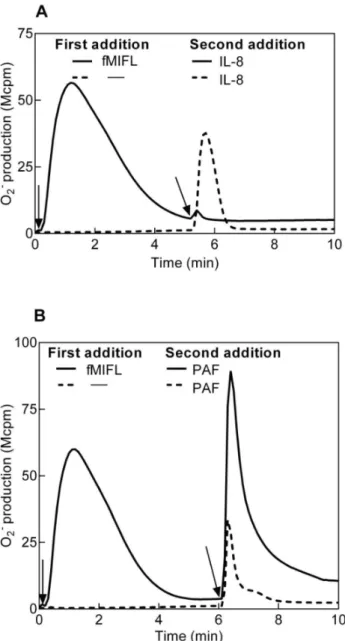
Figure 1. Receptor cross talk between neutrophil FPR1 and PAFR/CXCR1/2 determined as superoxide production.Human neutrophils desensitized with fMIFL were cross-desensitized to IL8 (A) but primed in their response to PAF (B). Neutrophils (105cells, 37
uC) were first activated by the FPR1 specific agonist fMIFL (0.1 nM, added at time indicated by the arrows to the left) leading to receptor desensitization (solid lines in A and B). A second stimulus (A; IL8, 100 ng/ml,B; PAF, 100 nM) was added to the cells (solid lines) at the time point indicated by the arrows to the right. Activation of naı¨ve (non-desensitized) neutrophils by IL8 (A) and PAF (B) was determined in parallel and is shown for comparison (broken lines). A representative experiment is shown, n.5. Abscissa, time of study (min); Ordinate, superoxide production (counts per minute6106; Mcpm).
Isolation of Human Neutrophils
Human peripheral blood neutrophils were isolated from buffy coats from healthy blood donors using dextran sedimentation and Ficoll-Paque gradient centrifugation as described [36]. The remaining erythrocytes were disrupted by hypotonic lysis, the neutrophils were washed twice, resuspended in KRG, and stored on melting ice until use. This isolation procedure permits cells to be purified with minimal granule mobilization.
Neutrophil NADPH-oxidase Activity
The NADPH-oxidase activity was determined using isoluminol-enhanced chemiluminescence (CL) [37,38]. The CL activity was measured in a six-channel Biolumat LB 9505 (Berthold Co., Wildbad, Germany), using disposable 4-ml polypropylene tubes with a 900ml reaction mixture containing 105 cells, isoluminol (261025
M) and HRP (2U). The tubes were equilibrated in the Biolumat for 5 min at 37uC, after which the stimulus (100ml) was added and the light emission was recorded continuously. Receptor desensitized cells are defined as naı¨ve (non-desensitized) cells that had first been stimulated with receptor-specific agonist and returned to baseline after the resulting release of superoxide. These cells were then stimulated a second time. When experiments were performed with antagonists, the antagonists were added to the CL reaction mixture 1 min before the second stimulation. Control cells received no treatment but were incubated at the same basal condition as stimulated cells.
Calcium Mobilization
Neutrophils at a density of 1–36106cells/ml were washed with Ca2+
-free KRG and centrifuged at 2206g. The cell pellets were
resuspended at a density of 26107cells/ml in KRG containing 0.1% BSA, and loaded with 2mM FURA 2-AM for 30 minutes at room temperature. The cells were then diluted to twice the original volume with RPMI 1640 culture medium without phenol red (PAA Laboratories GmbH, Pasching, Austria) and centrifuged. Finally, the cells were washed once with KRG and resuspended in the same buffer at a density of 26107/ml. Calcium measurements were carried out in a Perkin Elmer fluorescence spectrophotom-eter (LC50), with excitation wavelengths of 340 nm and 380 nm, an emission wavelength of 509 nm, and slit widths of 5 nm and 10 nm, respectively. The transient rise in intracellular calcium is presented as the ratio of fluorescence intensities (340 nm: 380 nm) detected. The measuring cuvette contained catalase (2000 U) to counteract inactivation of the chemoattractants by the MPO-H2O2-system [39].
The Cellular Content of F-actin
The F-actin content in neutrophils was analyzed by staining with FITC-phalloidin. The cells were fixed with equal volumes of paraformaldehyde (4% w/v in PBS), permeabilized with Triton X-100 (0.1% W/V in PBS), and incubated with FITC-phalloidin according to the manufacturer’s instructions. The cellular content of F-actin was determined by flow cytometry using an AccuriC6 cytometer (Becton Dickinson, Mountain View, CA, USA).
Results
Receptor Hierarchy between FPRs and the Receptors for PAF (PAFR) and IL8 (CXCR1/2)
Formylated peptides are potent activators of neutrophil granulocytes, binding to 7TMRs of the FPR family [13,14]. Neutrophils exposed to low nM concentrations of the FPR1-specific formylated peptide fMIFL respond by rapid activation of the NADPH-oxidase, resulting in release of superoxide anions (Fig. 1). The fMIFL-induced response is transient and terminates in less than 5 minutes after which the cells become non-responsive to a new challenge with the same agonist (data not shown and [39]). The fMIFL-stimulated cells have thus been transferred to an FPR1 desensitized state (FPR1des). The FPR1 has been shown to communicate with the IL8 receptors CXCR1/2 [40]. According-ly, FPR1 activation led to desensitization not only of FPR1 but also of CXCR1/2; no superoxide release was induced when IL8 was added to FPRdes neutrophils (Fig. 1A). This cross talk was hierarchial (uni-directional) shown by that FPR1 was not desensitized by pre-stimulation of cells with IL8 (data not shown). The FPRdes cells were desensitized also to the lipid chemoat-tractant LTB4(data not shown).
The molecular mechanism behind heterologous receptor de-sensitization between FPR1 and CXCR1/2 has been attributed to hierarchical signaling downstream of the two receptors [28]. Such hierarchical receptor desensitization is however not valid for the PAFR. When IL-8 was replaced by PAF as the trigger of superoxide anion release from FPR1des cells, the cells were fully responsive (Fig. 1B). In fact, the PAF response in the FPR1descells was actually primed; the superoxide response was stronger and more persistent than the PAF response in naı¨ve cells (Fig. 1B, 2B). Similar results were obtained with neutrophils desensitized to another FPR1 agonist (fMLF) or an FPR2 agonist (WKYMVM); also these cells were heterologously desensitized to IL8 but primed when challenged with PAF (data not shown).
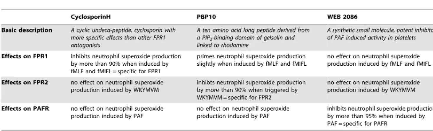
Table 1.Characteristics of the receptor antagonists used.
CyclosporinH PBP10 WEB 2086
Basic description A cyclic undeca-peptide, cyclosporin with more specific effects than other FPR1 antagonists
A ten amino acid long peptide derived from a PIP2-binding domain of gelsolin and linked to rhodamine
A synthetic small molecule, potent inhibitor of PAF induced activity in platelets
Effects on FPR1 inhibits neutrophil superoxide production by more than 90% when induced by fMLF and fMIFL = specific for FPR1
primes neutrophil superoxide production slightly when induced by fMLF and fMIFL
no effect on neutrophil superoxide production induced by fMLF and fMIFL
Effects on FPR2 no effect on neutrophil superoxide production induced by WKYMVM
inhibts neutrophil superoxide production by more than 90% when triggered by WKYMVM = specific for FPR2
no effect on neutrophil superoxide production induced by WKYMVM
Effects on PAFR no effect on neutrophil superoxide production induced by PAF
no effect on neutrophil superoxide production induced by PAF
inhibits neutrophil superoxide production by more than 95% when induced by PAF = specific for PAFR
We conclude that agonist binding to FPRs induced homologous desensitization of the occupied receptor as well as heterologous desensitization of the receptors for IL8 and LTB4. In contrast, agonist binding of FPRs potently primed the response to PAF.
Receptor Specific Antagonists Inhibit the Responses Induced by PAFR and FPR Agonists in Naı¨ve Neutrophils
To ellucidate the molecular basis for the cross talk between FPRs and PAFR described above, we used receptor specific inhibitors (Table 1). As expected, the PAFR antagonist WEB2086 completely and selectively abolished the release of superoxide upon PAF stimulation, demonstrating that PAFR is responsible for the PAF-induced activation of human neutrophils (Fig. S1). It should be noted that PAF is a fairly potent stimulus with an EC50 of<500 nM (for comparison, the fMLF EC50= 20 nM and the fMIFL EC50= 0.2 nM). The FPR1 specific antagonist cyclosporin H abolished the release of superoxide upon fMIFL (or fMLF) stimulation and the FPR2 specific inhibitor PBP10 totally inhibited the superoxide release induced by the FPR2 specific agonist WKYMVM (Fig. S1). At the concentrations used, there were no cross-inhibitory effects of the PAFR antagonist on the fMIFL- or WKYMVM-induced neutrophil responses, and the FPR blockers were without effects on the PAF-induced response.
PAF Triggers a Reactivation of FPR1desin Neutrophils
The antagonist effects were next determined in FPR1des cells activated by PAF. Addition of the PAFR antagonist WEB2086 to FPR1des neutrophils 1 min prior to PAF stimulation resulted, as expected, in a significant inhibition of the PAF response (Fig. 2A & B), showing that the response requires signaling through the PAFR. Unexpectedly, however, the PAF-induced response was largely inhibited also by the FPR1 specific antagonist cyclosporin H, when added 1 min prior to PAF stimulation (Fig. 2A & B). This implies that the PAF-triggered response in FPR1descells involves also activation of FPR1, i.e., there is a cross talk between the two receptors.
We next tested whether the reactivation effect was dependent on agonist occupancy of FPR1. When neutrophils were desensi-tized by 0.1 nM fMIFL at 15uC [41] and then diluted to a final concentration of 1 pM of the peptide, the cells could not be reactivated by PAF (data not shown). In contrast, if such FPRdes cells were diluted without reducing the fMIFL concentration, PAF-induced reactivation was intact (data not shown). This indicates that PAF-induced reactivation of FPR1des neutrophils relies on a continual occupancy of FPR1 by fMIFL present in the surrounding medium. Furthermore, a cross talk signal induced by PAF was evident even when the concentration of fMIFL (used to desensitize FPR1) was as low as 10 pM, a concentration that in it Figure 2. Receptor cross talk from the PAFR induces
reactiva-tion of FPR1des.Human neutrophils (105) were desensitized with the
FPR1 agonist fMIFL (0.1 nM) as described in Figure 1. (A) The FPR1des
neutrophils were activated with PAF (100 nM, added at time indicated by arrow; solid line). The involvment of FPR1 and PAFR in the PAF-induced response was examined by addition of cyclosporin H (1mM, FPR1 antagonist, broken line) or WEB2086 (1mM, PAFR antagonist, dotted line) at 3 min prior to PAF addition. For comparison, the
oxidative response to PAF in naı¨ve neutrophils is shown (inset). A representative experiment is shown, n.5. Abscissa, time of study (min); Ordinate, superoxide production (counts per minute6106; Mcpm). (B) Inhibition of the PAF-induced response in FPR1descells by cyclosporin H
(1mM, FPR1 specific antagonist) or WEB2086 (1mM, PAFR antagonist) shown as mean peak values6SEM of the responses (Mcpm, n = 5 for WEB2086, n = 19 for control, cyclosporine H). The PAF induced response in naı¨ve neutrophils is shown for comparison (n = 19). (C) Human neutrophils (105) were activated/desensitized with different
concentra-tions of the FPR1 agonist fMIFL (added at time indicated by arrow to the left). The neutrophils were then activated with PAF (100 nM final concentration, added at time indicated by arrow to the right). For comparison, a PAF-induced response in naı¨ve neutrophils is shown (solid line). A representative experiment is shown, n.5. Abscissa, time of study (min); Ordinate, superoxide production (counts per min-ute6106; Mcpm).
self is too low to induce any respiratory burst activity in naı¨ve neutrophils (Fig. 2C). Comparing the ‘‘pure’’ PAF response in FPR1descells, i.e., the response measured in the presence of the FPR1 antagonist cyclosporin H, with the PAF-induced response in naı¨ve neutrophils, revealed a substantially lower response in the FPR1descells (Fig. 2A inset and 2B). The EC50value for PAF was, however, the same (around 500 nM) between the naı¨ve and FPR1descells.
The PAF-induced reactivation phenomenon was not exclusive for FPR1 but was seen also for FPR2. The PAF induced response in FPR2descells (desensitized with WKYMVM) was blocked by the FPR2 specific inhibitor PBP10 (Fig. S2), in analogy with the results for FPR1descells. The reactivation of FPR2descells by PAF was FPR2 specific and did not engage FPR1 (cyclosporin H was without any effect; data not shown). Also desensitized C5aR could be reactivated by PAF, even though the response was very low, part of the PAF induced response in C5aRdescells was sensitive to a C5aR antagonist (data not shown).
We next reversed the order in which the stimuli were added. Cells were first stimulated with PAF to generate PAFRdes neutrophils, after which the cells were activated with FPR1 or FPR2 agonists. The PAFRdes cells were fully responsive to both FPR agonists, and both responses were completely inhibited by the specific inhibitors cyclosporin H and PBP10, respectively (Fig. S3 and data not shown). The PAFR antagonist WEB2086 was however completely without effect on the responses triggered by fMIFL or WKYMVM in PAFdes cells (Fig. S3 and data not shown). The receptor cross talk is, thus, highly regulated and restricted to one direction, i.e., reactivation signals are only transmitted from the PAFR to the FPRs and not vice versa.
In addition to PAF, the PAFR recognizes the more stable PAF analogue mcPAF as well as the PAF precursor lysoPAF [42], which were examined for capacity to trigger the cross talk and
reactivation of the FPRdes. The mcPAF and lysoPAF induced a similar receptor cross talk and FPR1desreactivation as PAF; i.e., the neutrophil NADPH-oxidase activity in FPR1descells triggered with mcPAF or lysoPAF was substantially inhibited by the FPR1 antagonist cyclosporin H (Fig. S4).
Taken together, our data clearly reveal a novel form of receptor cross talk from PAFR to FPR, leading to reactivation of desensitized FPRs.
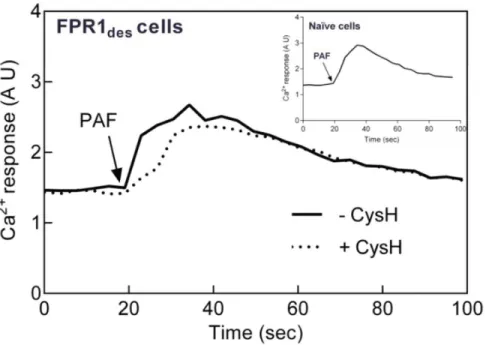
The PAF-induced Rise in Intracellular Ca2+in FPR des
Neutrophils is not Inhibited by Cyclosporin H
When 7TMR agonists bind their receptors, one of the very early signals generated is a rise in the cytosolic concentration of free Ca2+, achieved through emptying of intracellular Ca2+ stores.
Consequently, naı¨ve cells responded by transient increases in Ca2+
to both fMIFL (Fig. S6) and PAF (Fig. 3), effects that were completely blocked by cyclosporine H and WEB2086, respectively (data not shown). A rise in intracellular Ca2+was also induced by
PAF when added to FPRdes cells (Fig. 3). In contrast to the oxidative response, this Ca2+
response was not affected by cyclosporin H (Fig. 3), demonstrating that it is independent of FPR1.
When measuring activation of the NADPH-oxidase, the FPRdes cells were primed to PAF, giving a substantially increased oxidative response as compared to PAF-stimulated naı¨ve cells. With regard to the Ca2+
response induced by PAF in FPRdescells the magnitude was not elevated but rather decreased as compared to the PAF response in naı¨ve neutrophils (Fig. 3 inset).
Taking these data together, we conclude that two signaling pathways are triggered by PAF in FPRdesneutrophils, one FPR-dependent signal that triggers oxidase activation and another, FPR-independent signal that leads to an intracellular Ca2+
increase. Figure 3. Intracellular Ca2+response triggered upon reactivation of FPR1
desby PAF is not cyclosporin H sensitive.FPR1desneutrophils
(desensitized with 0.1 nM fMIFL) loaded with Fura-2 (26106/ml) were activated by PAF (1 nM final concentration) in the absence (solid line) or presence (broken line) of the FPR1 specific antagonist cyclosporin H (1mM added 30 sec before PAF). The changes in fluorescence were followed using dual excitation of Fura-2 at 340 and 380 nm, respectively, with an emission wavelength of 510 nm. For comparison, a PAF-induced intracellular Ca2+
response is shown for naı¨ve neutrophils (inset). A representative experiment is shown, n = 3. Abscissa, time of study (sec); Ordinate, relative change inhelloCa2+
]i(arbitrary units, AU).
Opposite Effects of CalyculinA on Naı¨ve and Desensitized Neutrophils
Phosphatase inhibition has been suggested to reduce binding of ligand-occupied FPRs to the actin cytoskeleton [32], a process known to limit/terminate the response triggered by FPRs [19]. Accordingly, phosphatase inhibitors have earlier been shown to prime cells to FPR1 agonists [32,43]. CalyculinA is a phosphatase inhibitor that selectively inhibits the serine/threonine phospha-tases PP1 and PP2A. We investigated the effect of CalyculinA on the PAF-induced oxidative responses of naı¨ve and FPRdes neutrophils. We first confirmed that CalyculinA primes naive cells to FPR1 stimulation and in addition we found that also the PAF induced response in naı¨ve neutrophils was primed (Fig. 4A).
CalyculinA had no direct effect on the oxidase activity in naı¨ve cells besides priming. We next investigated the effect of CalyculinA on the cross talk between the PAFR and FPR1. We found that CalyculinA blocked the PAF-induced reactivation of FPRdescells (Fig. 4B), suggesting that serine/threonine phosphatases are involved in the PAF-induced cross talk signaling leading to reactivation of FPRdes.
Cytoskeleton-disrupting Agents Trigger a Reactivation of FPRdesthat in Some Respects Resembles that of PAF
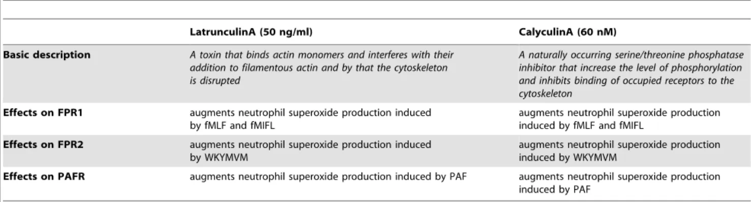
Agonist-binding rapidly transfers FPR to a non-signaling (FPRdes) state and as mentioned above, coupling of ligand-receptor complexes to the actin cytoskeleton has been suggested to play a major role in the termination of signaling and de-sensitization process [17,32]. The two drugs latrunculinA and cytochalasinB both disrupt the actin cytoskeleton in cells by interfering with the polymerization of filamentous (F-)actin during actin remodeling [44]. Accordingly, the presence of latrunculinA or cytochalasinB results in an increased and prolonged response when naı¨ve neutrophils are activated by formylpeptides [45] or PAF (Fig. S5; Table 2).
Similar to the reactivation of FPRdescells by PAF, addition of latrunculinA to these cells induced a pronounced, cyclosporin H-sensitive, reactivation of the NADPH-oxidase, although with a different time course (Fig. 5). LatrunculinA-induced reactivation was induced also in FPR2des cells, and PBP10 abolished this response completely (data not shown). Taken together, our data show that FPRdesreactivation can be achieved not only by PAF, but also by disruption of the actin cytoskeleton.
No direct activation was obtained by latrunculinA or cytocha-lasinB when added alone to naı¨ve neutrophils (data not shown), and no superoxide release was obtained from PAFRdescells upon the addition of the inhibitors (data now shown).
PAF- and latrunculinA-induced Reactivation of FPRs Display Similarities in Signaling
As stated above, the PAF-induced NADPH-oxidase activation in FPR1descells is not associated with a cytosolic Ca2+transient. Similarly, superoxide production induced by reactivation of FPRdes cells by latrunculinA occurred without any rise in intracellular Ca2+
(Fig. S6 inset). The FPRdesreactivation leading to superoxide production is thus not associated with any activation of the PLC/IP3signaling route that leads to an emptying of the intracellular Ca2+
stores.
Also in agreement with the PAF-induced reactivation of FPRdes, the latrunculinA-induced reactivation was inhibited by CalyculinA (Fig. 5). Taken together, these data indicate that similar signaling pathways are operating when FPRdesare reactivated by PAF and by disruption of the cytoskeleton.
PAF-induced Reactivation of FPRdesOccurs Regardless of
Receptor Uncoupling from the Cytoskeleton
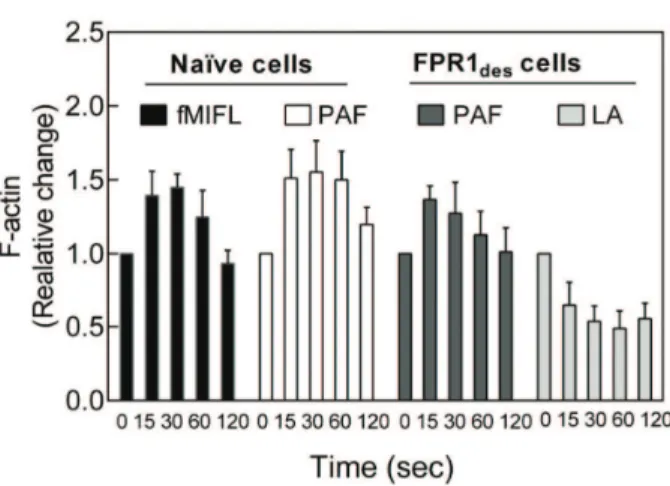
Separation of ligand-receptor complexes from signaling G-proteins through a direct interaction of the occupied receptors with the actin cytoskeleton could form the molecular basis for both receptor desensitization and reactivation (see the model presented in Fig. 6). The similarity between PAF and inhibitors of actin polymerization in reactivation of FPRdes promoted us to in-vestigate the effects of PAF on actin polymerization in FPRdescells. As measured by phalloidin staining, PAF induced a rapid and transient polymerization of actin in both naı¨ve and FPRdes neutrophils, and the levels were of similar magnitude (Fig. 7). The Figure 4. Phosphatase inhibition by CalyculinA has both
inhibitory and priming effects on the neutrophil NADPH-oxidase response.(A) Human neutrophils were incubated without or with CalyculinA (CA; 60 nM) at 37uC for 10 min prior to stimulation with PAF (100 nM) or fMIFL (0.1 nM), and the release of superoxide anions was recorded. The graph shows ratios of superoxide production induced by PAF or fMLF between samples with and without calyculin A (fold increase, mean6SEM; n = 5). (B) FPR1desneutrophils (desensitized
with 0.1 nM fMIFL) were incubated at 37uC for 10 min without (control and inset, solid line) or with CalyculinA (CA, 50 nM; inset, broken line). The cells were then stimulated with PAF (100 nM) or latrunculin A (100 ng/ml final concentration) and the release of superoxide anions was recorded. A representative experiment for PAF stimulation is shown in the inset. The stimulus-induced responses in the CalyculinA treated FPR1desneutrophils are expressed as percent of non-treated controls
reactivation of FPRdesneutrophils by latrunculinA was associated with reduced levels of actin polymerization, as expected (Fig. 7).
The oxidative reactivation response induced by latrunculinA in FPRdes cells declines slowly (Fig. 5) and when the activity has returned to basal level, the cells are refractory to further stimulation/reactivation by another dose of either fMIFL or latrunculinA (Fig. 8, inset, and data not shown). This suggests that the actin cytoskeleton is fully disrupted in the latrunculinA treated FPRdes cells. However, addition of PAF to latrunculinA-treated FPRdescells resulted in a new burst of superoxide, a response that was inhibited by cyclosporin H (Fig. 8). This strongly suggests that the cross talk signals generated by PAF to trigger reactivation of FPRdesis transmitted in an actin-independent manner.
In conclusion, although the reactivation of FPRdes cells by PAF and cytoskeleton-disrupting agents share signaling pathways, the disruption of actin per se is not part of the PAF-induced signaling leading to reactivation.
Discussion
Neutrophils as well as most other cell types express many different 7TMRs and one specific ligand–receptor pair does not generally or necessarily operate alone. On the contrary, co-expressed receptors have the ability to communicate with one another. Such receptor cross talk can involve i) a direct physical interaction between identical or different receptors, ii) receptor phosphorylation that ‘‘spills over’’ from one occupied receptor to another, and iii) cross talk of downstream signaling events [46]. We now describe a novel receptor cross talk mechanism in neutrophils, unique in that the signals generated by one 7TMR transfer another receptor from a desensitized (non-signaling) state back to an actively signaling state. To our knowledge, this is the first description of such a unique cross talk between two GPCRs. Our full understanding of the mechanisms behind the described receptor reactivation, is prohibited by the general lack in basic knowledge regarding termination of signaling from an occupied FPR. Although we have made several attempts to gain knowledge on the molecular mechanisms that underlie the discussed de-sensitization and reactivation phenomena in neutrophils, we can at present only speculate on their composition and function. Much work remains to be done before we can fully understand not only the cross talk at a molecular level but also its biological significance. Possible mechanisms, operating at multiple levels are discussed below and some of the ideas put forward should be regarded as mere speculations.
The FPRs and the PAFR share many features but there is at least one fundamental difference between the desensitized state of these two receptor types; the desensitized FPRs can be reactivated while the PAFR cannot. This suggests that different regulatory mechanisms for desensitization are operating. Reactivation of FPRdes is hardly directly linked to receptor internalization and recycling since reactivation can be achieved following an initial interaction of neutrophils and the FPR ligand at a temperature (15uC) that allows receptor desensitization but is too low to permit receptor internalization.
Currently the foremost accepted model for desensitization of GPCRs highlights the role ofb-arrestin-receptor binding as the basis for termination of signaling. Even though FPRs bind arrestin [15] this mechanism seeems to be of minor importance for the termination of FPR sigaling [16]. Instead we and others have proposed a direct binding of the signaling receptor-ligand complex to the actin cytoskeleton (Fig. 6) as the terminating event. According to this model, the cytoskeleton physically separates the ligand-receptor complex from the signaling G-protein, terminating downstream transduction of signals [18,47]. Experi-Table 2.Characteristics of cytoskeleton interfering drugs used.
LatrunculinA (50 ng/ml) CalyculinA (60 nM)
Basic description A toxin that binds actin monomers and interferes with their addition to filamentous actin and by that the cytoskeleton is disrupted
A naturally occurring serine/threonine phosphatase inhibitor that increase the level of phosphorylation and inhibits binding of occupied receptors to the cytoskeleton
Effects on FPR1 augments neutrophil superoxide production induced by fMLF and fMIFL
augments neutrophil superoxide production induced by fMLF and fMIFL
Effects on FPR2 augments neutrophil superoxide production induced by WKYMVM
augments neutrophil superoxide production induced by WKYMVM
Effects on PAFR augments neutrophil superoxide production induced by PAF augments neutrophil superoxide production induced by PAF
doi:10.1371/journal.pone.0060169.t002
Figure 5. The cytoskeleton disrupting agent latruculin A induces reactivation of FPR1des. Latrunculin A (100 ng/ml) was
added to FPR1desneutrophils (105cells; desensitized with 0.1 nM fMIFL)
in the absence (solid line) or presence (dotted line) of cyclosporin H (1mM, FPR1 specific antagonist, added 1 min before latrunculin A) and the release of superoxide anions was determined. For comparison, a PAF-induced reactivation of FPR1desneutrophils is included (dashed
line). A representative experiment is shown, n.5. Abscissa, time of
study (min); Ordinate, superoxide production (counts per minute6106; Mcpm).
mental support for this mechanism is based on pharmacological inhibition of actin polymerization which prolongs signaling from occupied FPRs, and our data on receptor reactivation induced by latrunculinA also fits this model like a glove. There must, however, be mechanism(s) apart from actin dynamics that terminate the signaling since, i) signaling from neutrophil GPCRs (including both FPRs and PAFR) is terminated also when the cytoskeleton is disrupted by inhibitors of actin polymerization (i.e., latrunculinA and cytochalasinB), and ii) the desensitized PAFR is not reactivated when the cytoskeleton is disrupted.
With regard to involvement of cytoskeleton uncoupling as basis for the PAF-induced reactivation of FPRdescells discussed in this study, this is an attractive hypothesis as there are valid similarities between the reactivation responses induced by latrunculinA and
PAF (e.g., both responses are inhibited by the phosphatase inhibitor CalyculinA), However, PAF reactivated FPRdes also when the actin cytoskeleton had been disrupted, and our data showing no net reduction of polymerized actin during PAF-induced FPR1des reactivation are also in opposition to such a model.
We show that FPR/PAFR activation as well as FPRdes reactivation depend on cellular phosphorylation levels. CalyculinA primed the direct activation of the FPRs in naı¨ve cells while reactivation induced by PAF in FPRdes cells was inhibited. Previous studies in naı¨ve neutrophils have shown that FPR1, as well as many other proteins, are phosphorylated upon agonist binding. This phosphorylation is thought critical for receptor internalization and desensitization, as well as forb-arrestin binding Figure 6. Model for FPR activation, desensitization and reactivation. A) The agonist-occupied FPR activates a G-protein and the second messengers generated activate the electron-transporting NADPH-oxidase that reduces oxygen to superopxide anion. The signaling state of the receptor is fairly short lived.B) The agonist-occupied receptor is desensitized and the functional response is terminated. This non-signaling state is hypothetically achieved through a physical separation of the receptor-ligand complex from the G-protein, made possible by binding of actin polymers and/or arrestin molecules to the receptor.C) The desensitized FPR is reactivated by signals generated when PAF binds to its neutrophil receptor (arrow, 1). Reactivation of the desensitized FPR is achieved also with cytoskeletal inhibitors, (shorter filaments, 2), suggesting a mechanism for reactivation that involves uncoupling of the receptor-ligand complex from the cytoskeleton. The described cross talk is hierarchial and unidirectional.
[10,48,49,50]. We have earlier suggested that the priming effect induced in naı¨ve neutrophils by phosphatase inhibition is due to decreased binding of occupied receptors to the cytoskeleton [32]. It is however hard to fully fit the results on both naı¨ve cells and FPRdesneutrophils into this model. Clearly, there might be several other basic mechanisms behind the phenomena described and at present we cannot distinguish whetherthe phosphorylation level affects one or the other of the two receptors involved, some of the unknown downstream signaling molecules, and/or the direct assembly and function of the NADPH-oxidase. Inhibition of phosphatases will lead to an increased level of phosphorylation irrespectively if the receptors trigger activation of CalyculinA sensitive phosphatases or not, and we know virtually nothing about the identity of the protein(s) that prime naı¨ve cells and inhibits desensitized cells.
The protein b-arrestin, initially identified as a mediator for GPCR desensitization and internalization, has not been studied in primary neutrophils. Recent research using other cell types has, however, drawn much attention to the very complex relationship between receptor binding of b-arrestin and downstream phos-phorylation reactions and receptor as well as to its roles in signaling achieved by scaffolding of signaling proteins following receptor recruitment [51]. It is of particular interest that b -arrestins bind a number of actin assembly proteins and thus may play a requisite role in reorganization of the actin cytoskeleton [52]. The precise mechanisms by which this regulation of actin reorganization is achieved, and the role this has as a regulatory pathway in neutrophils is not known. In our attempt to understand the signalings involved in FPRdesreactivation, we show that this Figure 7. PAF induces actin polymerization in both naı¨ve and
FPR1des neutrophils.Human neutrophils (naı¨ve or FPR1des) were
activated with a receptor agonist or latrunculin A and the change in polymerized actin was determined att different time points (15 to 120 sec) after activation. Naı¨ve neutrophils were activated by PAF (100 nM) or fMLF (0.1 nM) and FPR1desneutrophils were reactivated by
PAF (100 nM) or latrunculin A (200 ng/ml). The stimulation at indicated time points was terminated by adding ice cold paraformyldehyde (final concentration 2%) to the cells. The amount of polymerized actin was determined by flow cytometry after phalloidin staining and compared to the amount of actin at time zero before activation. The values are shown as mean ratio6SEM; n = 3.
doi:10.1371/journal.pone.0060169.g007
Figure 8. PAF activates FPR1desneutrophils also in the presence of latrunculinA.Human FPR1desneutrophils were incubated in the
absence or presence of latunculinA (LA, 50 ng/ml) and after return of the NADPH-oxidase activity to background levels (after around 20 min; not shown in the figure) the cells were activated with PAF (100 nM) and the measurement of oxidase activity was started. In some experiments, cyclosporinH (CA, 1mM) was added to the cells just prior to PAF. The response induced was sensitive to this FPR1 specific antagonist. The results are expressed as peak response (Mcpm, open bars) and total production (area under curve; AUC, filled bars) in percent of control (PAF-induced peak response in FPRdes in the absence of LA and CA; mean6SEM, n = 3). The FPR1desneutrophils treated with latrunculin A (50 ng/ml) could not be
reactivated by additional latrunculin A (100 ng/ml, inset, dotted line). For comparison, reactivation of control cells (FPR1desneutrophils without
process does not trigger a Ca2+
response, a feature necessary to the signaling pathways of most GPCRs. In relation to this it is interesting to note that many of the scaffold functions ofb-arrestin occurs without any involvement of classical signaling G-proteins. Whetherb-arrestins plays a role in FPR desensitization remains to be determined, together with the possible impact of multiple signalingb-arrestin scaffolds in FPRdes. The fact that the signaling route ultimately leading to reactivation of FPRdes bypasses the Ca2+
pathway will in the future direct our attention to cell models that express the two cross talking receptors in conjunction with a Ca2+
independent read-out system triggered by the reactivated receptor.
In summary, the data presented in this study provide evidence that PAF can modulate neutrophil functions, either directly or through a receptor cross talk with other receptors, and by this promote the neutrophil activation. These findings not only point to the possibility that PAF-mediated pathology may involve cross talk with other receptors that are reactivated by PAF stimulation, but also demonstrate that unique signaling pathways are utilized downstream of the PAFR, leading to priming and agonist-driven receptor reactivation. Clearly, more experiments are needed in the future in order to validate our hypothesis regarding the direct role of actin-depedent versusb-arrestin-mediated desensitization path-ways. Also the involvement of b-arrestin scaffold-mediated signaling, and of so far unidentified signaling pathway(s) that may be linked in one way or another to the cell cytoskeleton, requires further study. Our data showing that FPRdes can be reactivated by PAF also when the actin cytoskeleton has been disrupted, strongly support the concept that FPR can be desensitized through an actin-independent pathway.
Supporting Information
Figure S1 Characterization of receptor specific antago-nists for FPRs and PAFR in naı¨ve neutrophils. Naı¨ve neutrophils (105cells) were incubated in the absence (solid lines) or presence (broken lines) of antagonist (WEB2086, 1mM, a PAFR specific antagonist; cyclosporin H, 1mM an FPR1 specific antagonist; PBP10, 1mM an FPR2 specific antagonist) for 5 min at 37uC and were then activated with PAF (100 nM, upper panel), fMIFL (0.1 nM, middle panel), or WKYMVM (100 nM, lower panel). A representative experiment is shown, n.5. Abscissa, time of study (min); ordinate, superoxide production (counts per minute6106, Mcpm).
(TIF)
Figure S2 A PAFR-initiated cross talk induces reactiva-tion of FPR2 in desensitized neutrophils.Human neutro-phils (105) were desensitized with the FPR2 agonist WKYMVM (100 nM final concentration) and subsequently activated with PAF (100 nM final concentration, added at arrow). The involvment of FPR2 in the resulting PAF-induced superoxide production was examined by addition of the FPR2 antagonist PBP10 (1mM, dotted line) 1 min before the addition of PAF. For comparison, a PAF-induced response in naı¨ve neutrophils is shown (inset). Representative experiments are shown, n.5. Abscissa, time of study (min); Ordinate, superoxide production (counts per min-ute6106, Mcpm).
(TIF)
Figure S3 No reactivation is induced by fMIFL in PAFRdes neutrophils. Human neutrophils (105) were desensi-tized with PAF (100 nM final concentration). The desensidesensi-tized
neutrophils were activated with fMIFL (0.1 nM final concentra-tion, added arrow; solid line). The involvement of FPR1 and PAFR in fMIFL-induced superoxide production was examined by addition of cyclosporin H (1mM, FPR1 antagonist, dotted line) or WEB2086 (1mM, PAFR antagonist, broken line) 1 min before addition of fMIFL. For comparison, a fMIFL-induced response in naı¨ve neutrophils is shown (inset). A representative experiment is shown, n.5. Abscissa, time of study (min); Ordinate, superoxide production (counts per minute6106, Mcpm).
(TIF)
Figure S4 The PAF precursor lysoPAF and the stable analogue mcPAF both reactivate FPR1des neutrophils. Human neutrophils (105) were desensitized with the FPR1 agonist fMIFL (0.1 nM final concentration). The desensitized neutrophils were activated with lysoPAF (A; 1mM final concentration added at arrow; solid line) or mcPAF (B; 1mM final concentration added at arrow; solid line). The involvement of FPR1 in the responses was examined by the addition of cyclosporin H (1mM, FPR1 antagonist, broken lines) 1 min before addition of the agonist. For comparison, a lyso PAF- (A, inset) or mcPAF- (B, inset) induced response in naı¨ve neutrophils is shown. The figures show representative experiments, n.5. Abscissa, time of study (min); Ordinate, superoxide production (counts per minute6106, Mcpm). (TIF)
Figure S5 The PAF-induced neutrophil response is primed by inhibitors of actin polymerization. Naı¨ve human neutrophils were incubated at 37uC for 5 min with either Cytochalasin B (Cyt B, 5mg/ml; grey bars) or latrunculin A (LA, 50 ng/ml; white bars). Control cells were incubated at the same conditions but in the absence of actin polymerization inhibitor. The cells were then activated with PAF (100 nM) and the release of superoxide was recorded continuously. Data are expressed as fold increase of peak values in treated cells as compared to non-treated controls (mean6 SEM; n = 3). The dashed line denotes the value expected in the absence of effect.
(TIF)
Figure S6 Latrunculin A induces no increase in in-tracellular Ca2+
in FPR1desneutrophils.Intracellular Ca2+ canges was determined in Fura-2 loaded naı¨ve and FPR1des (0.1 nM fMIFL) neutrophils. Naı¨ve neutrophils were activated by fMIFL (1 nM; solid line), and FPR1des neutrophils were reactivated by latrunculin A (100 ng/ml; inset). The changes in fluorescence were followed using dual excitation at 340 nm and 380 nm, and an emission wavelength of 510 nm. Representative experiments are shown. Abscissa, time of study (min); Ordinate, relative change in [Ca2+
]i. (TIF)
Acknowledgments
The suggestions given by the other members of the Phagocyte Research Group at the Department of Rheumatology and Inflammation research have been very helpful and the whole group is acknowledged.
Author Contributions
References
1. Luttrell LM (2006) Transmembrane signaling by G protein-coupled receptors. Methods in molecular biology 332: 3–49.
2. Magalhaes AC, Dunn H, Ferguson SS (2011) Regulation of G Protein-Coupled Receptor Activity, Trafficking and Localization by Gpcr-Interacting Proteins. British journal of pharmacology.
3. Giniatullin R, Nistri A, Yakel JL (2005) Desensitization of nicotinic ACh receptors: shaping cholinergic signaling. Trends in neurosciences 28: 371–378. 4. Hendriks-Balk MC, Peters SL, Michel MC, Alewijnse AE (2008) Regulation of G protein-coupled receptor signalling: focus on the cardiovascular system and regulator of G protein signalling proteins. European journal of pharmacology 585: 278–291.
5. Kendall RT, Luttrell LM (2009) Diversity in arrestin function. Cellular and molecular life sciences : CMLS 66: 2953–2973.
6. Vroon A, Heijnen CJ, Kavelaars A (2006) GRKs and arrestins: regulators of migration and inflammation. Journal of leukocyte biology 80: 1214–1221. 7. Kenakin T (2010) G protein coupled receptors as allosteric proteins and the role
of allosteric modulators. Journal of receptor and signal transduction research 30: 313–321.
8. Rajagopal S, Rajagopal K, Lefkowitz RJ (2010) Teaching old receptors new tricks: biasing seven-transmembrane receptors. Nature reviews Drug discovery 9: 373–386.
9. Mundell SJ, Luo J, Benovic JL, Conley PB, Poole AW (2006) Distinct clathrin-coated pits sort different G protein-coupled receptor cargo. Traffic 7: 1420– 1431.
10. Potter RM, Maestas DC, Cimino DF, Prossnitz ER (2006) Regulation of N-formyl peptide receptor signaling and trafficking by individual carboxyl-terminal serine and threonine residues. Journal of immunology 176: 5418–5425. 11. Peeters MC, van Westen GJ, Li Q, AP IJ (2011) Importance of the extracellular
loops in G protein-coupled receptors for ligand recognition and receptor activation. Trends in pharmacological sciences 32: 35–42.
12. Urwyler S (2011) Allosteric modulation of family C G-protein-coupled receptors: from molecular insights to therapeutic perspectives. Pharmacological reviews 63: 59–126.
13. Fu H, Karlsson J, Bylund J, Movitz C, Karlsson A, et al. (2006) Ligand recognition and activation of formyl peptide receptors in neutrophils. J Leukoc Biol 79: 247–256.
14. Ye RD, Boulay F, Wang JM, Dahlgren C, Gerard C, et al. (2009) International Union of Basic and Clinical Pharmacology. LXXIII. Nomenclature for the formyl peptide receptor (FPR) family. Pharmacol Rev 61: 119–161. 15. Forsman H, Onnheim K, Andreasson E, Dahlgren C (2011) WHAT FORMYL
PEPTIDE RECEPTORS, IF ANY, ARE TRIGGERED BY COMPOUND 43 AND LIPOXIN A(4) ? Scand J Immunol.
16. Huet E, Boulay F, Barral S, Rabiet MJ (2007) The role of beta-arrestins in the formyl peptide receptor-like 1 internalization and signaling. Cellular signalling 19: 1939–1948.
17. Bylund J, Bjorstad A, Granfeldt D, Karlsson A, Woschnagg C, et al. (2003) Reactivation of formyl peptide receptors triggers the neutrophil NADPH-oxidase but not a transient rise in intracellular calcium. J Biol Chem 278: 30578– 30586.
18. Klotz KN, Jesaitis AJ (1994) Neutrophil chemoattractant receptors and the membrane skeleton. BioEssays : news and reviews in molecular, cellular and developmental biology 16: 193–198.
19. Omann GM, Swann WN, Oades ZG, Parkos CA, Jesaitis AJ, et al. (1987) N-formylpeptide-receptor dynamics, cytoskeletal activation, and intracellular calcium response in human neutrophil cytoplasts. Journal of immunology 139: 3447–3455.
20. Shenoy SK, Lefkowitz RJ (2011) beta-arrestin-mediated receptor trafficking and signal transduction. Trends in pharmacological sciences.
21. Bylund J, MacDonald KL, Brown KL, Mydel P, Collins LV, et al. (2007) Enhanced inflammatory responses of chronic granulomatous disease leukocytes involve ROS-independent activation of NF-kappa B. European journal of immunology 37: 1087–1096.
22. Segal BH, Grimm MJ, Khan AN, Han W, Blackwell TS (2012) Regulation of innate immunity by NADPH oxidase. Free radical biology & medicine 53: 72– 80.
23. Segal BH, Veys P, Malech H, Cowan MJ (2011) Chronic granulomatous disease: lessons from a rare disorder. Biology of blood and marrow transplantation : journal of the American Society for Blood and Marrow Transplantation 17: S123–131.
24. McDonald B, Pittman K, Menezes GB, Hirota SA, Slaba I, et al. (2010) Intravascular danger signals guide neutrophils to sites of sterile inflammation. Science 330: 362–366.
25. Powell WS, Rokach J (2005) Biochemistry, biology and chemistry of the 5-lipoxygenase product 5-oxo-ETE. Progress in lipid research 44: 154–183. 26. Stephens L, Milne L, Hawkins P (2008) Moving towards a better understanding
of chemotaxis. Current biology : CB 18: R485–494.
27. Wymann MP, Sozzani S, Altruda F, Mantovani A, Hirsch E (2000) Lipids on the move: phosphoinositide 3-kinases in leukocyte function. Immunology today 21: 260–264.
28. Heit B, Tavener S, Raharjo E, Kubes P (2002) An intracellular signaling hierarchy determines direction of migration in opposing chemotactic gradients. The Journal of cell biology 159: 91–102.
29. Fu H, Bylund J, Karlsson A, Pellme S, Dahlgren C (2004) The mechanism for activation of the neutrophil NADPH-oxidase by the peptides formyl-Met-Leu-Phe and Trp-Lys-Tyr-Met-Val-Met differs from that for interleukin-8. Immunology 112: 201–210.
30. Ali H, Richardson RM, Haribabu B, Snyderman R (1999) Chemoattractant receptor cross-desensitization. The Journal of biological chemistry 274: 6027– 6030.
31. Didsbury JR, Uhing RJ, Tomhave E, Gerard C, Gerard N, et al. (1991) Receptor class desensitization of leukocyte chemoattractant receptors. Proceed-ings of the National Academy of Sciences of the United States of America 88: 11564–11568.
32. Harbecke O, Liu L, Karlsson A, Dahlgren C (1997) Desensitization of the fMLP-induced NADPH-oxidase response in human neutrophils is lacking in okadaic acid-treated cells. Journal of leukocyte biology 61: 753–758. 33. Liu L, Harbecke O, Elwing H, Follin P, Karlsson A, et al. (1998) Desensitization
of formyl peptide receptors is abolished in calcium ionophore-primed neutrophils: an association of the ligand-receptor complex to the cytoskeleton is not required for a rapid termination of the NADPH-oxidase response. J Immunol 160: 2463–2468.
34. Tomhave ED, Richardson RM, Didsbury JR, Menard L, Snyderman R, et al. (1994) Cross-desensitization of receptors for peptide chemoattractants. Charac-terization of a new form of leukocyte regulation. Journal of immunology 153: 3267–3275.
35. Richardson RM, Haribabu B, Ali H, Snyderman R (1996) Cross-desensitization among receptors for platelet activating factor and peptide chemoattractants. Evidence for independent regulatory pathways. The Journal of biological chemistry 271: 28717–28724.
36. Boyum A, Lovhaug D, Tresland L, Nordlie EM (1991) Separation of leucocytes: improved cell purity by fine adjustments of gradient medium density and osmolality. Scandinavian journal of immunology 34: 697–712.
37. Dahlgren C, Karlsson A, Bylund J (2007) Measurement of respiratory burst products generated by professional phagocytes. Methods Mol Biol 412: 349–363. 38. Lundqvist H, Dahlgren C (1996) Isoluminol-enhanced chemiluminescence: a sensitive method to study the release of superoxide anion from human neutrophils. Free radical biology & medicine 20: 785–792.
39. Karlsson J, Bylund J, Movitz C, Bjorkman L, Forsman H, et al. (2010) A methodological approach to studies of desensitization of the formyl peptide receptor: Role of the read out system, reactive oxygen species and the specific agonist used to trigger neutrophils. J Immunol Methods 352: 45–53. 40. Jones SA, Wolf M, Qin S, Mackay CR, Baggiolini M (1996) Different functions
for the interleukin 8 receptors (IL-8R) of human neutrophil leukocytes: NADPH oxidase and phospholipase D are activated through IL-8R1 but not IL-8R2. Proceedings of the National Academy of Sciences of the United States of America 93: 6682–6686.
41. Lundqvist H, Gustafsson M, Johansson A, Sarndahl E, Dahlgren C (1994) Neutrophil control of formylmethionyl-leucyl-phenylalanine induced mobiliza-tion of secretory vesicles and NADPH-oxidase activamobiliza-tion: effect of an associamobiliza-tion of the ligand-receptor complex to the cytoskeleton. Biochimica et biophysica acta 1224: 43–50.
42. Welch EJ, Naikawadi RP, Li Z, Lin P, Ishii S, et al. (2009) Opposing effects of platelet-activating factor and lyso-platelet-activating factor on neutrophil and platelet activation. Molecular pharmacology 75: 227–234.
43. Garcia RC, Whitaker M, Heyworth PG, Segal AW (1992) Okadaic acid produces changes in phosphorylation and translocation of proteins and in intracellular calcium in human neutrophils. Relationship with the activation of the NADPH oxidase by different stimuli. The Biochemical journal 286 (Pt 3): 687–692.
44. Yarmola EG, Somasundaram T, Boring TA, Spector I, Bubb MR (2000) Actin-latrunculin A structure and function. Differential modulation of actin-binding protein function by latrunculin A. The Journal of biological chemistry 275: 28120–28127.
45. Jesaitis AJ, Tolley JO, Allen RA (1986) Receptor-cytoskeleton interactions and membrane traffic may regulate chemoattractant-induced superoxide production in human granulocytes. The Journal of biological chemistry 261: 13662–13669. 46. Vazquez-Prado J, Casas-Gonzalez P, Garcia-Sainz JA (2003) G protein-coupled receptor cross-talk: pivotal roles of protein phosphorylation and protein-protein interactions. Cellular signalling 15: 549–557.
47. Jesaitis AJ, Klotz KN (1993) Cytoskeletal regulation of chemotactic receptors: molecular complexation of N-formyl peptide receptors with G proteins and actin. European journal of haematology 51: 288–293.
48. Potter RM, Maestas DC, Cimino DF, Prossnitz ER (2006) Regulation of N-formyl peptide receptor signaling and trafficking by individual carboxyl-terminal serine and threonine residues. J Immunol 176: 5418–5425.
50. Tardif M, Mery L, Brouchon L, Boulay F (1993) Agonist-dependent phosphorylation of N-formylpeptide and activation peptide from the fifth component of C (C5a) chemoattractant receptors in differentiated HL60 cells. Journal of immunology 150: 3534–3545.
51. DeFea KA (2011) Beta-arrestins as regulators of signal termination and transduction: how do they determine what to scaffold? Cellular signalling 23: 621–629.