O R I G I N A L P A P E R
A new CuZ active form in the catalytic reduction of N
2O
by nitrous oxide reductase from
Pseudomonas nautica
Simone Dell’Acqua•Sofia R. Pauleta •
Patrı´cia M. Paes de Sousa•Enrico Monzani•
Luigi Casella• Jose´ J. G. Moura•Isabel Moura
Received: 19 January 2010 / Accepted: 4 April 2010 / Published online: 27 April 2010 ÓSBIC 2010
Abstract The final step of bacterial denitrification, the two-electron reduction of N2O to N2, is catalyzed by a multi-copper enzyme named nitrous oxide reductase. The catalytic centre of this enzyme is a tetranuclear copper site called CuZ, unique in biological systems. The in vitro recon-struction of the activity requires a slow activation in the presence of the artificial electron donor, reduced methyl viologen, necessary to reduce CuZ from the resting non-active state (1CuII/3CuI) to the fully reduced state (4CuI), in contrast to the turnover cycle, which is very fast. In the present work, the direct reaction of the activated form of Pseudomonas nauticanitrous oxide reductase with stoichi-ometric amounts of N2O allowed the identification of a new reactive intermediate of the catalytic centre, CuZ°, in the
turnover cycle, characterized by an intense absorption band at 680 nm. Moreover, the first mediated electrochemical study ofPs. nauticanitrous oxide reductase with its physi-ological electron donor, cytochromec-552, was performed. The intermolecular electron transfer was analysed by cyclic
voltammetry, under catalytic conditions, and a second-order rate constant of (5.5±0.9)9105M-1s-1 was
deter-mined. Both the reaction of stoichiometric amounts of substrate and the electrochemical studies show that the active CuZ°species, generated in the absence of reductants,
can rearrange to the resting non-active CuZ state. In this light, new aspects of the catalytic and activation/inactivation mechanism of the enzyme are discussed.
Keywords Nitrous oxide reductaseCatalytic mechanism DenitrificationBioelectrochemistry
Introduction
The multi-copper enzyme nitrous oxide reductase (N2OR) catalyzes the final step of bacterial dissimilatory denitrifica-tion (2NO3-?2NO2-?2NO?N2O?N2), namely
the two-electron reduction of the kinetically inert molecule nitrous oxide (N2O) to dinitrogen (N2) and water [1,2].
Recently, the structure of N2OR from Pseudomonas nautica [3], Paracoccus denitrificans [4] and Achromo-bacter cycloclastes [5] was solved. The crystal structure revealed that N2OR is a functional homodimer containing two different multi-copper centres per subunit, called CuA and CuZ.
The binuclear copper centre, CuA, is an electron transfer centre similar to the CuA found in cytochrome oxidases and its properties have been extensively studied [6–9]. CuZ is a novel mixed-valence copper centre (Cu4S) with a sulfide ion bridging a distorted tetrahedron of copper atoms, a unique structural feature in biology. This cluster is coordinated by seven histidines, and a water-derived ligand is proposed to bridge the two copper ions (CuI and CuIV), where the substrate binds to the enzyme.
Electronic supplementary material The online version of this article (doi:10.1007/s00775-010-0658-6) contains supplementary material, which is available to authorized users.
S. Dell’AcquaS. R. PauletaP. M. Paes de Sousa J. J. G. MouraI. Moura (&)
REQUIMTE/CQFB, Departamento de Quı´mica,
Faculdade de Cieˆncias e Tecnologia, Universidade Nova de Lisboa, 2829-516 Caparica, Portugal e-mail: [email protected]
S. Dell’AcquaE. MonzaniL. Casella Dipartimento di Chimica Generale, Universita` di Pavia,
Via Taramelli 12, 27100 Pavia, Italy
The CuZ from Ps. nautica N2OR, in the as-isolated state, contains a 1CuII/3CuIredox state with a total spin of , where the unpaired electron is delocalized between two or more copper atoms through the bridging sulfide ion [10–12]. This state, which has a typical electronic spectral band at 640 nm and a four-line splitting EPR signature [13,14], cannot be easily reduced or oxidized. The cata-lytically active, fully reduced form (4CuI) can be obtained only after a prolonged incubation with reduced methyl viologen (MV) [15,16].
In contrast, the catalytic centre, CuZ, ofPa. denitrifi-cansN2OR has been identified in two forms, named CuZ and CuZ*, with the relative proportion between them depending on the purification conditions [17]. In particular, the CuZ* form, characterized by an intense absorption band at 650 nm, is more abundant in the ‘aerobic’ prepa-ration, whereas the CuZ form, with an absorption band at 670 nm, is predominant in the ‘anaerobic’ preparation. CuZ* is redox-inert, whereas the CuZ midpoint potential was calculated to be E°0=60 mV (vs. the standard
hydrogen electrode). Despite this feature, the ‘aerobic’ preparation shows a slightly higher steady-state activity compared with the ‘anaerobic’ preparation. In addition, the CuZ form has an EPR spectrum similar to that of the CuZ* form, suggesting a 1CuII/3CuIredox state for both species [17].
Recently, the catalytic parameters ofPs. nauticaN2OR activity were determined to compare the physiological (Ps. nauticacytochromec-552) and artificial (MV) electron donors [18]. This study revealed that these two electron donors have a distinct mechanism of interaction towards the enzyme, whereas when cytochromec-552 was used as an electron donor, kcat=3.8 s
-1
, Km=50.2lM and pKa=8.3 were estimated, and when an artificial electron donor, MV, was used,kcat =320 s
-1
,Km=11.5lM and pKa=6.6 were obtained.
We present here the first mediated electrochemical study of N2OR with its physiological electron donor, cytochrome c-552. The electrochemical behaviour of this cytochrome has been well characterized [19], and it has already been shown to be the electron donor of other enzymes isolated fromPs. nautica, cytochromecperoxidase [20] and cyto-chrome cd1 nitrite reductase [21]. In the latter case the electron transfer reaction was also investigated using electrochemical techniques.
To understand the complex mechanism of activation and catalysis of N2OR CuZ, we have also studied the properties of the enzyme in the activated form by direct reaction of N2OR with stoichiometric amounts of substrate in the absence of reductants. The identification of a new active intermediate of the catalytic centre, CuZ°, led us to revise
both the catalytic and the activation mechanism of this challenging enzyme.
Materials and methods
Protein purification
N2OR was purified from Ps. nautica617 (also known as Marinobacter hydrocarbonoclasticus 617) cellular extract as previously described [13], with some minor modifica-tions. Enzyme concentrations were determined by the Lowry method [22].
Ps. nauticacytochromec-552 was purified as previously described [23], with some minor modifications. The con-centration of cytochrome c-552 was determined spectro-photometrically using the extinction coefficient at 552 nm,
e=19.3 mM-1cm-1[24], for the fully reduced form.
Enzyme activation and activity assay
Enzyme activation was performed in a glove box as pre-viously described [18]. The activity assay used was the one described in [18], using MV as the electron donor. Electrochemical methods
Voltammetric measurements were performed using an AUTOLAB/PSTAT 12 potentiostat/galvanostat from ECO Chemie (Utrecht, The Netherlands). Data were analysed with the GPES software package from ECO Chemie. A conventional three-electrode-configuration cell was used, with a platinum auxiliary electrode and a saturated calomel reference electrode [?244 mV vs. the standard hydrogen
electrode (SHE)]. The working electrode was a pyrolytic graphite electrode, with a diameter of 3 mm (surface area 0.07 cm2), which was used in a membrane configu-ration [19]. Throughout the paper, all potentials are refer-red to the SHE.
Before each experiment, the pyrolytic graphite electrode was polished by hand on a polishing cloth (Buehler 40-7212) using a water–alumina slurry (0.05lm, Buehler 40-6365-006), sonicated for 5 min and rinsed carefully with Milli-Q water. The membrane electrode was prepared by dropping a 5-ll drop, containing 2.5lM preactivated N2OR and 50lM oxidized cytochromec-552, on a small square (10 mm910 mm) of negatively charged Spectra/
Por MWCO 3500 dialysis membrane. The membrane was fitted tightly to the electrode with a rubber O-ring.
In typical experiments, the working solution contained 0.1 M phosphate buffer pH 7.0, the scan rate was 40 mV s-1, and cyclic voltammograms were obtained in the range from?0.6 to-0.25 V versus the SHE.
125, 330 and 1,000lM N2O were added as N2O-saturated water. In the pH-dependence studies of the catalytic cur-rent, the following buffers were used: 0.1 M potassium phosphate buffer at pH 5.9, 6.2, 6.4, 6.7 and 7.0, or 0.1 M tris(hydroxymethyl)aminomethane (Tris)–HCl at pH 7.3, 7.6, 8.0, 8.4 and 8.8. All the electrochemical experiments were performed inside a glove box filled with an argon-saturated atmosphere.
Direct reaction of N2OR with substrate
For the direct reaction of the enzyme with substrate, an equimolar amount of N2O was added to 35lM N2OR preactivated in 0.1 M Tris–HCl at pH 7.6. The reaction was followed using a TIDAS diode-array spectrophotometer. At corresponding incubation times, an aliquot of this solution was taken to determine the enzyme activity, using MV reduced with sodium dithionite as an electron donor, as previously described [18].
EPR spectroscopy
X-band EPR spectra were recorded using a Bruker EMX spectrometer equipped with a rectangular cavity (model ER4102ST) and an Oxford Instruments continuous-flow cryostat. EPR spectra were simulated using the program WINEPR Simfonia version 1.2 from Bruker Instruments.
The sample of the enzyme form characterized by the 680-nm absorption band was prepared under anaerobic conditions (glove box) by mixing 75lM preactivated N2OR and an equimolar amount of N2O, in 0.1 M Tris– HCl. The EPR tube was frozen with liquid nitrogen 1 min after the addition of N2O. The resting CuZ form, with absorption at 640 nm, was obtained by exposing the pre-vious sample to air for 30 min. The experimental condi-tions are described in the legend for Fig.6.
Redox titration
Preactivated N2OR (35lM) in 0.1 M Tris–HCl pH 7.6 was titrated in a 1-ml cuvette. The potential was measured with a platinum–silver/silver chloride combined electrode (Crison). The oxidation experiments were performed with addition of potassium ferricyanide (E= ?420 mV vs. SHE) and the reaction was followed by the UV–vis spectra. The reduction experiments were performed with addition of sodium dithionite or titanium(III) citrate and the reaction was also followed by the UV–vis spectra. The following mediators were added, each at 2lM concentration: 1,2-naphthoquinone-4-sulfonic acid, 1,2-naphthoquinone, phenazine methosulfate, resorufin (2,8-dihydroxyphenox-azine), indigodisulfonate, 2-hydroxy-1,4-naphthoquinone and MV. The titration was performed in a glove box.
Results and discussion
Electrocatalytic activity of Ps. nauticaN2OR with cytochromec-552 as an electron donor
The catalytic activity of Ps. nautica N2OR for N2O reduction was analysed for the first time by mediated electrochemistry using Ps. nautica cytochrome c-552, a small electron transfer protein that was established to be the physiological redox partner of this enzyme [18]. For this purpose, both redox partners were entrapped between the electrode surface and a dialysis membrane [25] and cyclic voltammetry was performed.
In the absence of cytochrome c-552, preactivated or non-pre-activated N2OR does not exhibit any electro-chemical signal, probably owing to the non-compatibility of the negatively charged graphite electrode surface and the negatively charged surface of Ps. nautica N2OR (pI=5.4). Moreover, no catalytic current was observed in these conditions upon addition of nitrous oxide to preac-tivated N2OR within the potential range used.
In previous reports, cytochrome c-552 was shown to exhibit a well-defined direct electrochemical signal at a car-bon membrane electrode [19]. In our experimental conditions, this reversible electrochemical behaviour was also verified and the calculated redox potential, E°0= ?(245±5) mV
versus the SHE at pH 7, is in agreement with the one reported in that study. In our work, although the membrane configu-ration was used, the peak currents of cytochromec-552 varied linearly with the square root of the scan rate, as expected for a diffusion-controlled process, a behaviour that has also been observed in other studies for this type of protein with this electrode configuration [26].
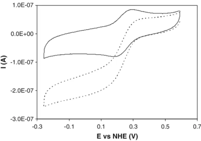
The cyclic voltammograms of cytochrome c-552 alone (data not shown) and the ones in the presence of preacti-vated N2OR are identical (Fig.1, continuous line). Upon addition of a saturating amount of substrate, N2O, the original peak-shaped voltammogram transforms into the characteristic sigmoid catalytic wave, with a steady-state current plateau (Fig.1, dashed line).
This behaviour can be interpreted with the reaction mechanism shown in Scheme1. An initial heterogeneous electron transfer reaction at the electrode (step 1) is fol-lowed by two homogeneous chemical reactions: the reduced form of cytochrome c-552 is oxidized by N2OR (step 2), which is then regenerated by N2O (step 3).
This mechanism can be simplified to
cytochromec552oxþecytochromec552red ð1Þ cytochromec552redþN2ORox
as long as four conditions are obeyed: (1) the heteroge-neous electron transfer (step 1) is a reversible reaction; (2) the homogeneous chemical reaction (step 2) is irreversible; (3) the reaction between cytochromec-552 and N2OR is pseudo-first order with the reaction rate constant given by k0 =k[N2OR], where k is the second-order rate constant; (4) reaction 3 is fast.
As reported in [19] and confirmed in this work, the first condition is verified. The experiments were performed under an excess of the substrate N2O; thus, the enzyme is reoxidized by the catalytic reaction (step 3), and not by transferring electrons back to cytochrome c-552. For this reason, the second condition is obeyed. Condition 3 implies that the enzyme concentration is much higher than that of cytochromec-552. Although this is not the case, the saturating concentration of N2O guarantees that the oxi-dized form of the enzyme is quickly restored by step 3, as long as the latter is not rate-limiting (condition 4). There-fore, N2OR will always be available to react with cyto-chromec-552 and pseudo-first-order conditions are met.
According to the theory of steady-state voltammetric catalysis, the rate constant can only be determined from the
steady-state catalytic current if the latter is not scan-rate-dependent [27,28]. For the mediated catalysis of N2OR by cytochrome c-552, the catalytic current increases linearly with the scan rate, between 5 and 40 mV s-1, and then becomes independent of this parameter, up to 100 mV s-1 (data not shown). Moreover, the catalytic current decreases after the first scan, until the initial cytochromec-552 signal is restored. This behaviour, corroborated by the fact that further addition of substrate does not restore the catalytic signal, suggests that the enzyme is being inactivated (see below).
The rate constant of the intermolecular electron transfer reaction between cytochrome c-552 and N2OR was esti-mated using two approaches, with the catalytic currents being measured at 40 mV s-1. In one of the approaches, the cyclic voltammetry data were processed according to the Nicholson and Shain theory [27], where the pseudo-first-order rate constant,k0, can be determined by plotting the ratio between the catalytic current (the plateau current value, icat) and the diffusion-controlled current (the cyto-chrome c-552 reduction peak current, ip) versus the reci-procal of the square root of the scan rate, v:
icat=ip¼2:241ðRT=nFÞ1=2k01=2ð1=mÞ1=2; ð1Þ
wherenis the number of electrons exchanged, andR,TandF are the universal gas constant, the temperature, and the Faraday constant, respectively. The cytochromec-552 redox reaction is a reversible one-electron process, and thus from the slope a k0 value of 1.4±0.2 s-1 was obtained. The
intermolecular rate constant, k, for the reaction between N2OR and cytochromec-552 at pH 7.0 was determined to be (5.4±0.8)9105M-1s-1, since the concentration of
enzyme entrapped in the membrane was 2.5lM.
In the other approach,kis calculated from the value of the N2O-saturated limiting current,icat[29–32]:
icat ¼nFACcytochromec Dcytochromeck0
1=2
; ð2Þ
where Ccytc is the concentration of cytochrome c-552 under the membrane, Dcytc is its diffusion coefficient (1.0910-6cm2s-1 [33]) and A is the electrode surface area (see ‘‘Materials and methods’’). Calculations with this equation gavek=(5.6±0.4)9105M-1s-1at pH 7.0, a
value identical to the one obtained with the previous approach (Eq. 1).
This value compares well with other intermolecular rate constants determined using cyclic voltammetry. Cyto-chromec-552 was used in mediated electrochemical exper-iments for the reduction of nitrite byPs. nauticacytochrome cd1 nitrite reductase [21] and a similar rate constant was calculated in that study [k=(4.1±0.1)9105M-1s-1at
pH 6.3]. Other examples are the electron transfer from A. cycloclastes pseudoazurin to its electron donor, nitrite
cyt ox
cyt red N2ORox
N2ORred N2O
N2
electr
ode e
-Step 3 Step 2
Step 1
Scheme 1 Mediation scheme for N2OR: the electrode reduces
cytochromec-552, which is immediately reoxidized by N2OR; the
level of oxidized N2OR is then restored by conversion of N2O to N2
-3.0E-07 -2.0E-07 -1.0E-07 0.0E+00 1.0E-07
-0.3 -0.1 0.1 0.3 0.5 0.7 E vs NHE (V)
I (A)
Fig. 1 Cyclic voltammograms (40 mV s-1) of 50
lM cytochromec
-552 entrapped in a membrane electrode with 2.5lM activated
Pseudomonas nautica nitrous oxide reductase (N2OR), in 0.1 M
phosphate buffer, pH 7. Thecontinuous lineis the cyclic voltammo-gram in the absence of substrate and thedashed lineis the cyclic voltammogram after addition of 1 mM N2O.NHEnormal hydrogen
reductase, which was determined to have a k value of 7.39105 M-1s-1 [34], and also the electron transfer
betweenParacoccus pantotrophus pseudoazurin and cyto-chromecperoxidase, withk=1.49105M-1s-1[25].
This voltammetric theory implies that the catalytic current should be directly proportional to the mediator concentration. This condition is verified because the electrochemical experiments were performed with 50lM, corresponding to the linear region of the Michaelis– Menten equation (Kmc552¼50lM [18]). However, investigation of the catalytic current at higher cytochrome c-552 concentrations revealed a non-linear behaviour (data not shown), confirming that also cytochrome c-552 con-tributes to the global rate with a Michaelis–Menten term [18].
It is possible to compare the second-order kinetic con-stant, k, calculated with the electrochemical approach [k=(5.5±0.9)9105M-1s-1] with the ratio kcat/Km determined by the steady-state kinetic study [(7.6±0.7)9
104M-1s-1; see the electronic supplementary material]. The difference between the two values can be attributed to the limitations of the steady-state kinetic study, wherekcat andKm were not determined accurately owing to experi-mental problems at high cytochromec-552 concentrations [18].
The dependence of the electrochemical activity on the nitrous oxide concentration was studied using constant concentrations of N2OR and cytochrome c-552 entrapped in the membrane. The catalytic currents calculated for each substrate concentration were fitted to the Michaelis– Menten equation (Fig.2), using aKmof (16±2) lM and anicatmaxof (3.9±0.1)910
-7 A.
KmN2O, with cytochrome c-552 as an electron donor, could not be estimated in the steady-state activity assay because the N2O reduction is involved in a fast step compared with the N2OR reduction by cytochrome c-552 [18]. The value obtained in that study using MV as an electron donor (ðKmN2O¼14:02:9lM)) is very similar
to the one calculated by steady-state kinetics, but it must be pointed out that when a stationary electrode is used, mass transport limitations are not avoided at low substrate con-centrations. Therefore, the Km value estimated from the fitting of the electrochemical assay should not be regarded as accurate, whereas from theicatmaxvalue it was possible to calculate a k0max value of (1.4±0.1) s-1. Since the
N2OR concentration used in this study was 2.5lM, akof (5.6±0.4)9105M-1s-1was estimated, a value that is identical to the one calculated using the approaches pre-sented above.
The effect of pH on the intermolecular rate constant was analysed, revealing a bell-shaped curve, that can be simu-lated using Eq.3 [35] and pKa values of 5.5±1.0 and 8.0±0.7 (Fig.3).
Act¼Actmax
.
1þ10ðpKa1pHÞþ10ðpHpKa2Þ
ð3Þ
The latter pKa matches the solution kinetic value (pKa of 8.3 [18]), which was attributed to a deprotonation occur-ring at the catalytic CuZ centre and identified as a key process in the reduction mechanism of CuZ. The more acidic pKa was not detected in the steady-state kinetic experiment, since it is outside the pH range studied in that work (pH between 6.2 and 8.7).
0.0E+00 1.0E-07 2.0E-07 3.0E-07 4.0E-07 5.0E-07
0 200 400 600 800 1000
[N2O] (µM)
i
ca
t
(A)
Fig. 2 Catalytic current from electrochemical assays ofPs. nautica N2OR using cytochromec-552 as a mediator versus N2O
concentra-tion. The assays were performed with 2.5lM activated N2OR and
50lM cytochromec-552 entrapped in the membrane electrode, in
0.1 M phosphate buffer at pH 7, and in the presence of 8, 17, 25, 33, 48, 125, 330 and 1,000lM N2O-saturated water. The experimental
data were fitted with the Michaelis–Menten equation, using aKmof
(16±2)lM and anicatmaxof (3.9±0.1)910
-7A
0.0 0.2 0.4 0.6 0.8 1.0 1.2 1.4 1.6
5.5 6.5 7.5 8.5 9.5
pH
k'
(s
-1 )
Fig. 3 Intermolecular rate constants ofPs. nauticaN2OR versus pH,
determined by electrochemical assays using cytochromec-552 as a mediator. The assays were performed with 2.5lM activated N2OR
and 50lM cytochromec-552 entrapped in the membrane electrode,
1 mM N2O-saturated water, in different buffer systems with pH
between 5.9 and 8.8. The data were non-linearly fitted using Eq.3, and pKavalues of 8.0±0.7 and 5.5±1.0 (solid line). Thedashed
As mentioned above, in the electrochemical studies of the mediated activity of N2OR by cytochromec-552 it was observed that the catalytic current, measured at successive scans, decreases after each scan (Fig.5b, filled circles). This decay can be attributed to an inactivation process that occurs with time (between each new scan). Moreover, in the experiments performed at different pH, it was observed that this inactivation process is lower at acidic pH than at basic pH (Fig.4), and a pKaof 7.1±0.8 was estimated. This is an indication that a deprotonation is involved in the inactivation of the enzyme (vide infra), as has been pro-posed before [18].
Direct reaction with substrate N2O
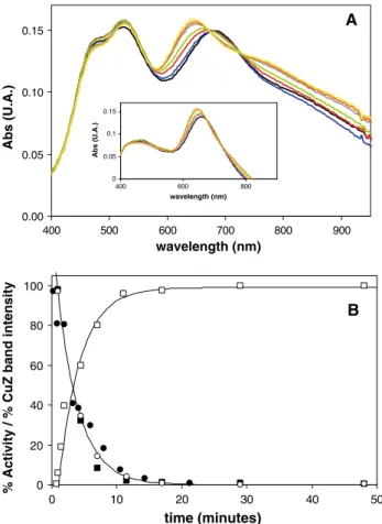
The reaction between preactivated N2OR and a stoichi-ometric amount of N2O, monitored by UV–vis spectros-copy, showed a rapid (within 1 s) oxidation of the CuA centre, through the increase of the two characteristic absorption bands at 480 and 540 nm [13], and the devel-opment of an absorption band at 680 nm, which has not been reported previously. Then, this 680-nm absorption band slowly (on the order of minutes) shifts to the usual position of 640 nm for the resting enzyme (Fig.5a). At times corresponding to those in the UV–vis monitoring, the enzyme activity was assayed to understand the contribution to catalysis of the N2OR forms characterized by the 680-nm absorption band and the form featuring the 640-680-nm absorption band (Fig.5b).
The increase of the intensity of 640-nm band in the UV– vis spectrum versus time (Fig.5b, open squares) can be fitted with a rate similar to the decay rate of the enzymatic activity (Fig.5b, filled squares) (k=0.3 min-1 for both
experimental data sets), suggesting that the two processes are directly correlated.
The maximum activity for this enzyme preparation determined in a separate activity assay was 133 lmol N2O/ min mg N2OR. The activity calculated 1 min after the reaction between N2OR and a stoichiometric amount of N2O is 130lmol N2O/min mg N2OR (98% with respect to the initial value). The activity decreases with time, reach-ing a value of 0.6lmol N2O/min mg N2OR (0.4% with respect to the initial value) after 48 min.
The direct correlation of the enzyme activity (Fig. 5b, filled squares) with the presence of a N2OR form exhibiting the 680-nm band (Fig.5b, open circles) indicates that CuZ is in a new redox-active form, which will be represented here as CuZ°. This also rules out that the broad 680-nm
0.0 0.2 0.4 0.6 0.8 1.0
6.0 6.5 7.0 7.5 8.0 8.5 9.0 pH
k
(min
-1 )
Fig. 4 Rate constant of the inactivation process detected by electro-chemical experiments versus pH. The k values were obtained by fitting the catalytic current decay with an exponential equation. The data were non-linearly fitted using Eq.3adapted for one pKaof 7.1
0 20 40 60 80 100
0 10 20 30 40 50
time (minutes)
% Activity / % CuZ band intensity
B 0.00
0.05 0.10 0.15
400 500 600 700 800 900
wavelength (nm)
Abs (U.A.)
A
0 0.05 0.1 0.15
400 600 800
wavelength (nm)
Abs (U.A.
)
Fig. 5 aSelected spectra of 35lMPs. nauticaN2OR after reaction
with an equimolar amount of N2O at the following times: 0.5 min
(black), 1 min (blue), 1.5 min (red), 2 min (green), 4.5 min (grey),
11 min (orange) and 45 min (yellow). Inset: Spectra obtained after subtraction of the oxidized CuA contribution at the following times: 0.5 min (black), 1 min (blue), 1.5 min (red), 2 min (green), 4.5 min (grey), 11 min (orange) and 45 min (yellow).bN2OR activity (filled
squares) versus time (100% corresponds to 133lmol N2O reduced
min-1mg-1 enzyme) and 640-nm band intensity (open squares)
versus time (100% corresponds to the final spectra att=48 min and 0% corresponds to the first spectrum att=1 min).Solid lines are exponential fits and k=0.3 min-1 was used for both fits. Filled
circles represent the percentage of electrocatalytic activity versus
time (100% corresponds to the maximum activity, characterized by an intermolecular rate constant for electron transfer,k0, of 1.4 s-1).Open
absorption band might have a contribution from the 640-nm absorption band.
Therefore, this is the first time that such an enzyme form has been shown to be directly involved in the turnover mechanism of N2OR. In fact, the two enzyme forms pre-viously identified for Pa. denitrificans N2OR, CuZ and CuZ*, which also have different absorption bands (670 and 650 nm, respectively), show very low specific activity when compared with the active CuZ°form [17].
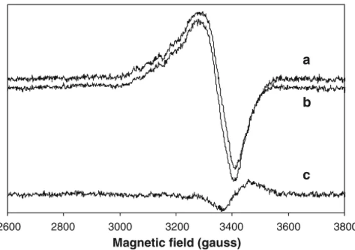
The EPR spectrum of the CuZ°form was obtained by
direct reaction of fully reduced N2OR with a stoichiometric amount of N2O for 1 min (Fig.6). The oxidized CuA centre contributes to the total EPR spectrum with its typical seven-line hyperfine splitting [7, 36], so this contribution was subtracted in the spectra shown in Fig.6. The resting, inactive CuZ form characterized by the band at 640 nm in the UV–vis spectrum, exhibits the following EPR param-eters:g|| =2.160 andg\=2.040 and a four-line hyperfine splitting [10] (Fig.6b).
The EPR signal of the active CuZ°form is very similar,
both in shape and intensity (Fig.6, spectrum a), indicating that it has the same paramagnetic state as the resting form (1CuII/3CuI), with a total spin S=. The g|| value is unchanged, whereasg\is only slightly lower (g\=2.037)
for CuZ°. In light of this similarity in the EPR spectrum,
the coordination environment of the coppers in the two forms has not changed drastically, and in particular we can assume that CuI remains in the formal Cu2? state within the cluster [10], whereas the small change in g\ may suggest that some minor local rearrangement is occurring at the site. Also the shift of the electronic band from CuZ to
CuZ°can be correlated to some minor change in the CuI
environment.
The high intensity of the 680-nm band in CuZ°confirms
that this absorption band remains a S2-?Cu ligand to
metal charge transfer (LMCT) transition, as for the 640-nm band in the resting CuZ, which is actually a multicompo-nent band with partially overlapping features at higher and lower energy [11, 12]. Indeed, looking carefully at the difference spectra reported in the inset in Fig.5a, it seems that the 680-nm absorption of CuZ° results from the
overlap between the high-intensity component, which is red-shifted from its original position at 640 nm, and a low-intensity component, blue-shifted from its position near 720 nm, in the CuZ spectrum, which is missing in the CuZ°
spectrum. The increased bandwidth of the 680-nm band with respect to the 640-nm band supports this interpreta-tion. We may therefore interpret the 640- to 680-nm shift of the LMCT band as a result of the reduction of the splitting between the components of the complex S2-?Cu LMCT band, indicating that in the CuZ°centre,
the CuI atom undergoes a small displacement in its relative position versus the l4–S2- ligand with respect to the
resting CuZ cluster.
A possible explanation for the spectral difference between CuZ and CuZ° is a protonation/deprotonation
process of the hydroxo ligand between CuI and CuIV. As a simple proton transfer would be faster than the timescale of conversion of CuZ°to the redox-equivalent, inactive form,
it is likely that the deprotonation of a bound water mole-cule in CuZ° (see below) occurs with formation of a l
-hydroxo-bridge between CuI and CuIV in the resting form [18]. This will likely involve some minor structural change in the cluster to meet the geometrical requirement needed to optimize the formation of the bridge.
In a recent crystallographic study of A. cycloclastes N2OR, it was shown that the CuZ cluster is flexible and can accommodate alternatively one water molecule and one hydroxyl group or even an iodide ion in a bridged mode [5]. In particular, it was shown that the CuZ cluster can rearrange its structure by changing the Cu–S distances and the orientation of some of the histidine residues. From our data we can suggest that the configuration with the l
-hydroxo bridge between CuI and CuIV prevents the CuZ centre from undergoing further catalysis, whereas a non-bridged water molecule would be more susceptible to be released, enabling the accommodation of a new substrate molecule at CuZ.
Mechanistic insight involving the new CuZ°
active form
On the basis of the identification of the fast-reacting CuZ°
N2OR form, it is possible to formulate a new mechanism of 2600 2800 3000 3200 3400 3600 3800
Magnetic field (gauss)
a
b
c
reduction, catalysis and inactivation of the catalytic N2OR centre, as shown in Scheme2. The resting CuZ state, characterized by the absorption band at 640 nm, does not participate in the catalytic cycle because of the slow reduction (activation process) required to obtain the fully reduced state [15]. The fast turnover cycle (kcat=320 s-1 and kcat=3.8 s
-1
for MV and cytochrome c-552, respectively [18]) implies that the re-reduction of the N2O-oxidized CuZ centre must be rapid in the enzymatic turn-over and excludes the involvement of the resting CuZ state. This is also in agreement with the fact that as-purified N2OR is not catalytically active, unless it is subjected to prolonged activation with reduced MV.
Therefore, the two-electron-oxidized form of CuZ is not detectable as it has a very short life and is very rapidly reduced by one electron coming from CuA, yielding the 1Cu2?3Cu? intermediate species CuZ° (Scheme2). The
intramolecular electron transfer producing CuZ°cannot be
analysed in our conditions but is signalled by the formation of the characteristic optical bands of oxidized CuA (on 0.1-s time0.1-scale). Then, during catalytic turnover, CuZ° is
directly reduced back to the fully reduced enzyme by another electron coming from CuA. As the enzymatic reaction produces a water molecule by consumption of two protons, we believe that this water molecule is bound to the oxidized CuI in CuZ°. The fast reduction of the latter
species during turnover prevents the rearrangement of CuZ° to the l-hydroxo-bridged, redox-equivalent resting
form. We already noticed that the Cu(II)/Cu(I) redox potential of the l-hydroxo-bridged dinuclear Cu(II)
com-plexes is lower than that of the corresponding aqua
complexes [18]. This is consistent with the formulation of the oxidized CuI–CuIV as a non-bridged Cu2?Cu2?–H2O species, as this would make the subsequent reduction by CuA easier.
In the typical activity assays, the strong absorption of the reductants (reduced MV or cytochromec-552) prevents the detection of the CuZ° intermediate form. Instead, as
shown here, in the absence of reductants, this form is slowly converted to the inactive resting state of CuZ. Moreover, the inactivation process does not involve any electron transfer reaction, because CuA remains in the oxidized state (Cu1.5?/Cu1.5?) during this process.
The inactivation process that was detected in the elec-trochemical experiments, where the activity of N2OR is mediated by cytochromec-552, has the same decay as the one observed in the UV–vis study, which suggests that the process that is occurring is the same in the two situations. In addition, the effect of pH on the inactivation process is consistent with the deprotonation of the water ligand between CuI and CuIV in the CuZ° species, as the pKa
calculated from the inactivation process (7.1±0.8) is
compatible with the values calculated through the elec-trochemical assay and the steady-state kinetic study (pKa=8.0±0.7 and pKa=8.3, respectively)
Redox titration
The titration of N2OR was performed in the oxidation direction since the activated form is in the fully reduced state. The CuA centre has a midpoint potential at
E&240 mV (Fig.7, filled squares in the oxidation
CuZ resting state 1Cu2+ 3Cu+
640-nm - INACTIVE
Activation(k= 0.07 min-1) MVredincubation
4 Cu+
Inactivation (k= 0.3 min-1)
N2O + 2H+
N2+ H2O Catalysis
(k= 3.8 s-1forc552; k= 320 s-1for MV)
CuAred CuZred
CuAred 2Cu+2Cu2+
CuAox
1Cu2+ 3Cu+ CuZ°680-nm
e- fast Reductant
MVred/c552
red No reductants e
-CuAox
MVred c552
red
Scheme 2 New mechanism of reduction, inactivation and catalysis of the catalytic N2OR
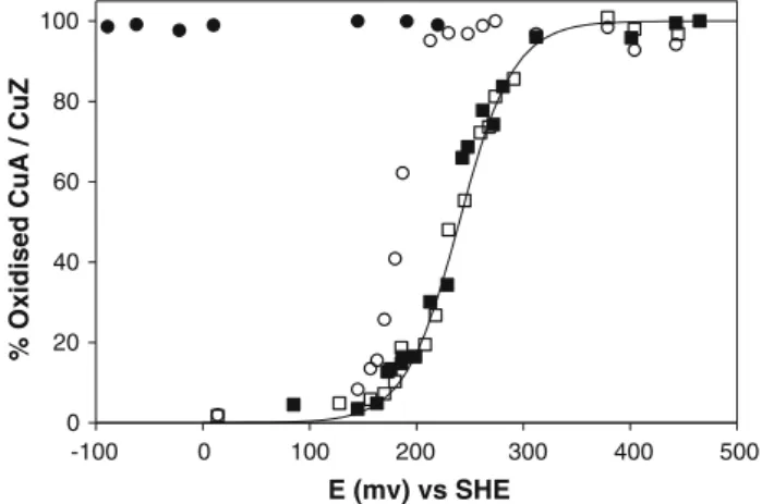
direction, open squares in the reduction direction). This midpoint potential is in full agreement with literature val-ues for this centre in N2OR [13,17,37]. The oxidation of CuZ brings this centre directly into the form characterized by the 640-nm band with no contribution at 680 nm (Fig.7, open circles, spectra not shown). The impossibility to reduce this form [titrations using either dithionite or titanium(III) citrate were not successful; Fig.7, filled cir-cles] does not allow the application of the Nernst equation, which can be used only for a reversible system, and does not enable the calculation of the midpoint redox potential for this centre. In fact, this data set does not fit with the Nernst equation for one electron.
The oxidation of the fully reduced CuZ promoted by ferricyanide brings CuZ directly into the inactive form, which is characterized by the 640-nm band. This form cannot be re-reduced again unless through an activation process with reduced MV (Scheme2).
The irreversible oxidation of CuZ is catalytically unproductive and this behaviour confirms the proposed mechanism in which CuZ° is the only active form in the
turnover cycle, where it is generated by the substrate and it is rapidly reduced by the electron flow from the CuA centre.
Conclusion
The first example of mediated electrochemistry of N2OR described here allowed the calculation of the rate constant of the intermolecular electron transfer to the physiological electron donor [kof (5.5±0.9)9105M-1s-1]. The rate
constant is similar to the values found for other intermo-lecular protein–protein electron transfers determined using cyclic voltammetry. Moreover, the study of the dependence of the rate on N2O concentration and pH confirms the recent results obtained in the kinetic analysis between N2OR and cytochromec-552 fromPs. nautica.
The reaction of the activated form of N2OR with a stoichiometric amount of substrate enabled the identifica-tion of a new active CuZ°form in the turnover cycle, which
is characterized by an absorption band at 680 nm and is different from the resting and inactive CuZ form, previ-ously observed. In the absence of reductants to complete the catalytic cycle, the CuZ°form rearranges to restore the
resting form in a slow process (k=0.3 min-1), compared
with the turnover rate. This inactivation process was also detected in the electrochemical studies, where the catalytic current decreased at a rate similar to the rate of formation of the inactive enzyme form.
A scheme that shows the activation required in vitro and the inactivation detected during these experiments was presented. In particular, the difference between the resting CuZ and the active CuZ° was discussed to clarify the
activation mechanism and the catalysis of N2OR.
Acknowledgements This research was supported by FCT (Fun-dac¸a˜o para a Cieˆncia e Tecnologia) grants PTDC/QUI/64638/2006 (to I.M.) and SFRH/BD/30414/2006 (to S.D.) and by the University of Pavia through FAR (to E.M. and L.C.). We thank Pablo J. Gonzalez for the help with EPR data collection.
References
1. Zumft WG, Kroneck PM (2007) Adv Microb Physiol 52:107–227 2. Tavares P, Pereira AS, Moura JJ, Moura I (2006) J Inorg
Bio-chem 100:2087–2100
3. Brown K, Tegoni M, Prudencio M, Pereira AS, Besson S, Moura JJ, Moura I, Cambillau C (2000) Nat Struct Biol 7:191–195 4. Haltia T, Brown K, Tegoni M, Cambillau C, Saraste M, Mattila
K, Djinovic-Carugo K (2003) Biochem J 369:77–88
5. Paraskevopoulos K, Antonyuk SV, Sawers RG, Eady RR, Hasnain SS (2006) J Mol Biol 362:55–65
6. Scott RA, Zumft WG, Coyle CL, Dooley DM (1989) Proc Natl Acad Sci USA 86:4082–4086
7. Antholine WE, Kastrau DH, Steffens GC, Buse G, Zumft WG, Kroneck PM (1992) Eur J Biochem 209:875–881
8. Gorelsky SI, Xie X, Chen Y, Fee JA, Solomon EI (2006) J Am Chem Soc 128:16452–16453
9. Kroneck PM, Antholine WA, Riester J, Zumft WG (1989) FEBS Lett 248:212–213
10. Chen P, DeBeer George S, Cabrito I, Antholine WE, Moura JJ, Moura I, Hedman B, Hodgson KO, Solomon EI (2002) J Am Chem Soc 124:744–745
11. Chen P, Cabrito I, Moura JJ, Moura I, Solomon EI (2002) J Am Chem Soc 124:10497–10507
12. Chen P, Gorelsky SI, Ghosh S, Solomon EI (2004) Angew Chem Int Ed 43:4132–4140
13. Prudencio M, Pereira AS, Tavares P, Besson S, Cabrito I, Brown K, Samyn B, Devreese B, Van Beeumen J, Rusnak F, Fauque G, 0
20 40 60 80 100
-100 0 100 200 300 400 500 E (mv) vs SHE
% Oxidised CuA / CuZ
Fig. 7 Potentiometric redox titration of fully reduced Ps. nautica N2OR following the characteristic absorption bands of the CuA and
CuZ centres in the oxidation and reduction directions. The CuA centre was monitored by following the absorption at 540 nm in the oxidative (filled squares) and in the reductive (open squares) titrations. The titration curve was fitted withE°0= ?240 mV (solid line). The CuZ centre was monitored by following the absorption at 640 nm in the oxidative (open circles) and in the reductive (filled
Moura JJ, Tegoni M, Cambillau C, Moura I (2000) Biochemistry 39:3899–3907
14. Oganesyan VS, Rasmussen T, Fairhurst S, Thomson AJ (2004) Dalton Trans 7:996–1002
15. Ghosh S, Gorelsky SI, Chen P, Cabrito I, Moura JJ, Moura I, Solomon EI (2003) J Am Chem Soc 125:15708–15709 16. Chan JM, Bollinger JA, Grewell CL, Dooley DM (2004) J Am
Chem Soc 126:3030–3031
17. Rasmussen T, Berks BC, Butt JN, Thomson AJ (2002) Biochem J 364:807–815
18. Dell’acqua S, Pauleta SR, Monzani E, Pereira AS, Casella L, Moura JJ, Moura I (2008) Biochemistry 47:10852–10862 19. Correia dos Santos MM, Paes de Sousa PM, Simoes Gonc¸alves
ML, Krippahl L, Moura JJG, Lojou E, Bianco P (2003) J Elec-troanal Chem 541:153–162
20. Alves T, Besson S, Duarte LC, Pettigrew GW, Girio FM, De-vreese B, Vandenberghe I, Van Beeumen J, Fauque G, Moura I (1999) Biochim Biophys Acta 1434:248–259
21. Lopes H, Besson S, Moura I, Moura JJ (2001) J Biol Inorg Chem 6:55–62
22. Lowry OH, Rosebrough NJ, Farr AL, Randall RJ (1951) J Biol Chem 193:265–275
23. Fauque G, Moura JJG, Besson S, Saraiva L, Moura I (1992) Oceanis 18:211–216
24. Saraiva LM, Besson S, Fauque G, Moura I (1994) Biochem Biophys Res Commun 199:1289–1296
25. de Sousa PM, Pauleta SR, Goncalves ML, Pettigrew GW, Moura I, Dos Santos MM, Moura JJ (2007) J Biol Inorg Chem 12:691– 698
26. Ferapontova EE, Ruzgas T, Gorton L (2003) Anal Chem 75:4841–4850
27. Nicholson RS, Shain I (1964) Anal Chem 36:706–723
28. Bard AJ, Faulkner LR (2001) Electrochemical methods, funda-mentals and applications. Wiley, New York
29. Saveant JM, Vianello E (1965) Electrochim Acta 10:905–920 30. Coury LA, Oliver BN, Egekeze JO, Sosnoff CS, Brumfield JC,
Buck RP, Murray RW (1990) Anal Chem 62:452–458
31. Coury LA, Yang Liu, Murray RW (1993) Anal Chem 65:242– 246
32. Yang L, Coury LA, Murray RW (1993) J Phys Chem 97:1694– 1700
33. Correia dos Santos MM, Paes de Sousa PM, Simoes Gonc¸alves ML, Lopes H, Moura I, Moura JJG (1999) J Electroanal Chem 464:76–84
34. Kataoka K, Yamaguchi K, Kobayashi M, Mori T, Bokui N, Suzuki S (2004) J Biol Chem 279:53374–53378
35. Cornish-Bowden A (2001) Fundamentals of enzyme kinetics. Portland Press, London, pp 179–192
36. Farrar JA, Thomson AJ, Cheesman MR, Dooley DM, Zumft WG (1991) FEBS Lett 294:11–15