Sensitivity and Specificity of a Urine
Circulating Anodic Antigen Test for the
Diagnosis of
Schistosoma haematobium
in
Low Endemic Settings
Stefanie Knopp1,2,3*, Paul L. A. M. Corstjens4, Artemis Koukounari5, Colin I. Cercamondi6, Shaali M. Ame7, Said M. Ali7, Claudia J. de Dood4, Khalfan A. Mohammed8,
Jürg Utzinger2,3, David Rollinson1, Govert J. van Dam9
1Wolfson Wellcome Biomedical Laboratories, Department of Life Sciences, Natural History Museum, London, United Kingdom,2Department of Epidemiology and Public Health, Swiss Tropical and Public Health Institute, Basel, Switzerland,3University of Basel, Basel, Switzerland,4Department of Molecular Cell Biology, Leiden University Medical Center, Leiden, The Netherlands,5Biostatistics Department, Institute of Psychiatry, Psychology and Neuroscience, Kings College London, London, United Kingdom,
6Laboratory of Human Nutrition, Institute of Food, Nutrition, and Health, Eidgenössische Technische Hochschule (ETH) Zurich, Zurich, Switzerland,7Public Health Laboratory-Ivo de Carneri, Chake Chake, Pemba, United Republic of Tanzania,8Helminth Control Laboratory Unguja, Ministry of Health, Zanzibar Town, Unguja, United Republic of Tanzania,9Department of Parasitology, Leiden University Medical Center, Leiden, The Netherlands
Abstract
Background
Elimination of schistosomiasis as a public health problem and interruption of transmission in selected areas are key goals of the World Health Organization for 2025. Conventional para-sitological methods are insensitive for the detection of light-intensity infections. Techniques with high sensitivity and specificity are required for an accurate diagnosis in low-transmis-sion settings and verification of elimination. We determined the accuracy of a urine-based up-converting phosphor-lateral flow circulating anodic antigen (UCP-LF CAA) assay for
Schistosoma haematobiumdiagnosis in low-prevalence settings in Zanzibar, Tanzania.
Methodology
A total of 1,740 urine samples were collected in 2013 from children on Pemba Island, from schools where theS.haematobiumprevalence was<2%, 2–5%, and 5–10%, based on a single urine filtration. On the day of collection, all samples were tested for microhematuria with reagent strips and for the presence ofS.haematobiumeggs with microscopy. Eight months later, 1.5 ml of urine from each of 1,200 samples stored at -20°C were analyzed by UCP-LF CAA assay, while urine filtration slides were subjected to quality control (QCUF). In the absence of a true‘gold’standard, the diagnostic performance was calculated using la-tent class analyses (LCA).
OPEN ACCESS
Citation:Knopp S, Corstjens PLAM, Koukounari A, Cercamondi CI, Ame SM, Ali SM, et al. (2015) Sensitivity and Specificity of a Urine Circulating Anodic Antigen Test for the Diagnosis ofSchistosoma haematobiumin Low Endemic Settings. PLoS Negl Trop Dis 9(5): e0003752. doi:10.1371/journal. pntd.0003752
Editor:Aysegul Taylan Ozkan, Hitit University, TURKEY
Received:February 9, 2015
Accepted:April 11, 2015
Published:May 14, 2015
Copyright:© 2015 Knopp et al. This is an open access article distributed under the terms of the Creative Commons Attribution License, which permits unrestricted use, distribution, and reproduction in any medium, provided the original author and source are credited.
Data Availability Statement:All relevant data are within the paper and its Supporting Information files.
Principal Findings
The‘empirical’S.haematobiumprevalence revealed by UCP-LF CAA, QCUF, and reagent strips was 14%, 5%, and 4%, respectively. LCA revealed a sensitivity of the UCP-LF CAA, QCUF, and reagent strips of 97% (95% confidence interval (CI): 91–100%), 86% (95% CI: 72–99%), and 67% (95% CI: 52–81%), respectively. Test specificities were consistently above 90%.
Conclusions/Significance
The UCP-LF CAA assay shows high sensitivity for the diagnosis ofS.haematobiumin low-endemicity settings. Empirically, it detects a considerably higher number of infections than microscopy. Hence, the UCP-LF CAA employed in combination with QCUF, is a promising tool for monitoring and surveillance of urogenital schistosomiasis in low-transmission set-tings targeted for elimination.
Author Summary
The World Health Organization aspires to eliminate snail fever (schistosomiasis) as a pub-lic health problem and to interrupt the transmission of this disease in selected areas by 2025. Efforts to achieve these goals are currently being intensified. As a result, the preva-lence and intensity of infection will decline in many parts of the world. To detect light-in-tensity infections, diagnostic tools with a high sensitivity and specificity are needed. We assessed the accuracy of a method that is able to diagnose schistosomiasis via the detection of circulating anodic antigen (CAA) in urine. We examined 1,200 urine samples from chil-dren living on Pemba Island, Tanzania, a low-endemic area targeted for schistosomiasis elimination. We found that the CAA-test had a considerably higher sensitivity than con-ventional urine filtration microscopy and reagent strips that are widely used in schistoso-miasis control programs. The empirical prevalence of infection with the parasite
Schistosoma haematobiumdetermined by the CAA-test was up to 10 times higher than that obtained by urine filtration. Our results suggest that the CAA-test—in combination with urine filtration—is a promising approach for the diagnosis ofS.haematobiumin low-transmission settings that are targeted for elimination.
Introduction
After many years of neglect, schistosomiasis and other parasitic worm infections are given con-siderable attention by the research community, non-governmental organizations, funding bod-ies, international organizations, policy makers, and disease control managers [1,2]. Indeed, drug donations, in conjunction with political and financial commitment for scaling up control interventions, have markedly expanded since the London Declaration on Neglected Tropical Diseases was launched in early 2012 [3,4]. For example, the World Health Organization (WHO) strategic plan 2012–2020 for schistosomiasis aims at morbidity control by 2020, and elimination of schistosomiasis as a public health problem and interruption of transmission in selected areas by 2025 [5]. The mainstay of morbidity control is preventive chemotherapy that is the large-scale administration of praziquantel to at-risk groups (e.g., school-aged children). To achieve elimination of schistosomiasis as a public health problem (i.e., the reduction of
Health Research (NIHR) Biomedical Research Centre at South London and Maudsley National Health System (NHS) Foundation Trust and King’s College London. The views expressed are those of the authors and not necessarily those of the NIHR, the NHS, or the UK Government Department of Health. The funders had no role in study design, data collection and analysis, decision to publish, or preparation of the manuscript.
heavy infection intensities in the at-risk population to below 1%), additional public health mea-sures are recommended, alongside intensified interventions in pockets of high transmission [5,6]. For interruption of transmission (i.e., reducing the incidence of infection to zero), it is es-sential to increase the frequency of preventive chemotherapy and to supplement it with addi-tional control measures, such as improved access to clean water and adequate sanitation, and controlling intermediate host snails [5–9]. Monitoring the progress of control programs and rigorous surveillance to identify remaining or reemerging transmission hotspots and individu-als with high infection levels (so-called superspreaders), will be relevant for tailoring an ade-quate response in close-to-elimination settings [5,6,10].
An accurate diagnosis is essential and should be adapted to the specific stage of a schistoso-miasis control program [11,12]. Parasitological methods to detectSchistosomaeggs in stool (e.g., Kato-Katz thick smear [13]) or urine (e.g., urine filtration [14]), or reagent strips to detect microhematuria in urine [15] are widely used in control programs. However, while these meth-ods are reasonably accurate in diagnosing moderate and heavy infection intensities, they show low sensitivity for detecting light-intensity infections [16,17]. For appropriate monitoring and surveillance in areas approaching schistosomiasis elimination, other, highly sensitive tools are needed [4,11,18–22]. Moreover, as the prevalence of infection decreases over the course of a control program, specificity becomes more important and will be an absolute requirement for certification of elimination [11,19].
A promising diagnostic approach that might be suitable for highly sensitive and specific di-agnosis of very lightSchistosomainfections is the detection ofSchistosomaadult worm circulat-ing anodic antigen (CAA) in serum and urine uscirculat-ing an up-convertcirculat-ing phosphor-lateral flow (UCP-LF) assay [20,23–25]. Concentration systems allowing the recognition of very low worm numbers have been developed, and currently four urine-based assays are available in a robust dry-reagent format [26,27]. These UCP-LF tests can detect 30 pg/ml, 3 pg/ml, 0.3 pg/ml, and 0.1 pg/ml CAA using 10μl (UCAA10), 250μl (UCAA250), 2 ml (UCAA2000), and 7.5 ml urine (UCAA7500), respectively [27].
The UCAA2000 (and perhaps the UCAA250) is currently considered to show the best trade-off between a high sensitivity and convenient field applicability [28]. However, the per-formance of different UCP-LF CAA assays as sensitive and specific diagnostic tools for non-in-vasive monitoring and surveillance remain to be determined in laboratories inSchistosoma
low-endemic settings. Recently, the UCAA2000, as well as a variant of the test conducted with 500μl serum, have been successfully applied for the diagnosis ofS.japonicuminfections in low transmission settings in the People’s Republic of China [29]. First validation attempts have also been made with the UCAA250 forS.japonicumandS.mekongidetection in banked urine sam-ples from the Philippines and Cambodia, respectively [30].
Here, we assess the accuracy of the UCAA2000 for the diagnosis ofS.haematobiumin three low-prevalence settings (<2%, 2–5%, and 5–10%), as determined with a single urine filtration.
Methods
Ethics Statement
The study was approved by the Zanzibar Medical Research Ethics Committee (ZAMREC) in Zanzibar, United Republic of Tanzania (reference no. ZAMREC 0003/Sept/011), the ethics committee of Basel, Switzerland (reference no. EKBB 236/11), and the Institutional Review Board of the University of Georgia in the United States of America (project no. 2012-10138-0) [31]. The study is registered with the International Standard Randomized Controlled Trial Number register (identifier: ISRCTN48837681).
The purpose of collecting urine samples and potentially storing them for examination with newly developed and more sensitive diagnostic techniques was outlined in the participant in-formation sheet that was explained in lay terms to the children in school and distributed to the parents for their information when asked to provide written informed consent on behalf of children’s participation in the study. All school-aged children were offered praziquantel (40 mg/kg) against schistosomiasis and albendazole (400 mg) against soil-transmitted helmin-thiasis free of charge in the frame of the island-wide MDA campaigns conducted in June 2013 and November 2013 as part of the elimination interventions.
Sample Size Calculation
The required number of individuals per prevalence setting to detect significant differences in the diagnostic outcome was calculated with an equation given by Fleiss [34]. Based on prelimi-nary laboratory findings, we estimated the‘empirical’prevalence outcomes with the
UCAA2000 to be three times higher than with a single urine filtration in prevalence settings below 5% and at least two times higher in prevalence settings of at least 5%. Using a signifi-cance level of 5% and a power of 80%, the minimum required number of individuals that had to be examined per prevalence setting was 867 for settings<2%, 235 for settings 2–5%, and
303 for settings 5–10%, according to a single urine filtration. Hence, at least 1,405 urine sam-ples were required from individuals stemming from the three respective prevalence settings.
Study Area
The Zanzibar archipelago consist of two main islands, Unguja and Pemba, which are located in the Indian Ocean, approximately 70 km East and 180 km North-East, respectively, from Dar es Salaam, the economic capital of the United Republic of Tanzania located on the mainland’s coast. According to the 2012 population and housing census, Unguja consists of 210 and Pemba of 121 administrative areas (shehias) with an approximate combined population of 1.3 million inhabitants [35]. Achieving elimination of urogenital schistosomiasis as public health problem on Pemba and interruption of transmission on Unguja are the goals of the Zanzibar president, the Ministry of Health, and an alliance of institutions, including the Schis-tosomiasis Consortium for Operational Research and Evaluation (SCORE), WHO, the Schisto-somiasis Control Initiative (SCI), the Natural History Museum (NHM), and the Swiss Tropical and Public Health Institute (Swiss TPH) [31,33]. Since early 2012, biannual MDA on the whole islands and additionally snail control and behavior change interventions in selected communi-ties have been implemented to achieve the primary goals and to learn lessons about which in-tervention combination works best for elimination.
aged 9–12 years visiting primary schools in 16 shehias on Pemba Island between March and May 2013, more than 3 months after the last round of MDA that had been carried out in early November 2012. All urine samples were examined in the laboratories of the Public Health Lab-oratory-Ivo de Carneri (PHL-IdC) in Chake Chake, Pemba.
Field Procedures
In each primary school, the headmaster and teachers were informed about the aims of the study. Classes of standards 3 and 4 were visited by the field team of the PHL-IdC and the pur-pose of the study was explained in lay terms to the children. All children aged 9–12 years were asked to line up, stratified by boys and girls, and every third child was selected to participate in the study until 130 children were reached. The name, age, sex, and additional demographic in-formation of these children were recorded and they received an inin-formation sheet and a con-sent form to bring to their parents. If the parents agreed that their child participated, the children were asked to return the signed consent form the following day. After collection of the signed consent forms by the field team, children received a urine collection container (120 ml) and were asked to fill it with their own urine (urine collection occurred between 10 a.m. and 12 a.m.) and to give the filled containers to the field team.
Laboratory Procedures
At the day of collection, between March and May 2013, all urine samples of sufficient amount (at least 10 ml) were examined by trained laboratory technicians for microhematuria using re-agent strips (Hemastix; Siemens Healthcare Diagnostics GmbH, Eschborn, Germany), and for the presence and number of eggs detected under a microscope using the urine filtration meth-od with polycarbonate filters (Sterlitech, Kent, WA, United States of America). All urine filters were covered with hydrophilic cellophane soaked in glycerol solution and the slides were stored for a potential second reading for quality control.
At the day of collection, if a sufficiently large amount of urine was submitted, 1.8 ml of urine was frozen and stored at -20°C from children with IDs 1–100 from each shehia for future examinations, before subjecting to reagent strip testing and urine filtration. The frozen samples from children from the 16 shehias selected for this study were examined with the UCAA2000 or UCAA250 assays in November 2013 at PHL-IdC. Four laboratory technicians received an in-depth training for preparation of samples and how to conduct the UCAA2000 and UCAA250 tests by two of the authors (PLAMC and GJvD) at PHL-IdC. Supervised by, and in collaboration with a trained post-doctoral fellow (CIC), the technicians examined the samples as described elsewhere [26,27] blinded to the reagent strip and initial urine filtration reading results.
3.03.05 (QIAGEN Lake Constance GmbH; Hilden, Germany). A similar procedure was fol-lowed for the UCAA250, except that the Amicon Ultra-0.5 Centrifugal Filter Devices were loaded two times, allowing a sample volume of 1 ml supernatant (representing 500μl urine) to be tested. The devices were centrifuged in a benchtop microcentrifuge (Eppendorf Mini Spin; Hamburg, Germany) at 14,000 rpm for 2 x 15 min.
The stored urine filtration slides from all individuals, whose urines were examined with a UCP-LF CAA test, were retrospectively re-read between November 2013 and January 2014 by a post-doctoral fellow (CIC) blinded to the reagent strip, initial urine filtration, and UCP-LF CAA results. This second reading is indicated as quality control urine filtration (QCUF).
Data Handling and Statistical Analysis
The results from the reagent strip testing for microhematuria, urine filtration, and QCUF re-sults were recorded on paper laboratory forms and subsequently double entered into a Micro-soft Excel 2010 electronic database (MicroMicro-soft Corporation 2010) and cleaned. Discrepant results in the double entry were traced back in the original paper record forms and corrected. Results of the UCAA2000 and UCAA250 tests were directly transferred from the UCP-Quant reader into an electronic format. Data were analyzed using STATA version 12 (StataCorp.; Col-lege Station, TX, United States of America) and Mplus V7 [36].
Microhematuria was graded into negative, trace, 1+, 2+, and 3+ according to the color chart provided by the manufacturer.S.haematobiumegg numbers were recorded per 10 ml of urine. The concentration of CAA in urine was calculated using standard curves derived from daily fresh-ly prepared concentration series of partfresh-ly purified antigen and expressed as pg/ml. High and low specificity cut-offs were determined as described elsewhere [24,27]. A sample was considered pos-itive at CAA values of>0.4 pg/ml, as indecisive at 0.2–0.4 pg/ml, and as negative at<0.2 pg/ml
for the UCAA2000 assay. Samples tested with the UCAA250 were considered as positive at CAA levels of>1.4 pg/ml, indecisive at 0.7–1.4 pg/ml, and as negative at<0.7 pg/ml. Of note, applied
cut-off values are slightly different from those described by Corstjenset al. (2014) [27], and direct-ly related to the (slightdirect-ly smaller) sample volume input and the concentration factor obtained with the Amicon concentration devices.
The selection of schools withS.haematobiumprevalences of<2%, 2–5%, and 5–10% for
in-clusion into the present study was based on results of the initial urine filtration examination performed on the day of sample collection and including all children with written informed consent, microhematuria, and urine filtration results. For assessing diagnostic accuracy, how-ever, we only included data from individuals with complete diagnostic results on (i) reagent strip testing; (ii) urine filtration reading; (iii) UCAA2000 testing (considering indecisive results either as positive (UCAA2000+) or as negative (UCAA2000-) or as missing, depending on the approach applied to calculate diagnostic accuracy described below); and (iv) QCUF reading into the final analysis. While urine samples stored for UCP-LF CAA examination were not se-lected fully at random (i.e., only urine samples of sufficient amount of the first 100 among 130 collected samples per school were stored), we nevertheless considered this approach as valid and assumed complete randomness of missing samples (and that missing values are unrelated to the status ofS.haematobiuminfection), since the overall percentage of positive individuals detected by the initial urine filtration did not differ between the initially sampled group (3.3%;
Table 1) and the group included into the final analysis (3.4%;Table 2).
an individual was regarded as true-positive when the QCUF and/or UCAA2000+ indicated aS.
haematobiuminfection. A test specificity of 100% was assumed for each method. The sensitivi-ty of each individual test was determined by calculating the proportion of positives that were correctly identified by the test when compared to the imperfect‘gold’standard. The sensitivity of all diagnostic tests was calculated for (i) combined data from all individuals included into the final analysis and (ii) stratified data according to the originally selected different prevalence levels (<2%, 2–5%, and 5–10%). To assess a correlation between CAA pg/ml levels and the
number of eggs detected in 10 ml urines or microhematuria grading identified with reagent strips, we applied the non-parametric Spearman’s rank correlation test.
In the second approach, in the absence of a true‘gold’standard, we used LCA to estimate the sensitivity, specificity, and model estimated prevalences for reagent strip, QCUF, and UCAA2000 [37–39]. Four LCA models were applied and validated. The exact procedure is pre-sented in a supplementary file (S1 Models) and model details have been described by Ibironke and colleagues (2012) [38]. The four LCA models were fitted using MPlus V7 [36] with full in-formation maximum likelihood estimation and assuming that data were missing at random. We included the indecisive results of the UCAA2000 in all LCA models by considering them as ‘missing’and not forcing them in a positive or negative category [40]. The four LCA models were evaluated according to the lowest Bayesian information criterion (BIC) and Akaike infor-mation criterion (AIC) as indications of the best model fit and parsimony in combination with different biological plausible scenarios and tests of assumptions. In the results section, we pres-ent results from LCA model 1 (Table S1, Model 1 inS1 Models).
Table 1. S.haematobiumprevalence results according to single urine filtration readings.
Initial urinefiltration reading
Prevalence level
School Number of children examined
Females Males Median (range) age (years)
S.haematobium
positive
%
<2% Ng'ombeni 105 53 52 10 (9–11) 1 1.0
Kengeja 91 52 39 10 (9–12) 1 1.1
Kangani 125 71 54 11 (10–12) 2 1.6
Wesha 116 65 51 11 (9–13) 2 1.7
Chanjamjawiri 112 60 52 11 (9–12) 2 1.8
Kwale 109 64 45 11 (9–12) 2 1.8
Mbuzini 109 50 59 11 (8–12) 2 1.8
Mtambile 109 70 39 10 (9–12) 2 1.8
2–5% Wawi 103 52 51 10 (9–12) 3 2.9
Ole 122 68 54 11 (8–12) 4 3.3
Makangale 90 57 33 11 (9–12) 3 3.3
Mkanyageni 123 74 49 11 (9–12) 6 4.9
5–10% Konde A 118 66 52 11 (9–12) 6 5.1
Ngwachani 93 48 45 11 (9–12) 6 6.5
Kinowe 121 60 61 11 (9–12) 8 6.6
Shungi 94 42 52 11 (9–12) 8 8.5
Total 1,740 952 788 11 58 3.3
Primary schools from Pemba selected to be included in the study according to the initialS.haematobiumprevalence results as determined with a single urinefiltration reading at the day of sample collection between March and May 2013.
Results
Operational Results
To meet the prevalence thresholds and sample size for the study, we selected eight primary schools with a prevalence ofS.haematobium<2%, four schools with a prevalence of 2–5%,
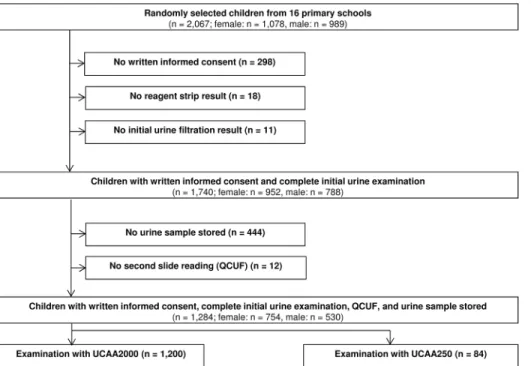
and four schools with a prevalence of 5–10% based on single urine filtration readings per child (Table 1). From the 16 selected schools, 2,067 children were randomly selected to participate in the annual parasitological survey in 2013. Among them, 298 did not provide written informed consent from their parents and were therefore not asked to submit a urine sample (Fig 1). An additional 29 children did not submit a urine sample of a sufficiently large amount to perform reagent strip and urine filtration examinations. Hence, the initialS.haematobiumprevalence at Table 2. S.haematobiumprevalences in children visiting 16 primary schools on Pemba in 2013, stratified by diagnostic approach.
Prevalence level School Number of children examined Micro-hematuria
UF QCUF UCAA2000- UCAA2000+ UCAA250- UCAA250
+ No. pos % No. pos % No. pos
% No. pos % No. pos % No.
pos
% No.
pos %
<2% Ng'ombeni 75 2 2.7 1 1.3 1 1.3 1 1.3 2 2.7
Kengeja 68 2 2.9 1 1.5 2 2.9 no
observation
no observation
10 14.7 21 30.9
Kangani 95 2 2.1 2 2.1 2 2.1 (10/79) 12.7 (16/79) 20.3 (0/
16)
0.00 (0/ 16)
0.00
Wesha 69 1 1.5 2 2.9 4 5.8 5 7.3 5 7.3 - - -
-Chanjamjawiri 80 0 0.0 1 1.3 4 5.0 14 17.5 24 30.0 - - -
-Kwale 86 1 1.2 0 0.0 0 0.0 5 5.8 6 7.0 - - -
-Mbuzini 76 1 1.3 1 1.3 1 1.3 5 6.6 8 10.5 - - -
-Mtambile 81 3 3.7 1 1.2 2 2.5 15 18.5 20 24.7 - - -
-Total setting*
546 10 1.8 8 1.5 14 2.6 55 10.1 81 14.8 - - -
-2–5% Wawi 76 6 7.9 2 2.6 4 5.3 5 6.6 7 9.2 - - -
-Ole 92 5 5.4 4 4.4 5 5.4 21 22.8 24 26.1 - - -
-Makangale 71 2 2.8 1 1.4 1 1.4 3 4.2 11 15.5 - - -
-Mkanyageni 87 6 6.9 4 4.6 5 5.8 10 11.5 14 16.1 - - -
-Total setting*
326 19 5.8 11 3.4 15 4.6 39 12.0 56 17.2 - - -
-5–10% Konde A 88 0 0.0 4 4.6 5 5.7 6 6.8 8 9.1 - - -
-Ngwachani 70 5 7.1 5 7.1 5 7.1 26 37.1 28 40.0 - - -
-Kinowe 90 5 5.6 5 5.6 10 11.1 21 23.3 30 33.3 - - -
-Shungi 80 10 12.5 8 10.0 8 10.0 12 15.0 15 18.8 - - -
-Total setting*
328 20 6.1 22 6.7 28 8.5 65 19.8 81 24.7 - - -
-Total* 1200 49 4.1 41 3.4 57 4.8 159 13.3 218 18.2 - - -
-UF: initial urinefiltration at the day of sample collection between March and May 2013; QCUF: second reading of the urinefiltration slide for quality control purposes between November 2013 and January 2014; UCP-LF CAA: up-converting phosphor-lateralflow assay detecting circulating anodic antigen in urine; UCAA2000+: UCP-LF CAA prepared with 1.5 ml of urine, indecisive results were considered as positive; UCAA2000-: UCP-LF CAA prepared with 1.5 ml of urine, indecisive results were considered as negative; UCAA250+: UCP-LF CAA prepared with 250μl of urine, indecisive results were considered as positive; UCAA250-: UCP-LF CAA prepared with 250μl of urine, indecisive results were considered as negative.
*Only participants with a UCAA2000 were included in analysis.
the unit of the school was calculated from single urine filtration results of 1,740 children. Among the 1,740 children examined with the initial urine filtration and reagent strip, 444 had no urine sample stored for future analysis, and 12 urine filtration slides were not available for reexamination by the quality control reader. Finally, UCP-LF CAA and QCUF readings were available from 1,284 children. The UCAA2000 and UCAA250 were applied on 1,200 and 84 urine samples, respectively.
The sample sizes of the UCAA2000 tests for prevalence settings<2%, 2–5%, and 5–10%
were 546, 326, and 328, respectively. While the numbers for the latter two prevalence settings are in line with our initial sample size calculation, there were fewer UCAA2000 tests performed for the lowest prevalence settings. This is due to interim analyses, which revealed that the prev-alence outcomes obtained with the UCAA2000 in these very low prevprev-alence settings were seven times higher than those based on a single urine filtration, and hence the sample size could be lowered to around 500 UCAA2000 tests. The UCAA250 was performed to gather new information on a different approach, which is potentially less complicated to perform in re-source-constrained settings.
Empirical
S.
haematobium
Prevalences According to Diagnostic
Approach
Fig 2shows that there were considerable differences in the empiricalS.haematobium preva-lences at the unit of the school and according to endemicity level, depending on the diagnostic approach applied. The thresholds of<2%, 2–5%, and 5–10% were only met with the initial
urine filtration reading, indicating an average prevalence of 1.5%, 3.4%, and 6.7% for the Fig 1. Flowchart detailing study participation and urine sampling procedures.Flowchart indicating the inclusion and exclusion of data for determining the accuracy of different methods for the diagnosis of Schistosoma haematobiumin children from Pemba, United Republic of Tanzania, in 2013. UCP-LF CAA: up-converting phosphor-lateral flow assay detecting circulating anodic antigen in urine; UCAA2000: UCP-LF CAA prepared with 1.5 ml of urine; UCAA250: UCP-LF CAA prepared with 250μl of urine.
schools stratified to each endemicity level (Table 2). The QCUF reading revealed slightly higher average prevalences of 2.6%, 4.6% and 8.5%, respectively, and reagent strips of 1.8%, 5.8%, and 6.1%, respectively. Considerably higher prevalences for individual schools and average preva-lences according to endemicity level were revealed by the UCAA2000. Considering indecisive results as negative, the UCAA2000- indicated average prevalences of 10.1%, 12.0% and 19.8%, respectively, for the three endemicity levels. Considering indecisive results as positive, the UCAA2000+ indicated average prevalences of 14.8%, 17.2% and 24.7%, respectively. Fig 2. Maps indicatingSchistosoma haematobiumprevalence levels according to different diagnostic tests.The two maps indicate differentS. haematobiumprevalence levels as identified with a single urine filtration method (A) and a urine-based up-converting phosphor-lateral flow circulating anodic antigen (UCAA2000) assay (B) in 16 schools on Pemba island, United Republic of Tanzania, in 2013. UCAA2000: up-converting phosphor-lateral flow assay detecting circulating anodic antigen in urine and prepared with 1.5 ml of urine; green spot: school with a prevalence of<2%; yellow spot: school with a prevalence of 2–5%; orange spot: school with a prevalence of 5-<10%; red spot: school with a prevalence of10%.
Sensitivity Estimates of Diagnostic Methods Using an Imperfect
‘
Gold
’
Standard
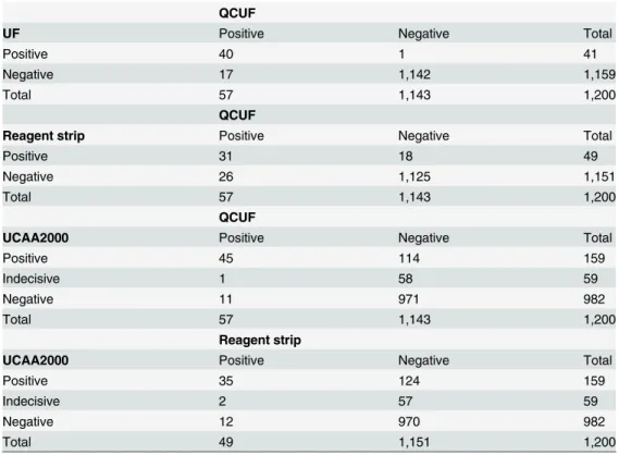
The numbers of positive, negative, and indecisive results for each diagnostic method combina-tion are shown in 6-cell matrixes inTable 3. Since QCUF revealed moreS.haematobium -in-fected individuals than the initial urine filtration microscopy, only QCUF results were considered for the calculation of sensitivity and specificity. Applying a combination of the QCUF and UCAA2000+ as imperfect diagnostic‘gold’standard, the UCAA2000+ had the highest overall sensitivity of 95.2%, followed by the UCAA2000- with a sensitivity of 69.4% (Table 4). The QCUF and reagent strips showed very low sensitivities (24.9% and 16.6%, re-spectively). While the UCAA2000+ showed stable sensitivity across the threeS.haematobium
prevalence settings (<2%, 2–5%, and 5–10%), a decreasing trend in sensitivity with lower
prev-alence was observed for the UCAA2000- and particularly for the QCUF. A considerable drop in the sensitivity of reagent strip results only occurred in the<2% prevalence setting. Changes
in sensitivity were, however, not statistically significant. Noteworthy, the geometric mean egg count decreased significantly from highest to lowest prevalence settings from 0.22 eggs/10 ml urine to 0.05 eggs/10 ml urine. As shown inFig 3, we found a significant relationship between CAA pg/ml levels andS.haematobiumegg counts (Spearman’s rho = 0.24; p<0.001), between
Table 3. Agreement between the different diagnostic approaches.
QCUF
UF Positive Negative Total
Positive 40 1 41
Negative 17 1,142 1,159
Total 57 1,143 1,200
QCUF
Reagent strip Positive Negative Total
Positive 31 18 49
Negative 26 1,125 1,151
Total 57 1,143 1,200
QCUF
UCAA2000 Positive Negative Total
Positive 45 114 159
Indecisive 1 58 59
Negative 11 971 982
Total 57 1,143 1,200
Reagent strip
UCAA2000 Positive Negative Total
Positive 35 124 159
Indecisive 2 57 59
Negative 12 970 982
Total 49 1,151 1,200
Four-cell and six-cell-matrixes showing the agreement of the number of positive, negative, and indecisive results of the initial urinefiltration (UF), the quality control slide reading (QCUF), the reagent strips, and the up-converting phosphor-lateralflow assay detecting circulating anodic antigen in urine (UCAA2000) methods for the diagnosis ofS.haematobiumin urine samples from children from 16 primary schools in Pemba, United Republic of Tanzania in 2013.
CAA pg/ml levels and microhematuria grading (Spearman’s rho = 0.23; p<0.001), and
be-tween egg counts and microhematuria grading (Spearman’s rho = 0.57; p<0.001).
Accuracy Estimates of Diagnostic Methods Using LCA
Statistical information criteria (i.e., AIC and BIC) indicated that no random effects were need-ed at the school level, suggesting that neither the diagnostic performance of reagent strip, QCUF, or UCAA2000 tests, nor the model estimatedS.haematobiumprevalence varied signifi-cantly between the surveyed schools. The assumption of conditional independence between the three diagnostic tests was considered as valid, since inspection of the standardized results from the final selected model (S1 Model1) did not show extreme values (i.e., residuals for all response patterns were between -2 and 2). Furthermore, when we allowed for partial condition-al independence between reagent strip and QCUF results, the model fit was not improved (S1 Model4), which further strengthened the argument for conditional independence between the tests.
Our final LCA model (S1 Model1, with the lowest AIC and BIC) revealed a sensitivity of 97.0% (95% CI: 90.5–100%), 85.5% (95% CI: 72.2–98.8%), and 66.7% (95% CI: 52.4–81.0%) for UCAA2000, QCUF, and reagent strip, respectively. The highest specificity was obtained for QCUF (99.1%, 95% CI: 98.5–99.7%), followed by reagent strip (98.9%, 95% CI: 98.3–99.5%), Table 4. Diagnostic accuracy of the tests used to detectS.haematobiuminfections stratified by prevalence setting.
‘Gold’standard Prevalence n GM eggs/10 ml Test Sensitivity % [95% CI]
Combination of QCUF and UCAA2000+ as‘gold’standard* all 1,200 Reagent strip 16.6 [12.0–22.1]
0.12 QCUF 24.9 [19.4–31.0]
CAA2000- 69.4 [63.0–75.3] CAA2000+ 95.2 [91.6–97.6] Combination of QCUF and UCAA2000+ as‘gold’standard* <2% 546 Reagent strip 8.3 [3.4–16.4]
0.05 [0.02–0.09] QCUF 16.7 [9.4–26.4] CAA2000- 65.5 [54.3–75.5] CAA2000+ 96.4 [89.9–99.3] Combination of QCUF and UCAA2000+ as‘gold’standard* 2–5% 326 Reagent strip 24.1 [13.9–37.2] 0.13 [0.05–0.23] QCUF 25.9 [15.3–39.0] CAA2000- 67.2 [53.7–79.0] CAA2000+ 96.6 [88.1–99.6] Combination of QCUF and UCAA2000+ as‘gold’standard* 5–10% 328 Reagent strip 19.5 [11.8–29.4] 0.22 [0.13–0.33] QCUF 32.2 [22.6–43.1] CAA2000- 74.7 [64.3–83.4] CAA2000+ 93.1 [85.6–97.4]
Diagnostic accuracy of reagent strips, quality control urinefiltration slide reading (QCUF), and the up-converting phosphor-lateralflow assay detecting circulating anodic antigen in urine methods (UCAA2000+, and UCAA2000-) forS.haematobiumdetection as calculated by comparison against an imperfect‘gold’standard (i.e., the combination of UCAA2000+ and QCUF results), stratified by prevalence setting in our study conducted in Pemba, United Republic of Tanzania in 2013.
QCUF: second reading of the urinefiltration slide for quality control purposes between November 2013 and January 2014; UCP-LF CAA: up-converting phosphor-lateralflow assay detecting circulating anodic antigen in urine; UCAA2000+: UCP-LF CAA prepared with 1.5 ml of urine, indecisive results were considered as positive; UCAA2000-: UCP-LF CAA prepared with 1.5 ml of urine, indecisive results were considered as negative; UCAA250+: UCP-LF CAA prepared with 250μl of urine, indecisive results were considered as positive; UCAA250-: UCP-LF CAA prepared with 250μl of urine, indecisive results were considered as negative; GM: geometric mean
*: Test specificity was assumed to be 100%.
and UCAA2000 (90.1%, 95% CI: 88.3–91.9%). The model estimatedS.haematobium preva-lence including all schools was 4.5%.
Discussion
Enhanced efforts to achieve the schistosomiasis control and elimination goals put forth by WHO for the years 2020 and 2025 will likely reduce theSchistosomaprevalence and infection intensities in targeted populations. To discover and investigate continuing transmission and to reliably confirm schistosomiasis elimination without missing very light infection intensities, di-agnostic tools with high sensitivity and specificity are needed [11,19–22,41].
We assessed the accuracy of the UCAA2000 assay forS.haematobiumdiagnosis in low-en-demicity settings on Pemba Island. Based on a single urine filtration, we selected schools with a prevalence ofS.haematobium<2%, 2–5%, and 5–10%. LCA revealed an overall sensitivity and
specificity of a single UCAA2000 of 97.0% and 90.1%, single QCUF of 85.5% and 99.1%, and single reagent strips of 66.7% and 98.9%, respectively, and a model estimated prevalence of 4.5%. No significant drop in the empirical sensitivity from highest to lowest investigated en-demicity scenario was revealed, but we observed a clear tendency of decreasing sensitivity of particularly the QCUF and reagent strip test results with lower prevalence and geometric mean egg count levels. The overallS.haematobiumprevalence empirically determined with the UCAA2000+, UCAA2000-, urine filtration, and reagent strips were 18.2%, 13.3%, 4.8%, and 4.1%, respectively.
Our results show that empirically, a single UCAA2000 test detects a considerably higherS.
haematobiumprevalence than microscopy or reagent strips. Even if indecisive results obtained with the UCAA2000 were considered as negative, the overallS.haematobiumprevalence was almost three times higher than that elucidated by QCUF. Particularly evident were the differ-ences in the lowest endemicity setting, where UCAA2000+ and UCAA2000- revealed preva-lences of 14.8% and 10.1%, respectively, while QCUF and reagent strips found considerably lower prevalences of 2.6% and 1.8%, respectively. If indeed correct, this finding would have im-portant ramifications for the schistosomiasis elimination program in Zanzibar. Since egg out-put and microhematuria were reasonably low, according to the current definitions, elimination of schistosomiasis as a public health problem had been reached and the setting was on the way toward interruption of transmission. The UCAA2000 revealed, however that more than 10% of the surveyed population excreted CAA and thus harbored living worms, which potentially could produce eggs at some point passed in urine. Given these persistent very low intensity infections in the presence of ongoing control interventions and the potential for disease recru-descence or the parasite’s reintroduction into parasite-free environments also by very modest external inputs, the situation would require an adequate response in terms of more effective lo-cally targeted control strategies [42].
Fig 3. Correlation of circulating anodic antigen (CAA) levels andS.haematobiumegg counts or microhematuria grading.(A) Correlation of CAA levels (pg/ml) in 1.5 ml of urine and the number ofS. haematobiumeggs detected in 10 ml of urine (Spearman’s rho = 0.24; P<0.001); (B) correlation of CAA levels (pg/ml) and the microhematuria grading (Spearman’s rho = 0.23; P<0.001); and (C) correlation ofS. haematobiumeggs detected and microhematuria grading (Spearman’s rho = 0.57; p<0.001), in urine samples from children from Pemba, United Republic of Tanzania, collected in 2013. The horizontal
continuous red line indicates the cut-off value of>0.4 pg/ml for samples clearly indicated asS.haematobium -positive by the UCAA2000 (A and B). The horizontal dotted red line indicates the cut-off value of<0.2 pg/ml
for samples clearly indicated asS.haematobium-negative by the UCAA2000 (A and B). Values right from the vertical continuous red line (A) and above the horizontal continuous red line (C) indicate egg-positive urine filtration tests
The model estimated overallS.haematobiumprevalence, in contrast, was only 4.5%, taking into account an imperfect specificity of the UCAA2000, which was estimated by LCA at 90.1%. Considering this imperfect specificity, a considerable amount ofS.haematobiumcases detected by the UCAA2000 might have been false-positives. However, a specificity of 90.1% is below the specificity of circulating antigen assays and the UCP-LF CAA test as postulated elsewhere [27,29,43,44]. Moreover, studies with the UCAA2000 in non-endemic African settings using samples from previous studies, banked at Leiden University Medical Center revealed that no false-positives were detected by the method. One also has to note that CAA is released by living worms that might or might not produce eggs. In case children were reached by the latest MDA conducted in November 2012 and knowing that CAA clears within a few days or weeks after successful treatment [45,46], it might be that the CAA-positive results in the egg-negative urines collected between March and May 2013 indicate worms that survived but were sterilized by the previous praziquantel treatment, or worms that were schistosomula at the time of treat-ment and not affected by praziquantel, or new infections with schistosomes that were not yet producing eggs. Since we were working in an elimination setting where transmission is mostly low, it might also be that CAA-positive but egg-negative individuals were infected with single worms rather than worm pairs, and hence no eggs were produced. The sensitivity of the UCAA2000 of 97.0% determined with the LCA and of the UCAA2000+ of 95.2% determined with an imperfect‘gold’standard in our study, is in line with findings from the People’s Repub-lic of China, where the UCAA2000+ sensitivity forS.japonicumdetection was 93% [29].
A limitation of our study and approaches to estimate sensitivity and specificity is that the UCAA2000 was only compared to two diagnostic techniques with a limited accuracy, particu-larly for examining samples from a low-endemic area and when only a single urine sample was examined. Clearly, the UCAA2000 has a very high sensitivity and a sufficient specificity to serve as a tool for diagnosing urogenital schistosomiasis in low-endemicity settings targeting elimination. Whether its specificity is high enough to deserve also the title“confirmation of elimination tool”remains to be elucidated in future studies comparing its performance not only with microscopy and reagent strips but also with more accurate methods, for example with polymerase chain reaction (PCR) [47], in a similar close-to-elimination setting.
The considerable number of“potentially positive”individuals determined by“indecisive” UCAA2000 results is another limitation of our study. Indecisive results appeared due to the se-lection of a higher specificity and a lower specificity cut-off. As described in Corstjenset al. (2014), the cut-off thresholds may be influenced by technical factors such as batch-to-batch variation, as well as (immuno-)epidemiological settings (e.g., co-infections, age, and geogra-phy) [27]. In the current setting, suboptimal sample volume and centrifugation capacities were considered the main reason for the need of a higher cut-off threshold. Moreover, the definition of a precise cut-off for any UCP-LF CAA approach is pending and can only be developed for large reagent strip batches and by testing a large number of clearly uninfected people from en-demic and non-enen-demic settings. Currently, to obtain a clear result for potentially positive in-dividuals, their urine sample would either need to be repeatedly tested with the UCAA2000, or larger amounts of their urines would need to be examined, for example with the UCAA7500, which has a lower detection limit of 0.03 pg/ml [27], or their CAA level could be retested after praziquantel treatment to investigate whether it decreased.
for studies on diagnostic accuracy as well as for prevalence estimates in schistosomiasis control and elimination programs.
Our study shows that the UCAA2000 is a highly sensitive diagnostic tool that is able to diag-noseS.haematobiuminfections in very low endemicity settings. The dry format allows conve-nient transport of dry reagents without a cold chain to third-party laboratories [27]. The assay can be implemented by trained local technicians in laboratories in endemic settings, given they are adequately equipped such as the PHL-IdC in Pemba. When sufficient centrifugation capac-ities and a UCP-Quant reader are available, up to 100 samples can be processed by one techni-cian per day, and hence, the test has a higher throughput than parasitological approaches requiring microscopy. However, in the current format, the UCAA2000 cannot be applied in field laboratories without centrifugation and pipetting capacities.
Moreover, the costs for a single UCAA2000 are high and the test is not commercialized. Hence, at the time being, this test is out of reach for most control programs and large-scale studies in endemic areas, but is only used in collaborative projects. Efforts to develop a simple-to-use but still highly sensitive point-of-care (POC)-CAA rapid test for commercialization at an affordable price are underway.
One also has to consider that the UCAA2000 is a highly sensitive test to reflect active infec-tion, but that it does not encapsulate morbidity. In low endemic settings, where the primary aim is to assess transmission, this test holds particular potential. For indicating morbidity caused by urogenital schistosomiasis, rapid tests such as reagent strips showing the grade of he-maturia, and detecting proteinuria and leukocyturia or urine albumin and creatinine have clear advantages [22,48,49].
Once standardized, commercialized, and widely available at reasonable costs, we consider the UCAA2000 as a suitable tool for large-scale monitoring of urogenital schistosomiasis in control programs in low-endemicity settings targeting elimination and for surveillance in areas that achieved elimination. For surveillance at a smaller scale, including testing of suspected cases in remote public health care centres without laboratory equipment, a simple-to-use but still highly sensitive POC-CAA rapid test is highly desirable.
Supporting Information
S1 Checklist. STARD checklist. (PDF)
S1 Data. Dataset used for analysis of the data presented in this manuscript. (PDF)
S1 Models. Latent class analysis (LCA) modeling approaches for sensitivity and specificity estimation.
(PDF)
S1 Translation. Translation of abstract into Dutch, French, and German. (PDF)
Acknowledgments
Abdalla M. Omar who prepared and analyzed the stored urine samples with the UCP-LF CAA tests. We would also like to thank Muriel Rabone for creating the maps forFig 2. Last but not least, we acknowledge the children visiting primary schools in Pemba for their participation in the study.
Author Contributions
Conceived and designed the experiments: SK PLAMC SMAl KAM JU DR GJvD. Performed the experiments: SK PLAMC CIC SMAm CJdD DR GJvD. Analyzed the data: SK AK GJvD. Contributed reagents/materials/analysis tools: SK PLAMC AK SMAl CJdD KAM DR GJvD. Wrote the paper: SK PLAMC AK CIC SMAm SMAl CJdD KAM JU DR GJvD.
References
1. Utzinger J, Keiser J. Research and development for neglected diseases: more is still needed, and faster. Lancet Glob Health. 2013; 1: e317. doi:10.1016/S2214-109X(13)70148-7PMID:25104587
2. Molyneux DH. Neglected tropical diseases: now more than just 'other diseases'—the post-2015 agen-da. Int Health. 2014; 6: 172–180. doi:10.1093/inthealth/ihu037PMID:24969646
3. Multiple Partners. Uniting to combat neglected tropical diseases. Ending the neglect & reaching 2020 goals. Table of commitments. London, United Kingdom. 30 January 2012. Available:https://www.gov. uk/government/uploads/system/uploads/attachment_data/file/67442/NTD_20Event_20-_20Table_ 20of_20Commitments.pdf. Accessed: 14 April 2015.
4. Stothard JR, Stanton MC, Bustinduy AL, Sousa-Figueiredo JC, van Dam GJ, Betson M, et al. Diagnos-tics for schistosomiasis in Africa and Arabia: a review of present options in control and future needs for elimination. Parasitology. 2014; 141: 1947–1961. doi:10.1017/S0031182014001152PMID:25158604
5. WHO. Schistosomiasis: progress report 2001–2011 and strategic plan 2012–2020. Geneva: World Health Organization; 2013. pp 1–80.
6. Rollinson D, Knopp S, Levitz S, Stothard JR, Tchuem Tchuenté LA, Garba A, et al. Time to set the agenda for schistosomiasis elimination. Acta Trop. 2013; 128: 423–440. doi:10.1016/j.actatropica. 2012.04.013PMID:22580511
7. Utzinger J, Raso G, Brooker S, de Savigny D, Tanner M, Ornbjerg N, et al. Schistosomiasis and ne-glected tropical diseases: towards integrated and sustainable control and a word of caution. Parasitolo-gy. 2009; 136: 1859–1874. doi:10.1017/S0031182009991600PMID:19906318
8. WHO. Neglected tropical diseases. Prevention, control, elimination and eradication. Provisional agen-da item 9.2. Geneva: World Health Organization; 2012. pp 1–10.
9. Grimes JET, Croll D, Harrison WE, Utzinger J, Freeman MC, Templeton MR. The relationship between water, sanitation and schistosomiasis: a systematic review and meta-analysis. PLoS Negl Trop Dis. 2014; 8: e3296. doi:10.1371/journal.pntd.0003296PMID:25474705
10. King CH. Toward the elimination of schistosomiasis. N Engl J Med. 2009; 360: 106–109. doi:10.1056/ NEJMp0808041PMID:19129524
11. Bergquist R, Johansen MV, Utzinger J. Diagnostic dilemmas in helminthology: what tools to use and when? Trends Parasitol. 2009; 25: 151–156. doi:10.1016/j.pt.2009.01.004PMID:19269899
12. Lustigman S, Prichard RK, Gazzinelli A, Grant WN, Boatin BA, McCarthy JS, et al. A research agenda for helminth diseases of humans: the problem of helminthiases. PLoS Negl Trop Dis. 2012; 6: e1582. doi:10.1371/journal.pntd.0001582PMID:22545164
13. Katz N, Chaves A, Pellegrino J. A simple device for quantitative stool thick-smear technique in schisto-somiasis mansoni. Rev Inst Med Trop São Paulo. 1972; 14: 397–400.
14. Peters PA, Mahmoud AA, Warren KS, Ouma JH, Siongok TK. Field studies of a rapid, accurate means of quantifyingSchistosoma haematobiumeggs in urine samples. Bull World Health Organ. 1976; 54: 159–162. PMID:1088097
15. King CH, Bertsch D. Meta-analysis of urine heme dipstick diagnosis ofSchistosoma haematobium in-fection, including low-prevalence and previously-treated populations. PLoS Negl Trop Dis. 2013; 7: e2431. doi:10.1371/journal.pntd.0002431PMID:24069486
17. Koukounari A, Donnelly CA, Moustaki I, Tukahebwa EM, Kabatereine NB, Wilson S, et al. A latent Mar-kov modelling approach to the evaluation of circulating cathodic antigen strips for schistosomiasis diag-nosis pre- and post-praziquantel treatment in Uganda. PLoS Comput Biol. 2013; 9: e1003402. doi:10. 1371/journal.pcbi.1003402PMID:24367250
18. McCarthy JS, Lustigman S, Yang GJ, Barakat RM, García HH, Sripa B, et al. A research agenda for hel-minth diseases of humans: diagnostics for control and elimination programmes. PLoS Negl Trop Dis. 2012; 6: e1601. doi:10.1371/journal.pntd.0001601PMID:22545166
19. Solomon AW, Engels D, Bailey RL, Blake IM, Brooker S, Chen JX, et al. A diagnostics platform for the integrated mapping, monitoring, and surveillance of neglected tropical diseases: rationale and target product profiles. PLoS Negl Trop Dis. 2012; 6: e1746. doi:10.1371/journal.pntd.0001746PMID: 22860146
20. Knopp S, Becker S, Ingram K, Keiser J, Utzinger J. Diagnosis and treatment of schistosomiasis in chil-dren in the era of intensified control. Expert Rev Anti Infect Ther. 2013; 11: 1237–1258. doi:10.1586/ 14787210.2013.844066PMID:24127662
21. Bergquist R, Yang GJ, Knopp S, Utzinger J, Tanner M. Surveillance and response: tools and ap-proaches for the elimination stage of neglected tropical diseases. Acta Trop. 2015; 141: 229–234. doi: 10.1016/j.actatropica.2014.09.017PMID:25301340
22. Utzinger J, Becker SL, van Lieshout L, van Dam GJ, Knopp S. New diagnostic tools in schistosomiasis. Clin Microbiol Infect. 2015 Apr 2. pii: S1198-743X(15)00378-X. doi:10.1016/j.cmi.2015.03.014
23. Polman K, de Vlas SJ, Gryseels B, Deelder AM. Relating serum circulating anodic antigens to faecal egg counts inSchistosoma mansoniinfections: a modelling approach. Parasitology. 2000; 121 Pt 6: 601–610. PMID:11155931
24. Corstjens PLAM, van Lieshout L, Zuiderwijk M, Kornelis D, Tanke HJ, Deelder AM, et al. Up-converting phosphor technology-based lateral flow assay for detection ofSchistosomacirculating anodic antigen in serum. J Clin Microbiol. 2008; 46: 171–176. PMID:17942645
25. Downs JA, van Dam GJ, Changalucha JM, Corstjens PLAM, Peck RN, de Dood CJ, et al. Association of schistosomiasis and HIV infection in Tanzania. Am J Trop Med Hyg. 2012; 87: 868–873. doi:10. 4269/ajtmh.2012.12-0395PMID:23033399
26. van Dam GJ, de Dood CJ, Lewis M, Deelder AM, van Lieshout L, Tanke HJ, et al. A robust dry reagent lateral flow assay for diagnosis of active schistosomiasis by detection ofSchistosomacirculating anodic antigen. Exp Parasitol. 2013; 135: 274–282. doi:10.1016/j.exppara.2013.06.017PMID:23850995
27. Corstjens PLAM, de Dood CJ, Kornelis D, Fat EM, Wilson RA, Kariuki TM, et al. Tools for diagnosis, monitoring and screening ofSchistosomainfections utilizing lateral-flow based assays and upconvert-ing phosphor labels. Parasitology. 2014; 141: 1841–1855. doi:10.1017/S0031182014000626PMID: 24932595
28. Corstjens PLAM, Nyakundi RK, de Dood CJ, Kariuki TM, Ochola E, Karanja DM, et al. Improved sensi-tivity of the urine CAA lateral-flow assay for diagnosing activeSchistosomainfections by using larger sample volumes. Parasit Vectors. 2015; In press.
29. van Dam GJ, Xu J, Bergquist R, de Dood CJ, Utzinger J, Qin ZQ, et al. An ultra-sensitive assay target-ing the circulattarget-ing anodic antigen for the diagnosis ofSchistosoma japonicumin a low-endemic area, People's Republic of China. Acta Trop. 2015; 141: 190–197. doi:10.1016/j.actatropica.2014.08.004 PMID:25128703
30. van Dam GJ, Odermatt P, Acosta L, Bergquist R, de Dood CJ, Kornelis D, et al. Evaluation of banked urine samples for the detection of circulating anodic and cathodic antigens inSchistosoma mekongi andS.japonicuminfections: a proof-of-concept study. Acta Trop. 2015; 141: 198–203. doi:10.1016/j. actatropica.2014.09.003PMID:25225158
31. Knopp S, Mohammed KA, Ali SM, Khamis IS, Ame SM, Albonico M, et al. Study and implementation of urogenital schistosomiasis elimination in Zanzibar (Unguja and Pemba islands) using an integrated multidisciplinary approach. BMC Public Health. 2012; 12: 930. doi:10.1186/1471-2458-12-930PMID: 23110494
32. WHO. Accelerating work to overcome the global impact of neglected tropical diseases—a roadmap for implementation. Geneva: World Health Organization; 2012. pp 1–42.
33. Knopp S, Person B, Ame SM, Mohammed KA, Ali SM, Khamis IS, et al. Elimination of schistosomiasis transmission in Zanzibar: baseline findings before the onset of a randomized intervention trial. PLoS Negl Trop Dis. 2013; 7: e2474. doi:10.1371/journal.pntd.0002474PMID:24147165
34. Fleiss JL. Statistical methods for rates and proportions. New York: Wiley; 1981. pp 212–236.
36. Muthén LK, Muthén BO. Mplus user's guide. Los Angeles: Muthén & Muthén; 2012. pp 1–856.
37. Rindskopf D, Rindskopf W. The value of latent class analysis in medical diagnosis. Stat Med. 1986; 5: 21–27. PMID:3961312
38. Ibironke O, Koukounari A, Asaolu S, Moustaki I, Shiff C. Validation of a new test forSchistosoma hae-matobiumbased on detection of Dra1 DNA fragments in urine: evaluation through latent class analysis. PLoS Negl Trop Dis. 2012; 6: e1464. doi:10.1371/journal.pntd.0001464PMID:22235360
39. van Smeden M, Naaktgeboren CA, Reitsma JB, Moons KG, de Groot JA. Latent class models in diag-nostic studies when there is no reference standard—a systematic review. Am J Epidemiol. 2014; 179: 423–431. doi:10.1093/aje/kwt286PMID:24272278
40. Simel DL, Rennie D, Bossuyt PM. The STARD statement for reporting diagnostic accuracy studies: ap-plication to the history and physical examination. J Gen Intern Med. 2008; 23: 768–774. doi:10.1007/ s11606-008-0583-3PMID:18347878
41. Colley DG, Bustinduy AL, Secor WE, King CH. Human schistosomiasis. Lancet. 2014; 383: 2253–
2264. doi:10.1016/S0140-6736(13)61949-2PMID:24698483
42. Spear RC, Seto EYW, Carlton EJ, Liang S, Remais JV, Zhong B, et al. The challenge of effective sur-veillance in moving from low transmission to elimination of schistosomiasis in China. Int J Parasitol. 2011; 41: 1243–1247. doi:10.1016/j.ijpara.2011.08.002PMID:21920366
43. Polman K, Diakhate MM, Engels D, Nahimana S, van Dam GJ, Falcão Ferreira ST, et al. Specificity of
circulating antigen detection for schistosomiasis mansoni in Senegal and Burundi. Trop Med Int Health. 2000; 5: 534–537. PMID:10995094
44. Midzi N, Butterworth AE, Mduluza T, Munyati S, Deelder AM, van Dam GJ. Use of circulating cathodic antigen strips for the diagnosis of urinary schistosomiasis. Trans R Soc Trop Med Hyg. 2009; 103: 45–
51. doi:10.1016/j.trstmh.2008.08.018PMID:18951599
45. de Jonge N, de Caluwe P, Hilberath GW, Krijger FW, Polderman AM, Deelder AM. Circulating anodic antigen levels in serum before and after chemotherapy with praziquantel in schistosomiasis mansoni. Trans R Soc Trop Med Hyg. 1989; 83: 368–372. PMID:2515625
46. van Lieshout L, de Jonge N, el-Masry N, Mansour MM, Bassily S, Krijger FW, et al. Monitoring the effi-cacy of different doses of praziquantel by quantification of circulating antigens in serum and urine of schistosomiasis patients. Parasitology. 1994; 108: 519–526. PMID:8052507
47. Vinkeles Melchers NV, van Dam GJ, Shaproski D, Kahama AI, Brienen EA, Vennervald BJ, et al. Diag-nostic performance ofSchistosomareal-time PCR in urine samples from Kenyan children infected with Schistosoma haematobium: day-to-day variation and follow-up after praziquantel treatment. PLoS Negl Trop Dis. 2014; 8: e2807. doi:10.1371/journal.pntd.0002807PMID:24743389
48. Stothard JR, Sousa-Figueiredo JC, Khamis IS, Garba A, Rollinson D. Urinary schistosomiasis-associ-ated morbidity in schoolchildren detected with urine albumin-to-creatinine ratio (UACR) reagent strips. J Pediatr Urol. 2009; 5: 287–291. doi:10.1016/j.jpurol.2008.09.010PMID:19168393