Cerrena
sp. HYB07 with Dye Decolorizing Ability
Jie Yang1,2, Qi Lin1, Tzi Bun Ng3, Xiuyun Ye1,2, Juan Lin1,2*
1College of Biological Sciences and Technology, Fuzhou University, Fuzhou, Fujian, China,2National Engineering Laboratory for Enzyme Expression, Fuzhou, Fujian, China,3School of Biomedical Sciences, Faculty of Medicine, The Chinese University of Hong Kong, Shatin, New Territories, Hong Kong, China
Abstract
Laccases (EC 1.10.3.2) are a class of multi-copper oxidases with important industrial values. A basidiomycete strainCerrena sp. HYB07 with high laccase yield was identified. After cultivation in the shaking flask for 4 days, a maximal activity of 210.8 U mL21 was attained. A 58.6-kDa laccase (LacA) with 7.2% carbohydrate and a specific activity of 1952.4 U mg21 was purified. 2,29-Azino-bis (3-ethylbenzothiazoline-6-sulfonic acid) was the optimal substrate, withKmandkcatbeing 93.4mM and 2468.0 s21, respectively. LacA was stable at 60uC, pH 5.0 and above, and in organic solvents. Metal ions Na+, K+, Ca2+,
Mg2+, Mn2+, Zn2+enhanced LacA activity, while Fe2+and Li+inhibited LacA activity. LacA decolorized structurally different
dyes and a real textile effluent. Its gene and cDNA sequences were obtained. Putativecis-acting transcriptional response elements were identified in the promoter region. The high production yield and activity, robustness and dye decolorizing capacity make LacA andCerrenasp. HYB07 potentially useful for industrial and environmental applications such as textile finishing and wastewater treatment.
Citation:Yang J, Lin Q, Ng TB, Ye X, Lin J (2014) Purification and Characterization of a Novel Laccase fromCerrenasp. HYB07 with Dye Decolorizing Ability. PLoS ONE 9(10): e110834. doi:10.1371/journal.pone.0110834
Editor:Marco Rito-Palomares, Tecnologico de Monterrey, Mexico
ReceivedJuly 25, 2014;AcceptedSeptember 25, 2014;PublishedOctober 30, 2014
Copyright:ß2014 Yang et al. This is an open-access article distributed under the terms of the Creative Commons Attribution License, which permits unrestricted use, distribution, and reproduction in any medium, provided the original author and source are credited.
Data Availability:The authors confirm that all data underlying the findings are fully available without restriction. All relevant data are within the paper and its Supporting Information files.
Funding:This study was funded by Natural Science Foundation of China (41306120), Oceanic Public Welfare Industry Special Research Project of China (201305015), State Key Laboratory of Pollution Control and Resource Reuse Foundation (PCRRF12011), Fujian Provincial Development and Reform Commission Bio-industry Special Project ([2011]1598) and Fuzhou Science and Technology Bureau (2012-G-125). The funders had no role in study design, data collection and analysis, decision to publish, or preparation of the manuscript.
Competing Interests:The authors have declared that no competing interests exist. * Email: [email protected]
Introduction
Dyestuffs are lost in industrial effluents, posing health and environmental threats [1,2]. Traditional processes cannot remove all dyes or are costly [3]. Microbial treatments suggested for degradation of azo and triphenylmethane dyes often result in mutagenic or carcinogenic products under anaerobic conditions [1,4]. In contrast, laccases may provide a green and efficient alternative for decolorizing dyes before discharge into sewage treatment systems or the environment. By oxidation, laccases catalyze dye transformation, leading to dye decolorization and often detoxification as well [3–7].
Laccases are copper-containing oxidases catalyzing oxidation of phenolic/non-phenolic lignin-related compounds and recalcitrant environmental pollutants [8]. Artificial or natural redox mediators
such as 2,29-azino-bis(3-ethylbenzothiazoline-6-sulfonic acid)
(ABTS) and acetosyringone (ACE) broaden the range of laccase substrates [4]. Because laccases have low substrate specificity, utilize oxygen as final electron acceptor and produce water as only by-product, they find applications in paper pulping and bleaching, textile refining, dye decolorization, bioremediation, organic synthesis, juice and wine clarification, etc. [3].
White-rot fungi are the most efficient laccase producers and most intensively studied. Proposed roles of fungal laccases include morphogenesis, plant pathogenesis, pigment production and lignin degradation [8].
Although laccases have been studied for decades, their applications are hampered by low production yields and reduced performance under industrial conditions. Work is under way to explore sources of laccases with easy availability, high catalytic efficiency, broad substrate specificity, tolerance to alkaline conditions, high temperatures and organic solvents, etc. [5,9–11]
Pleurotus ostreatus and Trametes versicolor are two model organisms in laccase research [12,13]. Other fungi, such as those
from the genera Cerrena and Ganoderma, may still provide
laccases with remarkable qualities [5,9].
To date, publications on laccase-manufacturingCerrenaspecies
mainly dealt with their isolation from the environment, fermen-tation medium and condition optimization, purification and biochemical characterization of laccases and potential applications in bioremediation and biodegradation [9,14–20]. In the present
work, a novel Cerrena sp. strain HYB07 with strong laccase
activity was identified. A laccase, designated as LacA, was purified from the fermentation broth of HYB07. The biochemical characteristics, kinetic properties and dye/effluent decolorizing potentials of LacA were investigated, and the gene and cDNA
sequences ofLacA were cloned. The research presented herein
Materials and Methods
2.1 Organisms and media
The strainCerrena sp. HYB07 [21] was kept in the culture
collection of College of Biological Sciences and Technology, Fuzhou University and maintained through periodic transfers on potato dextrose agar (PDA) (Difco, Franklin Lakes, New Jersey,
USA) at 4uC. For laccase fermentation, five mycelial plugs (1 cm
diameter) were removed from the peripheral region of 3-d-old PDA plates and inoculated in potato dextrose broth (PDB) (Difco,
Franklin Lakes, New Jersey, USA). After growing for 3 d at 28uC
and 150 rpm, an aliquot was taken to inoculate PDB medium
supplemented with 0.5% yeast extract (PDY) and 0.4 mM CuSO4.
2.2 Phylogenetic analysis
Phylogeny of the strain was identified by 18S rDNA sequencing. Genomic DNA was extracted with a DNA Quick Plant System (TIANGEN, Beijing, China), and universal primers NS1 and NS8 (Table 1) were used for amplification of 18S rDNA [22]. The PCR product was ligated with the pMD18-T vector (Takara, Dalian,
China), and the ligation products were transformed into E. coli
TOP10 competent cells (Life Technologies, Grand Island, New York, USA). Four individual clones were sequenced. The 18S rDNA sequence has been submitted to GenBank with the accession number KM233493. The phylogenetic analysis (with 1,000 bootstraps) was performed with MEGA version 5.0 (http:// www.megasoftware.net/) by the neighbor-joining method. Other fungal 18S rDNA sequences used in this study were from GenBank.
2.3 Enzyme activity assay
Laccase activity was assayed according to a previously published method [23] with slight modifications. The reaction contained 25 mM citrate-phosphate buffer (pH 3.0) and 0.5 mM ABTS (Sigma-Aldrich, St. Louis, Missouri, USA), and increase in OD at
420 nm (e= 36,000 M21cm21) was monitored for 5 min. One
unit of enzyme activity (U) was defined as the amount of laccase
required to oxidize 1mmol ABTS per min. All assays were carried
out in triplicate.
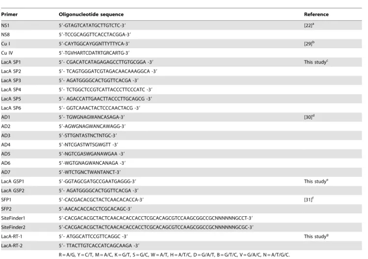
Table 1.Primers used in this study.
Primer Oligonucleotide sequence Reference
NS1 59-GTAGTCATATGCTTGTCTC-39 [22]a
NS8 59-TCCGCAGGTTCACCTACGGA-39
Cu I 59-CAYTGGCAYGGNTTYTTYCA-39 [29]b
Cu IV 59-TGVHARTCDATRTGRCARTG-39
LacA SP1 59- CGACATCATAGAGAGCCTTGTGCGGA -39 This studyc
LacA SP2 59- TCAGTGGGATCGTAGACAACAAAGGCA -39 LacA SP3 59- AGATGGGGCACTGGTTCACGA -39 LacA SP4 59- TCTGGCTCCGTCATTACCCTTCCCATC -39 LacA SP5 59- AGACCATTGAACTTACCCTTGCAGCG -39 LacA SP6 59- GGTCAAACTACTCCCAACTACG -39
AD1 59- TGWGNAGWANCASAGA-39 [30]d
AD2 59-AGWGNAGWANCAWAGG-39
AD3 59-STTGNTASTNCTNTGC-39
AD4 59-NTCGASTWTSGWGTT -39
AD5 59-NGTCGASWGANAWGAA -39
AD6 59-WGTGNAGWANCANAGA -39
AD7 59-WTCTGNCTWANTANCT-39
LacA GSP1 59-GGTAGCGATGCCGAATGAGGG-39 This studye
LacA GSP2 59- AGATGGGGCACTGGTTCACGA -39
SFP1 59-CACGACACGCTACTCAACACACCA-39 [31]f
SFP2 59-AACACACCACCTCGCACAGC-39
SiteFinder1 59-CACGACACGCTACTCAACACACCACCTCGCACAGCGTCCAAGCGGCCGCNNNNNNGCCT-39 SiteFinder2 59-CACGACACGCTACTCAACACACCACCTCGCACAGCGTCCAAGCGGCCGCNNNNNNGCGC-39
LacA-RT-1 59- ATGGCATTCCGTTCAGGC -39 This studyg
LacA-RT-2 59- TTACTTGTCACCATCAGCAAGA -39
R = A/G, Y = C/T, M = A/C, K = G/T, S = G/C, W = A/T, H = A/T/C, D = G/A/T, B = G/T/C, V = G/A/C, N = A/T/G/C.
aUniversal primers for amplification of 18S rDNA.
bDegenerate primers for amplification of the laccase sequence spanning the first and the fourth copper-binding motifs.
cGene-specific primers for TAIL-PCR to amplify the 59- and 39-flanking sequences of the laccase fragment obtained with primers Cu I and Cu IV. dArbitrary degenerate primers for TAIL-PCR.
eGene-specific primers for SiteFinding PCR to amplify the
LacApromoter sequence.
fSiteFinders and their primers (SFP1 and SFP2) for SiteFinding PCR. gPrimers for amplification of the cDNA sequence ofLacA.
2.4 Protein purification
After cultivation in PDY medium for 4 d, the fermentation broth was harvested by paper filtration and then centrifuged (10,000 g, 20 min). The precipitate formed using 40 to 60%
(NH4)2SO4 was collected by centrifugation (20,000 g, 20 min),
resuspended in buffer A (25 mM Tris-HCl buffer, pH 7.5), and centrifuged (12,000 g, 5 min). The supernatant was desalted with a HiPrep 26/10 desalting column (GE Healthcare,
Buckingham-shire, UK) and applied at 5 mL min21to a HiTrap DEAE column
(5 mL) pre-equilibrated with buffer A. Adsorbed proteins were sequentially eluted with 0.1 M, 0.2 M and 1 M NaCl in buffer A.
Fractions with laccase activity were pooled. (NH4)2SO4was added
until the final concentration was 1 M. The resulting sample was
loaded at 1.0 mL min21onto a HiTrap Phenyl FF column (5 mL)
pre-equilibrated with buffer A containing 1 M (NH4)2SO4.
Adsorbed proteins were eluted with a linear 1.0–0 M (NH4)2SO4
gradient in buffer A. Fractions with laccase activity were collected,
examined by SDS-PAGE and zymography and stored at 4uC.
Deglycosylation of laccase by peptide N-glycosidase F (Takara,
Dalian, China) was carried out. Protein identification with MALDI-TOF MS/MS was performed. Protein concentration
Figure 1. Phylogenetic relationships of Cerrenasp. HYB07 and related species based on 18S rDNA sequences.The numbers in parentheses are accession numbers of 18S rDNA sequences. Bootstrap values at nodes are percentages of 1,000 replicates. Scale bar indicates base substitutions/100 bases.
doi:10.1371/journal.pone.0110834.g001
Table 2.Purification of LacA from the culture supernatant ofCerrenasp. HYB07.
Purification step
Total protein (mg)
Total activity (U)
Specific activity
(U mg21) Purificationfold Yield(%)
Culture supernatant 283.2 178,361.3 629.8 1.0 100
40–60% (NH4)2SO4
precipitation
113.5 131,095.6 1155.0 1.8 73.5
DEAE FF 45.8 80,619.3 1760.2 2.8 45.2
Phenyl FF (High Sub) 36.4 71067.6 1952.4 3.1 39.8
was quantified by the Bradford method with bovine serum albumin as the standard [24].
2.5 SDS-PAGE analysis
SDS-PAGE [25] was conducted for molecular weight determi-nation. For zymography analysis, nonreducing loading buffer
(containing SDS but not DTT or b-mercaptoethanol) was
employed, and the sample was not boiled before loading. After electrophoresis the gel was incubated in 10 mL sodium phosphate buffer (pH 6.0) containing 1% (v/v) Triton X-100 for 15–30 min for SDS removal [26] and then immersed in 10 mL fresh sodium
phosphate buffer supplemented with 1 mM guaiacol for direct visualization of laccase activity on the gel.
2.6 Ultraviolet-visible (UV-Vis) absorption spectrum
The UV-Vis absorption spectrum of the purified laccase was recorded between 280 and 800 nm in buffer A.
2.7 Effects of pH and temperature on the activity and stability of purified LacA
The effect of pH was determined between pH 2.0 to 7.0 at
25uC. pH stability was studied by incubating the enzyme at pH
2.0–10.0 at 25uC for 105 h. Residual laccase activity was
quantified with ABTS as substrate. 50 mM glycine-HCl buffer (pH 2.0), citrate-phosphate buffer (pH 2.5–6.5), sodium phosphate buffer (pH 7.0–8.0) and glycine-NaOH buffer (pH 9.0–10.0) were used.
For ascertaining optimum temperature, laccase activity was
measured at optimum pH and temperatures from 25 to 80uC.
Thermostability was analyzed by incubating the enzyme at different temperatures over a duration of 150 min, and residual activity was assayed with substrate ABTS at optimum pH and temperature. All experiments were performed in triplicate.
2.8 Effects of inhibitors and metal ions on the activity of purified LacA
Inhibitors includingL-cysteine, DTT, EDTA, NaN3, SDS and
kojic acid, and metal ions including Na+
, K+
, Li+ , Cu2+
, Ca2+ , Mg2+, Mn2+, Zn2+, Fe2+in form of sulfate, Cr2+and Co2+in form
of nitrate, and Pb2+
in form of subacetate were examined. Individual inhibitor or metal ion was incorporated in the enzyme assay, and activity was determined at optimal temperature and pH. Enzyme activity in absence of inhibitors or metal ions was regarded as 100%.
2.9 Effects of various organic solvents on the activity and stability of LacA
5%, 10% or 25% of the individual solvents, namely methanol,
ethanol, acetonitrile, dimethyl sulfoxide (DMSO) and N,N
-dimethylformamide (DMF) was added to standard enzyme activity assay. For enzyme stability in presence of organic solvents, LacA
was pre-incubated with 5%, 10% or 25% of each solvent at 25uC
for 4 h, and residual enzyme activity was determined by using enzyme assay with ABTS as substrate.
2.10 Kinetic studies
Substrate specificity of LacA was determined in triplicate by
using 1–1500mM ABTS, and 25–2500mM guaiacol,
2,6-dimethoxyphenol (2,6-DMP) and catechol at the respective optimum temperature and pH. Oxidation of the four substrates were measured for 5 min [27,28]. The kinetic parameters were estimated based on nonlinear regression of the Michaelis-Menten equation using GraphPad Prism version 5.0 (GraphPad Software, San Diego, California, USA).
2.11 Decolorization
Dye decolorizing ability was evaluated using 25 dyes. The reaction mixture in 20 mL was composed of (in final
concentra-tion): 0.1 M citrate-phosphate buffer (pH 6.0), 0.2 or 2 U mL21
laccase, 0.1 mM ACE or 12.5 mM kojic acid (if needed) and dyes.
Decolorization was carried out in the dark at 28uC and 150 rpm
for 24 h. The negative control had no enzyme. Decolorization was followed at the wavelength for each dye, determined from its absorbance spectrum in 0.1 M citrate-phosphate buffer between
Figure 2. Purification and characterization of LacA fromCerrena
sp. HYB07.(A) Time course of laccase activity and secreted protein levels produced byCerrenasp. HYB07. Error bars represent standard deviations of triplicate experiments. (B) SDS-PAGE analysis of LacA. Lane M1, Protein molecular weight marker (Low) by Takara; lane 1, purified LacA; lane 2, purified LacA after peptideN-glycosidase F treatment; lane M2, Blue Plus II Protein Marker (Transgen, China); lane 3, purified LacA. Lanes 1 and 2 were stained with Coomassie Brilliant Blue R-250 after reducing electrophoresis; lane 3 was stained with guaiacol after non-reducing electrophoresis, and the sample was not heated before loading. (C) UV-Vis absorbance spectrum of purified LacA.
280 and 800 nm. Decolorization (%) = [(Ai–At)/Ai]6100, where
Ai is the initial absorption of the dye, and At is the absorption at the time of measurement.
The pH, conductivity and chemical oxygen demand of the textile effluent sample from Septwolves Group Co. in Fujian,
China were 6.3, 537.2mS cm21and 880.3 mg L21, respectively.
LacA was added to the sample at 20 U mL21, and the mixture
Figure 3. Effects of pH and temperature on activity and stability of LacA.(A) Effect of pH on LacA activity with ABTS, guaiacol, 2,6-DMP and catechol as substrates. (B) Effect of pH on LacA stability with ABTS as substrate. (C) Effect of temperature on LacA activity with ABTS, guaiacol, 2,6-DMP and catechol as substrate. (D) Effect of temperature on LacA stability with ABTS as substrate. Bars indicate standard deviations of triplicate determinations.
doi:10.1371/journal.pone.0110834.g003
Table 3.Effect of inhibitors and metal ions on LacA activity.
Inhibitor Concentration (mM) Relative activity (%) Metal ion Concentration (mM) Relative activity (%)
L-Cysteine 0.01 87.561.7 Na+ 10 115.160.9
0.1 5.560.1 K+
10 109.861.7
DTT 0.01 38.467.1 Li+ 10 48.8
60.9
0.1 5.960.7 Cu2+
10 101.964.3
EDTA 1 95.463.2 Ca2+
10 113.662.0
10 82.462.4 Mg2+ 10 111.6
63.2
NaN3 0.01 30.262.5 Mn2+ 10 117.362.3
0.1 9.261.5 Zn2+
10 111.063.9
SDS 0.01 80.860.9 Fe2+ 10 3.8
60.8
0.1 11.461.2 Cr2+ 10 99.0
61.1
Kojic acid 12.5 49.361.8 Co2+
10 99.864.1
50 31.763.2 Pb2+ 10 93.8
63.7
incubated at 28uC and 150 rpm for 6 d. Decolorization of the effluent was monitored every 24 h by measuring absorbance between 325 and 800 nm. All experiments were conducted in triplicate.
2.12 Cloning of theLacA sequences
All primers used are listed in Table 1. Mycelia were collected
from 4-d-oldCerrenasp. HYB07 cultivated in PDY broth. Total
RNA was extracted with TRIzol (Life Technologies, Grand Island, New York, USA). The RevertAid First Strand cDNA Synthesis Kit (Thermo Scientific, Waltham, Massachusetts, USA) was used to synthesize the first strands of cDNA. The degenerate primers Cu I and Cu IV (Table 1) designed according to the conserved amino acid sequences of the first and fourth copper-binding motifs in fungal laccases [29] were used to amplify laccase gene fragments from cDNA with high-fidelity DNA polymerase (Thermo Scientific, Waltham, Massachusetts, USA). PCR prod-ucts were purified and inserted into pMD18-T vector.
Recombi-nant vectors were transformed intoE. coliTOP10 competent cells.
Twenty clones were randomly selected and submitted to sequencing analysis (Life Technologies, Grand Island, New York, USA). All 20 clones corresponded to the same laccase gene, confirmed to be the gene encoding LacA by MALDI-TOF MS/
MS analysis. The 59- and 39-flanking genetic sequences of the
amplified laccase fragment were obtained by thermal asymmetric interlaced PCR (TAIL-PCR) [30] with HYB07 genomic DNA as the template, LacA SP1-6 as gene-specific primers and AD1-7 as arbitrary degenerate primers (Table 1) [30]. The full-length cDNA sequence was amplified with primers LacA-RT-1 and LacA-RT-2 (Table 1). The sequence has been submitted to GenBank with the accession number KF317949. SiteFinding PCR [31] was
per-formed to further extend the promoter region ofLacA, in which
the gene-specific primers were LacA GSP1 and GSP2 (Table 1).
2.13 Bioinformatic analysis
The LacA sequences were analyzed using the Vector NTI
program (Life Technologies, Grand Island, New York, USA) and BLAST (http://blast.ncbi.nlm.nih.gov/Blast.cgi) [32]. Signal pep-tide was predicted with SignalP 3.0 (http://www.cbs.dtu.dk/ services/SignalP/) [33]. The theoretical isoelectric point (pI) and molecular weight (MW) were predicted by the Compute pI/Mw
tool (http://web.expasy.org/compute_pi/) [34]. Potential N
-glycosylation sites (Asn-X-Ser/Thr) were identified with ScanPro-site (http://proScanPro-site.expasy.org/scanproScanPro-site/) [35]. Alignments of laccase proteins were generated with Clustal Omega (http://www. ebi.ac.uk/Tools/msa/clustalo/) [36]. The identities between
Figure 4. LacA-mediated decolorization of a real textile effluent.(A) Absorption spectra of the real textile effluent during LacA-mediated decolorization recorded every 24 h for 6 d. (B) Absorption spectra of the real textile effluent without LacA.
selected fungal laccases were calculated in MEGA version 5.0 (http://www.megasoftware.net/) [37].
Results
3.1 Species identification
A fragment of the 18S rDNA gene of the target fungal strain HYB07 was amplified by PCR and sequenced, and HYB07 was classified based on its 18S rDNA sequence. A phylogeny tree
(Fig. 1) was constructed, indicating HYB07 was closest toCerrena
unicolor (GenBank accession No. AY850007), sharing 99%
sequence identity. The strain was namedCerrenasp. HYB07.
3.2 Production and purification of LacA
Laccase production by Cerrena sp. HYB07 in PDY liquid
medium was monitored for 6 d in the shaking flask. With Cu2+as
inducer, maximum laccase activity (210.8 U mL21) was reached
on day 4. Specific activity (806.3 U mg21) peaked on day 3
(Fig. 2A).
After (NH4)2SO4precipitation, anion exchange and
hydropho-bic interaction chromatography, a monomeric laccase, designated as LacA, was purified 3.1-fold with 39.8% yield from the
fermentation broth. The specific activity was 1952.4 U mg21
(Table 2). Deglycosylation with peptideN-glycosidase F indicated
N-glycosylation. The molecular weight before and after
deglyco-sylation was 58.6 and 54.5 kDa, respectively, thus its glycodeglyco-sylation
level was 7.2%. The molecular weight of,70 kDa on zymograph
exceeded 58.6 kDa in SDS-PAGE (Fig. 2B).
The laccase solution was blue. UV-Vis absorption spectrum (Fig. 2C) showed a peak at 610 nm and a shoulder at 330 nm. The A280:A610 ratio was 20.6.
3.3 Effects of temperature and pH
The pH optimum was 3.0 for ABTS and 2,6-DMP, 4.0 for guaiacol and 4.5 for catechol (Fig. 3A). After storage at pH 4.0 for
105 h, 56% residual enzyme activity was retained. At pH 5.0 or
higher, .80% activity remained. In contrast, LacA activity
decreased to only approximately 1% and 20% at the end of
105-h incubation at pH 2.0 and 3.0, witht1/2being 18 h and 43 h,
respectively (Fig. 3B).
Optimal temperature was 45uC with ABTS, 50uC with
2,6-DMP and 45–50uC with guaiacol and catechol. The enzyme
displayed.50% of maximal activity between 25 and 65uC against
the four substrates except that at 65uC for ABTS, the relative
activity of LacA was only 45% (Fig. 3C). After incubation for
150 min at 50 and 60uC, approximately 85% and 56% of the
original enzyme activity was retained, respectively. LacA was
inactivated after 20 min at 70uC and 2 min at 80uC (Fig. 3D).
3.4 Effects of inhibitors and metal ions
Relative laccase activity was dramatically reduced in the
presence of L-cysteine (to 5.5%), DTT (to 5.9%), sodium azide
(to 9.2%) and SDS (to 11.4%) at the concentration of 0.1 mM. The laccase-specific inhibitor kojic acid at 12.5 mM reduced LacA activity by 50%. No significant activity loss was observed with 10 mM EDTA (Table 3).
At 10 mM, Fe2+exerted the strongest inhibition, followed by
Li+, while Na+, K+, Ca2+, Mg2+, Mn2+ and Zn2+ were slightly
stimulatory. Cu2+, Cr2+, Co2+and Pb2+had no significant effect
(Table 3).
3.5 Effects of various organic solvents
When individual water-miscible organic solvent was added at 5 and 10% final concentration, 102.5–72.7% activity was retained.
Activity was compromised in all solvents at 25%;.70% activity
remained with DMF and ethanol, approximately 60% activity remained with other three organic solvents (Table 4).
LacA was generally stable in the presence of the organic solvents. Residual activity was determined after incubation with
each solvent at 5, 10 or 25% for 4 h at 25uC. An exception was
Table 4.Effect of organic solvents on the activity and stability of purified LacA.
Organic solvent Concentration (%) Relative Activitya(%) Residual Activityb(%)
Methanol 5 92.965.3 92.761.1
10 72.766.7 107.763.1
25 57.366.9 116.565.9
Ethanol 5 101.064.7 94.067.2
10 83.162.9 101.064.5
25 70.963.5 100.263.0
Acetonitrile 5 102.564.2 105.062.8
10 96.861.9 95.965.1
25 66.262.0 50.462.6
DMSO 5 91.162.1 98.164.7
10 77.364.4 116.863.1
25 58.761.8 124.962.4
DMF 5 95.163.7 95.962.1
10 82.562.5 100.760.9
25 78.962.8 94.461.3
Values represent mean6standard deviation (n= 3).
aEnzyme activity was measured with individual organic solvent included in the enzyme activity assay mixture at the concentration indicated. bPurified LacA was incubated with individual organic solvent at the concentration indicated for 4 hours at 25
uC before the standard enzyme activity assay was conducted.
exposure to 25% acetonitrile which was detrimental to activity. LacA was more stable in methanol and DMSO than in aqueous solution.
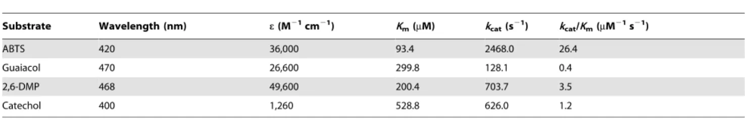
3.6 Kinetic properties of purified LacA
Initial rate kinetic analyses were performed with ABTS, guaiacol, 2,6-DMP and catechol at the respective optimal
temperature and pH values (Table 5). The lowestKmvalue was
found for ABTS (93.4mM), so were the highest turnover rate (kcat)
and catalytic efficiency (kcat/Km) values (2468.0 s21 and
26.4mM21s21, respectively). Catalytic efficiency on ABTS was
7.5 times of that on 2,6-DMP, 22 times of that on catechol and 65 times higher than that on guaiacol. Catechol had the highest
Km value (528.8mM), followed by guaiacol (299.8mM) and
2,6-DMP (200.4mM).
3.7 Dye and real textile effluent decolorization by LacA
Decolorizing capability of purified LacA on industrial and laboratory dyes was evaluated at a low enzyme activity of 0.2 U
mL21(Table 6). Without the help of small redox mediators, LacA
was able to effectively decolorize dyes of different classes, some of which were notoriously recalcitrant to biodegradation, e.g., the indigo dye carmine, triphenylmethane dye malachite green, anthraquinone dye Remazol Brilliant Blue R (RBBR), and azo dyes acid violet 7 and orange II. Among the 25 dyes studies, 13
could be oxidized by 0.2 U mL21LacA with efficiencies higher
than 50%. It was worth pointing out that although 24 h was arbitrarily selected for decolorization measurements, decoloriza-tion of some dyes proceeded faster than others. For example, decolorization of RBBR, indigo carmine and Evans blue was completed within 30 min, but took approximately 2 hours for malachite green and brilliant green (data not shown). A higher
enzyme activity of 2.0 U mL21helped improve decotablorization
efficiencies to different extents. For instance, the decolorization rate increased from 28.8% to 92.4% for Coomassie Brilliant Blue R-250, whereas for Congo red, only a modest improvement (from 33.0% to 57.2%) was seen. On the contrary, for four dyes, namely basic fuchsin, crystal violet, methylene blue and Rhodamine B, there were no significant changes. Therefore, LacA alone at 2.0 U
mL21could decolorize 19 dyes with efficiencies higher than 50%.
For the nine dyes with decolorization efficiencies lower than 60%, we further evaluated their removal by LacA in the presence of 0.1 mM ACE, a natural laccase mediator. Seven of the nine dyes
were efficiently decolorized by the LacA-ACE system except for Rhodamine B and methylene blue. For dyes such as methyl orange and neutral red which could already be partially decolorized by LacA, their decolorization rates were enhanced by ACE (from 53.2% and 28.0% to 92.3% and 80.1%, respectively). In the cases of crystal violet and basic fuchsin, however, ACE was required for their decolorization, since neither
was decolorized by LacA alone at 2.0 U mL21. Even for dyes with
over 80% decolorization efficiencies, inclusion of the laccase-specific inhibitor kojic acid in reactions decreased decolorization efficiencies to 4.2–28.4%, suggesting that LacA was responsible for dye decolorization.
Decolorization of the real textile effluent by LacA predomi-nantly occurred within the first 3 d, accompanied by flattening of the absorption spectrum (Fig. 4A). A negative control was conducted in parallel, and there was little decrease in the absorbance in the absence of LacA (Fig. 4B).
3.8LacAgene cloning and analysis
A fragment ofLacAbetween its first and fourth copper-binding
motifs was amplified fromCerrenasp. HYB07 cDNA. TAIL-PCR
was adopted to clone the 59 and 39-flanking genetic sequences of
the fragment, based on which the full-length gene sequence of
LacAwas obtained. The 3,685-bp DNA sequence consisted of an
892 bp 59-uncoding region, a 2168-bp gene sequence and a
625-bp 39-uncoding region. The full-length cDNA sequence (1,551 bp)
was subsequently obtained. The LacA cDNA corresponded to
purified LacA, as confirmed by MALDI-TOF MS/MS analysis (Fig. S1). The coding region was interrupted by 11 introns between 50–65 bp, and all splicing junctions adhered to the GT-AG rule. LacA consisted of 516 amino acids, with the first 21 residues
being the signal peptide, and 3 putative N-glycosylation sites at
positions 453, 489 and 495. The molecular weight of mature LacA was predicted to be 52.9 kDa, close to the observed 54.5 kDa. Calculated pI was 5.6.
The deduced amino acid sequence of LacA was aligned with
other fungal laccases, including 8Cerrenalaccases (Fig. 5). The
LacA protein possessed four conserved copper-binding motifs typical of fungal laccases, Cu I (HWHGFFQ), Cu II (HSHLSTQ), Cu III (HPFHLHGH) and Cu IV (HCHIDWHL), as well as 10 conserved His involved in copper atom coordination and 5 conserved Cys residues. LacA was most similar to and exhibited
86% identity to the laccase 1 precursor from Spongipellis sp.
Figure 5. Alignment of deduced amino acid sequence of LacA with other laccases (indicated by the GenBank accession numbers).
Laccases used in alignment are:Cerrenasp. HYB07 LacA: KF317949;Cerrenasp. WR1 Lcc1: ACZ58367;Cerrenasp. WR1 Lcc2: ACZ58368;Cerrenasp. WR1 Lcc3: ACZ58369;Cerrena maximalaccase chain A: 2H5U_A;Cerrena maximalaccase chain A: 3DIV_A;Cerrenasp. CTL-2011 laccase: AEL16568;
Cerrena unicolorlaccase: AEQ35306;Cerrena unicolorLac1: ACL93462;Spongipellissp. FERM P-18171 laccase 1 precursor: BAE79811;Panus rudis
laccase A: AAW28932; Rigidoporus microporuslaccase: ACL93333; Meripilus giganteuslaccase: CBV46340;Steccherinum murashkinskyi laccase 2: AFI41889;Trametessp. 420 laccase C: AAW28938;Coriolopsis trogiilaccase: CAC13040;Pleurotus eryngiilaccase: ACI62809. Four conserved copper binding domains are underlined. Conserved His residues are numbered, and conserved Cys residues are labeled.
doi:10.1371/journal.pone.0110834.g005
Table 5.Substrate specificity of LacA.
Substrate Wavelength (nm) e(M21cm21) K
m(mM) kcat(s21) kcat/Km(mM21s21)
ABTS 420 36,000 93.4 2468.0 26.4
Guaiacol 470 26,600 299.8 128.1 0.4
2,6-DMP 468 49,600 200.4 703.7 3.5
Catechol 400 1,260 528.8 626.0 1.2
FERM P-18171 (BAE79811), followed by Lac1 from Cerrena unicolor (ACL93462) with 81% identity, Lcc3 fromCerrenasp.
WR1 (ACZ58369) with 78% identity and laccase fromCerrenasp.
CTL-2011 (AEL16568) with 74% identity. On the other hand,
LacA was more distantly related to Trametes and Pleurotus
laccases (with no higher than 70% identity).
SiteFinding PCR was adopted to further extend the 59-flanking
region ofLacA, rendering a region of 1,544 bp upstream of the
start codon, referred to as the LacA promoter. Bioinformatics
analysis revealed multiple putativecis-acting transcription
regula-tion sites within theLacApromoter sequence in both orientations
(Fig. 6). A TATA box was located 92 bp upstream from the start codon ATG, and three CCAAT boxes were found at positions
2324, 2405 and 21134. The LacA promoter contained two
metal response elements (MREs) with the consensus sequence
TGCRCNC at positions21013 and21227 and one xenobiotic
response element (XRE) with the core sequence TNGCGTG [38]
at21223. Apart from MREs and XREs,LacAhad many ACE1
copper-responsive transcription factor binding sites, consisting of the HWHNNGCTGD or NTNNHGCTGN core [39], at
positions211,2456,2774 and21336, respectively. In addition,
one antioxidant response element (ARE) adhering to the consensus
sequence TGACNNNGC [40] was present at21035 in theLacA
promoter. Putative response elements (PREs) [38], often found in basidiomycete laccase promoter sequences, were also present
within the 59-flanking sequence ofLacA: a TGGGT was located at
position21360, an inverted one at21377, and two ATATC at
2122 and2795. Furthermore, two heat shock response elements
(HSEs) composed of alternately oriented NGAAN repeats [41]
were found at2422 and2857 in theLacApromoter.LacAalso
contained multiple transcription factor binding sites involved in nitrogen and carbon regulation. Two putative CreA-binding sites
(SYGGRG) were found at 2648 and 2706, and five NIT2
binding sites adhering to sequence TATCDH [5] were scattered at
2120, 2525, 2793, 21139 and 21286. No stress response
elements (STREs) with the consensus sequence of CCCCT or
Sp-1 transcription factor recognition site (GGGCGG) [39] were
identified in theLacApromoter sequence.
Discussion
Although laccases have industrial and environmental health implications, commercial applications are limited by low enzyme yields, high costs and tolerance to extreme conditions. Herein, a new Cerrena sp. strain HYB07 produced over 200 U mL21
laccase activity after cultivation for only 3 d with Cu2+ as the
inducer. The high laccase yield and short production period of HYB07 would be advantageous for application and commercial-ization, since many fungal laccase producers require longer
production periods [14]. For C. unicolor VKMF-3196, 15 U
mL21was obtained on the 8thday of cultivation [20]. Enhanced
laccase production byCerrena unicolorstrain MTCC 5159 was
observed with inducers, effluents and synthetic dyes, and a textile
effluent was the most effective inducer, resulting in 85.8 U mL21
after 12 d [18]. ForCerrenasp. WR1, maximal production was
202 U mL21after 13-d cultivation under Cu2+and 2,5-xylidine
induction [9]. ForCerrena unicolorC-139 [15,16], a production
of 250.0 U mL21 was reached after submerged culture for 7 d,
Table 6.Decolorization of the dyes by LacA.
Concentration
(mg L21) lmax
Decolorization (%)
0.2 U mL21 2 U mL21 2 U mL21 +ACE
Acid Chrome Blue K 25 547 41.369.1 93.5±2.8 ND
Acid Fuchsin 50 545 10.361.2 25.566.6 64.1±2.3
Acid Violet 7 25 519 87.8±2.1 96.4±0.9 ND
Aniline Blue 100 585 78.2±1.8 78.2±1.7 ND
Basic Fuchsin 25 543 6.962.1 9.962.6 81.4±0.7
Brilliant Green 25 622 90.6±4.3 91.7±4.7 ND
Bromocresol Green 25 614 12.462.7 58.3±2.5 96.8±0.3
Bromocresol Purple 25 588 78.5±2.4 89.7±1.6 ND
Bromothymol Blue 200 427 73.7±0.8 68.4±4.6 ND
Congo Red 50 478 33.061.4 57.2±4.5 80.6±1.9
Coomassie Brilliant Blue R-250
25 559 28.865.8 92.4±4.7 ND
Crystal Violet 25 554 5.862.1 6.462.9 87.0±1.7
Eriochrome Black T 100 576 84.2±1.5 85.3±1.7 ND
Evans Blue 25 597 89.3±1.6 94.0±0.6 ND
Indigo Carmine 50 609 95.3±2.1 97.7±0.4 ND
Malachite Green 25 618 94.1±7.4 96.3±1.2 ND
Methylene Blue 25 597 4.161.9 6.760.7 5.662.2
Methyl Orange 50 463 18.662.9 53.2±3.7 92.3±0.9
Methyl Red 25 429 25.262.9 62.8±0.8 ND
Neutral Red 25 517 13.461.9 28.062.4 80.1±3.8
Orange II 25 483 79.4±3.3 87.3±1.4 ND
RBBR 100 594 80.8±2.0 96.9±0.3 ND
Rhodamine B 25 546 3.861.6 6.761.3 5.561.2
Sudan IV 200 536 97.9±1.2 98.4±0.9 ND
Victoria Blue B 25 612 59.0±1.4 92.3±1.2 ND
Values represent mean6standard deviation (n= 3).
Decolorization efficiencies higher than 50% are highlighted in bold. ND, not determined.
and activity culminated at 450 U mL21after 14 d. We aim at optimizing the fermentation medium and conditions of HYB07, and even higher laccase yields are anticipated.
Apart from easy production, the novel laccase LacA purified from the fermentation broth of HYB07 showed low substrate specificity and strong decolorizing ability. LacA is a blue multicopper oxidase; the absorption peak at 610 nm and shoulder around 330 nm indicated the presence of type I (responsible for the blue color) and type III copper ions. The A280/A610 ratio of 20.6 is similar to other laccases [42].
LacA manifested 60–81% identity to other Cerrenalaccases.
Comparison with reported fungal laccases enabled speculation on five conserved Cys residues: (1) Cys-106 and Cys-505 formed a disulfide; (2) Cys-138 and Cys-226 formed another disulfide; and (3) Cys-470 was a ligand to type I copper domain [43]. Phe located ten residues downstream of Cys-470, indicating LacA is a Class 3
laccase likely with a high redox potential (E0) [44,45]. This
inference is reinforced by ‘‘Leu-Glu-Ala’’ located at positions+6 to
+8 of Cys-470, also adjacent to His-475. Site-directed mutagenesis on fungal laccases disclosed ‘‘Leu-Glu-Ala’’ and ‘‘Val-Ser-Gly’’ ensue in high and low redox potential, respectively [43,45].
Like other laccase promoters [5,39,41,46], putative regulatory elements found in the promoter sequence suggest regulation of
LacAtranscription by metal ions, aromatic compounds, phenolic
antioxidants, etc. Nitrogen and carbon may play a role inLacA
expression, as inferred from CreA and NIT2 binding sites in the
promoter region. LacA expression may be repressed by glucose
since CreA is a major regulator in carbon catabolite repression and NIT2 activates gene expression when nitrogen is limiting
[39,41].LacApromoter is reminiscent of reported laccases whose
transcription is regulated by metal ions, aromatic compounds related to lignin or lignin derivatives and nutrient nitrogen and carbon [18,39,41] and forms the basis for elucidating the functional correlation between possible regulatory elements and
LacAexpression.
LacA had a broad substrate range, as manifested by the kinetic and decolorization studies. Among the four compounds used for kinetic experiments, ABTS was the preferred substrate for LacA, and LacA had the lowest affinity for catechol. The specific activity of LacA against ABTS was 1.9–13.9 times as high as the specific
activity of laccases fromCerrena unicolor(CFC-120) [17],Cerrena
unicolor strain 137 [14] andCerrenasp. WR1 [9]. In addition, compared with an alkali-resistant and metal-tolerant laccase Tplac from Trametes pubescens [11], the specific activity of LacA was over 100-fold higher, along with a higher affinity and catalytic efficiency towards ABTS. Despite the common use of ABTS in laccase assays, LacA also reacted with guaiacol and 2,6-DMP, two lignin building blocks. Though the catalytic efficiency of LacA on
ABTS was only 1/10 of Lcc3 fromCerrenasp. WR1 (which has a
smallKmvalue), its catalytic efficiencies on guaiacol, 2,6-DMP and
catechol were approximately 2.4, 12.5 and 9.2-fold of the corresponding efficiencies of Lcc3 [9]. Furthermore, the catalytic efficiency of LacA on 2,6-DMP was also higher than those of both
laccases from Cerrena unicolor strain 137 [14]. The speculated
high redox potential and low substrate specificity of LacA can be beneficial for its practical application, which was corroborated by LacA-catalyzed breakdown of structurally different dyes. Since
dyes in the same class may not respond equally to the same laccase [47], it may be necessary to test the decolorizing efficiencies of individual dyes of interest with specific laccases. This was also true
for LacA. LacA alone decolorized malachite green with .90%
efficiency, whereas LacA degraded crystal violet only in presence of a laccase mediator. Besides degrading a variety of dyestuffs, LacA also decolorized a real textile effluent. Hence, LacA holds great promise for applications in biodegradation and bioremedia-tion, especially treatment of dye effluents.
The activity of LacA over 25–65uC helps reducing application
costs. LacA displayed strong decolorizing ability at 28uC. LacA
was more thermostable than alkali-resistant and metal-tolerant
Trametes pubescensTplac [11] whose unusual instability at 10/
20uC causes handling and storage problems. LacA was also more
robust at 60uC than Cerrena sp. WR1 Lcc3 [9], Shiraia sp.
SUPER-H168 laccase [10] and Streptomyces sviceus Ssl1 [27],
whoset1/2at 60uC are 40, 120 and 88 min, respectively. LacA was
more stable at pH 4.0 than Cerrena sp. WR1 Lcc3 [9] and
Trametes pubescens Tplac [11] and at various pH values than
laccases fromCerrena unicolorC-139[16] and MTCC5159 [19].
Furthermore, LacA was highly tolerant of organic solvents, a quality valuable for industrial processes such as organic synthesis. The high production yield, robustness, broad substrate specificity, and remarkable dye decolorizing capacity make LacA and
fermentation broth of Cerrena sp. HYB07 an attractive and
affordable candidate for applications in a diversity of industries including biodegradation and bioremediation, textile, paper and pulp.
Conclusion
A newCerrenasp. strain HYB07 was reported, which produced
over 200 U mL21 laccase activity after 3-d cultivation in the
shaking flask. The high laccase yield and short production period of HYB07 are advantageous for application and commercializa-tion. A major laccase, namely LacA, was purified from the fermentation broth of HYB07 and showed sequence homology to
otherCerrenalaccases. The promoter sequence ofLacAcontained
many putative regulatory elements. Biochemical characterization revealed that LacA had high specific activity, low substrate specificity, strong decolorizing ability, thermo- and pH-stability, and tolerance of organic solvents. Further work to explore LacA’s application potentials in various industrial processes, such as biodegradation, dye effluent decolorization and detoxification, textile finishing and organic synthesis, is warranted.
Supporting Information
Figure S1 The result of the MALDI-TOF MS/MS analysis of the purified LacA protein.
(DOCX)
Author Contributions
Conceived and designed the experiments: XY JL. Performed the experiments: JY QL. Analyzed the data: JY QL TBN XY JL. Contributed reagents/materials/analysis tools: TBN XY JL. Contributed to the writing of the manuscript: JY QL TBN XY JL.
References
1. Chung K-T, Stevens SE Jr (1993) Degradation azo dyes by environmental microorganisms and helminths. Environ Toxicol Chem 12: 2121–2132. 2. Wong Y, Yu J (1999) Laccase-catalyzed decolorization of synthetic dyes. Water
Res 33: 3512–3520.
3. Couto SR, Herrera JLT (2006) Industrial and biotechnological applications of laccases: A review. Biotechnol Adv 24: 500–513.
4. Murugesan K, In-Hee Y, Young-Mo K, Jong-Rok J, Yoon-Seok C (2009) Enhanced transformation of malachite green by laccase ofGanoderma lucidum
5. Zhuo R, Ma L, Fan F, Gong Y, Wan X, et al. (2011) Decolorization of different dyes by a newly isolated white-rot fungi strainGanodermasp.En3 and cloning and functional analysis of its laccase gene. J Hazard Mater 192: 855–873. 6. Abadulla E, Tzanov T, Costa S, Robra K-H, Cavaco-Paulo A, et al. (2000)
Decolorization and detoxification of textile dyes with a laccase fromTrametes hirsuta. Appl Environ Microbiol 66: 3357–3362.
7. Arora DS, Sharma RK (2010) Ligninolytic fungal laccases and their biotechnological applications. Appl Biochem Biotechnol 160: 1760–1788. 8. Baldrian P (2006) Laccases-occurrence and properties. FEMS Microbiol Rev 30:
215–242.
9. Chen S-C, Wu P-H, Su Y-C, Wen T-N, Wei Y-S, et al. (2012) Biochemical characterization of a novel laccase from the basidiomycete fungusCerrenasp. WR1. Protein Eng Des Sel 25: 761–769.
10. Yang Y, Ding Y, Liao X, Cai Y (2013) Purification and characterization of a new laccase fromShiraiasp.SUPER-H168. Process Biochem 48: 351–357. 11. Si J, Peng F, Cui B (2013) Purification, biochemical characterization and dye
decolorization capacity of an alkali-resistant and metal-tolerant laccase from
Trametes pubescens. Bioresour Technol 128: 49–57.
12. Piontek K, Antorini M, Choinowski T (2002) Crystal structure of a laccase from the fungusTrametes versicolorat 1.90-A˚ resolution containing a full complement of coppers. J Biol Chem 277: 37663–37669.
13. Pezzella C, Autore F, Giardina P, Piscitelli A, Sannia G, et al. (2009) The
Pleurotus ostreatus laccase multi-gene family: isolation and heterologous expression of new family members. Curr Genet 55: 45–57.
14. Michniewicz A, Ullrich R, Ledakowicz S, Hofrichter M (2006) The white-rot fungusCerrena unicolorstrain 137 produces two laccase isoforms with different physico-chemical and catalytic properties. Appl Microbiol Biotechnol 69: 682– 688.
15. Janusz G, Rogalski J, Szczodrak J (2007) Increased production of laccase by
Cerrena unicolorin submerged liquid cultures. World J Microbiol Biotechnol 23: 1459–1464.
16. Songulashvili G, Jimene´z-Tobo´n GA, Jaspers C, Penninckx MJ (2012) Immobilized laccase ofCerrena unicolorfor elimination of endocrine disruptor micropollutants. Fungal Biology 116: 883–889.
17. Kim Y, Cho N-S, Eom T-J, Shin W (2002) Purification and characterization of a laccase fromCerrena unicolorand its reactivity in lignin degradation. Bull Korean Chem Soc 23: 985–989.
18. D’Souza DT, Tiwari R, Sah AK, Raghukumar C (2006) Enhanced production of laccase by a marine fungus during treatment of colored effluents and synthetic dyes. Enzyme Microb Technol 38: 504–511.
19. D’Souza-Ticlo D, Sharma D, Raghukumar C (2009) A thermostable metal-tolerant laccase with bioremediation potential from a marine-derived fungus. Mar Biotechnol 11: 725–737.
20. Lisova ZA, Lisov AV, Leontievsky AA (2010) Two laccase isoforms of the basidiomyceteCerrena unicolorVKMF-3196. Induction, isolation and proper-ties. J Basic Microbiol 50: 72–82.
21. Lin J, Lan S, Yang J, Li R, Ye X (2013) Screening of fungal strains with high yield of laccase and studying on the laccase-producing conditions. J Chin Inst Food Sci Technol 13: 110–116.
22. White TJ, Bruns T, Lee S, Taylor J (1990) Amplification and direct sequencing of fungal ribosomal RNA genes for phylogenetics. In: Innis MA, Gelfand DH, Sninsky JJ, White TJ, editors. PCR protocols: A guide to methods and applications. New York, N. Y.: Academic Press. 315–322.
23. Kwang-Soo S, Chang-Jin K (1998) Properties of laccase purified from nitrogen limited culture of white-rot fungusCoriolus hirsutus. Biotechnol Tech 12: 101– 104.
24. Bradford MM (1976) A rapid and sensitive method for the quantitation of microgram quantities of protein utilizing the principle of protein-dye binding. Anal Biochem 72: 248–254.
25. Sambrook J, Russell D (2001) Molecular cloning: A laboratory manual. Cold Spring Harbor, New York: Cold Spring Harbor Laboratory Press.
26. Bischoff KM, Shi L, Kennelly PJ (1998) The detection of enzyme activity following sodium dodecyl sulfate-polyacrylamide gel electrophoresis. Anal Biochem 260: 1–17.
27. Gunne M, Urlacher V (2012) Characterization of the alkaline laccase Ssl1 from
Streptomyces sviceus with unusual properties discovered by genome mining. PLoS ONE 7: e52360.
28. Matijosˇyte˙ I, Arends IW, de Vries S, Sheldon RA (2010) Preparation and use of cross-linked enzyme aggregates (CLEAs) of laccases. J Mol Catal B: Enzym 62: 142–148.
29. D’Souza TM, Boominathan K, Reddy CA (1996) Isolation of laccase gene-specific sequences from white rot and brown rot fungi by PCR. Appl Environ Microbiol 62: 3739–3744.
30. Liu Z, Sun X, Qu Y (2008) Cloning Cellobiohydrolase I fromPenicillium decumbens114-2 with TAIL-PCR and comparing with its derepressed mutant JU-A10. Acta Microbiol Sin 48: 667–671.
31. Tan G, Gao Y, Shi M, Zhang X, He S, et al. (2005) SiteFinding-PCR: a simple and efficient PCR method for chromosome walking. Nucleic Acids Res 33: e122. 32. Altschul SF, Gish W, Miller W, Myers EW, Lipman DJ (1990) Basic local
alignment search tool. J Mol Biol 215: 403–410.
33. Bendtsen JD, Nielsen H, von Heijne G, Brunak S (2004) Improved prediction of signal peptides: SignalP 3.0. J Mol Biol 340: 783–795.
34. Gasteiger E, Hoogland C, Gattiker A, Duvaud S, Wilkins MR, et al. (2005) Protein identification and analysis tools on the ExPASy server. In: Walker JM, editor. The Proteomics Protocols Handbook. Totowa, NJ: Humana Press. 571– 607.
35. De Castro E, Sigrist CJA, Gattiker A, Bulliard V, Langendijk-Genevaux PS, et al. (2006) ScanProsite: detection of PROSITE signature matches and ProRule-associated functional and structural residues in proteins. Nucleic Acids Res 34: W362–W365.
36. Sievers F, Wilm A, Dineen D, Gibson TJ, Karplus K, et al. (2011) Fast, scalable generation of high-quality protein multiple sequence alignments using Clustal Omega. Mol Syst Biol 7: 539.
37. Tamura K, Peterson D, Peterson N, Stecher G, Nei M, et al. (2011) MEGA5: molecular evolutionary genetics analysis using maximum likelihood, evolution-ary distance, and maximum parsimony methods. Mol Biol Evol 28: 2731–2739. 38. Soden DM, Dobson ADW (2003) The use of amplified flanking region-PCR in the isolation of laccase promoter sequences from the edible fungusPleurotus sajor-caju. J Appl Microbiol 95: 553–562.
39. Janusz G, Kucharzyk KH, Pawlik A, Staszczak M, Paszczynski AJ (2013) Fungal laccase, manganese peroxidase and lignin peroxidase: Gene expression and regulation. Enzyme Microb Technol 52: 1–12.
40. Rushmore TH, Morton MR, Pickett CB (1991) The antioxidant responsive element: activation by oxidative stress and identification of the DNA consensus sequence required for functional activity. J Biol Chem 266: 11632–11639. 41. Piscitelli A, Giardina P, Lettera V, Pezzella C, Sannia G, et al. (2011) Induction
and transcriptional regulation of laccases in fungi. Curr Genomics 12: 104–112. 42. Palmieri G, Giardina P, Bianco C, Scaloni A, Capasso A, et al. (1997) A novel
white laccase fromPleurotus ostreatus. J Biol Chem 272: 31301–31307. 43. Liu W, Chao Y, Liu S, Bao H, Qian S (2003) Molecular cloning and
characterization of a laccase gene from the basidiomyceteFome lignosusand expression inPichia pastoris. Appl Microbiol Biotechnol 63: 174–181. 44. Eggert C, LaFayette PR, Temp U, Eriksson K-EL, Dean JFD (1998) Molecular
analysis of a laccase gene from the white rot fungusPycnoporus cinnabarinus. Appl Environ Microbiol 64: 1766–1772.
45. Xu F, Berka RM, Wahleithner JA, Nelson BA, Shuster JR, et al. (1998) Site-directed mutations in fungal laccase: effect on redox potential, activity and pH profile. Biochem J 334: 63–70.
46. Fan F, Zhuo R, Sun S, Wan X, Jiang M, et al. (2011) Cloning and functional analysis of a new laccase gene fromTrametessp. 48424 which had the high yield of laccase and strong ability for decolorizing different dyes. Bioresour Technol 102: 3126–3137.