Marta Alexandra Fernandes Silva
Licenciatura em Biotecnologia
Characterization of novel
heme-containing sensor proteins from
Geobacter sulfurreducens
Dissertação para obtenção do Grau de Doutor em Bioquímica, ramo Biofísica
Orientador: Prof. Carlos A. Salgueiro, Professor Auxiliar,
Faculdade de Ciências e Tecnologia, Universidade Nova de Lisboa
Presidente: Prof. Doutor Jorge Orestes Lasbarrères Cerdeira Arguentes: Doutor Douglas Vinson Laurents
Doutor Filipe dos Santos Folgosa
Vogais: Doutor Vítor Manuel Bordona de Sousa Paixão Prof. Doutor Carlos Alberto Salgueiro
Universidade Nova de Lisboa
Marta Alexandra Fernandes Silva
Licenciatura em Biotecnologia
Characterization of novel
heme-containing sensor proteins from
Geobacter sulfurreducens
Dissertação para obtenção do Grau de Doutor em Bioquímica, ramo Biofísica
Orientador: Prof. Carlos A. Salgueiro, Professor Auxiliar,
Faculdade de Ciências e Tecnologia, Universidade Nova de Lisboa
iii
Characterization of novel
heme-containing chemotaxis sensor proteins from
Geobacter sulfurreducens
“Copyright”
Marta Alexandra Fernandes Silva Faculdade de Ciências e Tecnologia Universidade Nova de Lisboa
Todos os capítulos foram parcialmente reproduzidos de artigos publicados sob permissão dos editores originais e sujeitos às restrições de cópia impostos pelos mesmos.
v
A
GRADECIMENTOS
Apesar das dificuldades inerentes a todos os trabalhos científicos, sem o apoio de diversas pessoas este teria sido impraticável. Nesse sentido não posso deixar de agradecer à equipa que me acompanhou durante este percurso que metaforicamente irei comparar a uma maratona.
Em primeiro lugar, quero agradecer ao meu orientador Prof. Doutor Carlos Salgueiro por ter confiado nas minhas capacidades numa altura em que ninguém acreditou. Correu incansável e continuamente ao meu lado desde o início e orientou-me da melhor maneira que encontrou. Agradeço a sua dedicação, disponibilidade, amizade e por vezes paciência nas inúmeras e infindáveis discussões que assumo por vezes serem exasperantes. Na minha opinião, o resultado final compensou em larga escala as adversidades. Aprendi a correr mesmo quando o corpo e a mente se encontram debilitados.
Quero agradecer à Prof. Doutora Teresa Catarino, por me ter recebido por diversas vezes no seu laboratório no Instituto de Tecnologia Quimica e Biológica (ITQB), por me ter orientado e iniciado nos estudos cinéticos e por ter cuidado da logística necessária durante a minha estadia no ITQB. A sua simpatia, as suas sugestões e debates científicos foram fundamentais para o desenvolvimento deste trabalho. Obrigado pelo tempo e conhecimento que partilhou comigo.
Agradeço também ao Prof. Doutor David L. Turner pelo seu contributo e confiança no estudos termodinâmicos e cinéticos.
Ao Doutor Cláudio M. Gomes agradeço a orientação, ajuda e transmissão de conhecimento no “folding” das proteínas. O seu dinamismo e confiança são admiráveis e entusiasmantes. Á Tânia Lucas, agradeço a disponibilidade e acompanhamento nesta maratona. A excelente forma que ambos apresentam é inspiradora.
Agradeço à Prof. Doutora Isabel Couto não só pela enorme ajuda científica, mas principalmente pela amizade, disponibilidade e por todos os bons conselhos. A iniciação científica que me proporcionou foi uma excepcional mais valia para me manter nesta corrida.
Agradeço à Prof. Doutora Marianne Schiffer, ao Doutor P. Raj Pokkuluri e ao Doutor. Yuri Y. Londer do Argonne National Laboratory pela disponibilização das proteínas e dos plasmídeos e às discussões científicas.
Agradeço à Prof. Doutora Marta Bruix, por me ter recebido no seu laboratório no Instituto de Química-Física “Rocasolano”, Consejo Superior de Investigaciones Científicas em Madrid e por ter estimulado o gosto pelo mundo do RMN. A sua sabedoria e entusiasmo são contagiantes e motivadores.
Agredeço a todos os que passaram pelo Laboratório 611 e que tornaram esta corrida dinâmica, divertida e principalmente com partilhas preciosas e inestimáveis. Agradeço em especial à Raquel Valente e ao Tiago Pereira por terem enfrentado comigo, sempre com entusiamo e dedicação, as adversidades na obtenção de dados. Agradeço à Ana Fernandes, pela ajuda nos crescimentos e purificações, à Leonor Morgado por me iniciar no trabalho anaeróbio e RMN, à Joana Dantas pela disponibilidade e apoio nas inúmeras “situações” que precisaram de resolução e ao Filipe Folgosa pelos valiosos conselhos e praticabilidade com que ultrapassa os problemas. Ao Eduardo Calçada e à Telma Santos agradeço o contributo em alguns apontamentos gráficos e à boa disposição e positivismo que os rodeia. Sem vocês seria tudo mais complicado.
vi
Quero deixar um agradecimento especial para a minha familia, racional e irracional. Aos meus Pais e Irmão, agradeço o carinho incondicional, preocupação e amparo. Com eles aprendi a andar, a correr, a permanecer teimosamente no meu caminho até chegar à meta proposta.
Ao Pedro e à Alexandra, obrigada por estarem sempre comigo. Mesmo quando o caminho se torna escuro vocês têm a capacidade de o iluminar. São a razão pela qual não desisto mesmo que as lesões sejam mais que as forças.
Agradeço à Rede Nacional de RMN financiada pela Fundação para a Ciência e Tecnologia através do Projecto “NMR-Net Centro Nacional de Ressonância Magnética Nuclear: Desde a Estrutura e Dinâmica Moleculares à Função Proteica, Fisiológica Celular e Metabolómica” (RECI/BBB-BQB/0230/2012), pelo acesso aos espectrómetros de RMN. Agradeço a disponibilidade do Prof. Doutor Eurico Cabrita e do Aldino Viegas, durante a utilização dos equipamentos.
vii
A
BSTRACT
The focus of this Thesis was the study of the sensor domains of two heme-containing methyl-accepting chemotaxis proteins (MCP) from Geobacter sulfurreducens: GSU0582 and GSU0935. These domains contain one c-type heme, form swapped dimers with a PAS-like fold and are the first examples of a new class of heme sensors.
NMR spectroscopy was used to assign the heme and polypeptide signals in both sensors, as a first step to probe conformational changes in the vicinity of the hemes. However, the presence of two conformations in solution impaired the confident assignment of the polypeptide signals.
To understand how conformational changes and swapped dimerization mechanism can effectively modulate the function of the two sensor domains and their signal transduction process, the sensor domains folding and stability were studied by circular dichroism and UV-visible spectroscopy. The results showed differences in the thermodynamic stability of the sensors, with GSU0582 displaying higher structural stability. These studies also demonstrated that the heme moiety undergoes conformational changes matching those occurring at the global protein structure and that the content of intrinsically disordered segments within these proteins (25% for GSU0935; 13% for GSU0582) correlates with the stability differences observed.
The thermodynamic and kinetic properties of the sensor domains were determined at different pH and ionic strength by visible spectroscopy and stopped-flow techniques. Despite the remarkably similar spectroscopic and structural features of the two sensor domains, the results showed that their properties are quite distinct. Sensor domain GSU0935 displayed more negative reduction potentials and smaller reduction rate constants, which were more affected by pH and ionic strength. The available structures were used to rationalize these differences.
Overall, the results described in this Thesis indicate that the two G. sulfurreducens MCP sensor domains are designed to function in different working potential ranges, allowing this bacterium to trigger an adequate cellular response in distinct anoxic subsurface environments.
ix
R
ESUMO
O trabalho desenvolvido no âmbito desta Tese incide no estudo dos domínios sensoriais de duas proteínas quimiotáxicas metil-aceitadoras (MCP) de Geobacter sulfurreducens: GSU0582 e GSU0935. Estes domínios contêm um hemo do tipo c, formam dímeros invertidos com um enrolamento do tipo PAS e são os primeiros exemplos de uma nova classe de sensores hémicos.
A espectroscopia de RMN foi usada para a atribuição dos sinais dos substituintes hémicos e das cadeias polipeptídicas de ambos os sensores como primeiro passo para estudar as alterações conformacionais na vizinhança do hemo. No entanto, a presença de duas conformações em solução prejudicou a atribuição dos sinais das cadeias polipeptídicas.
De forma a compreender como as alterações conformacionais e o mecanismo de dimerização podem regular a função destes domínios sensoriais, a sua estabilidade foi monitorizada através de espectroscopia de dicroísmo circular e UV-visível. Foi possível detectar diferenças na estabilidade dos dois sensores, sendo GSU0582 aquele que exibe uma maior estabilidade estrutural. Estes estudos demonstraram também que a região do hemo sofre alterações conformacionais consistentes com as que ocorrem na estrutura global da proteína e que o seu teor intrínseco de segmentos desordenados (25% para GSU0935; 13% para GSU0582) se correlaciona com as diferenças observadas ao nível da estabilidade.
As propriedades termodinâmicas e cinéticas dos domínios sensoriais foram determinadas a diferentes valores de pH e força iónica utilizando espectroscopia de visível e técnicas de cinética de fluxo interrompido. Apesar do elevado grau de semelhança a nível espectroscópico e estrutural dos dois domínios sensoriais, os resultados obtidos indicam que as suas propriedades são bastante distintas. O domínio sensorial GSU0935 possui potenciais de redução mais negativos e constantes de redução menores, que são mais afectados pelo pH e força iónica. As estruturas disponíveis foram usadas para racionalizar estas diferenças.
Em suma, os resultados descritos neste Tese indicam que os dois domínios sensoriais de G. sulfurreducens estudados são capazes de funcionar em diferentes gamas de potencial redox permitindo a esta bactéria desencadear uma resposta celular adequada em diferentes ambientes anóxicos.
xi
T
ABLE OF CONTENTS
AGRADECIMENTOS………...V
ABSTRACT………...…VII
RESUMO………..IX
ABBREVIATIONS, SYMBOLS AND CONSTANTS………...…XXI
1. INTRODUCTION ... 1
1.1 Two-component signal transduction systems ... 3
1.1.1 A specialized two-component system: chemotaxis ... 6
1.1.2 Methyl-accepting chemotaxis proteins ... 7
1.1.3 Effector domains: diversity of sensing domains ... 11
1.1.3.1 Cytoplasmic sensor domains ... 13
1.1.3.2 Membrane-embedded sensor domains ... 16
1.1.3.3 Extracytoplasmic sensor domains ... 16
1.2 Signaling Mechanisms ... 18
1.3 Heme-based sensors ... 20
1.3.1 b-type heme sensors ... 21
1.3.2 c-type heme sensors ... 22
1.4 The multiple chemosensory systems in Geobacter bacteria ... 22
Topology of heme methyl-accepting chemotaxis proteins in G. sulfurreducens ... 25
1.4.1 Structural features of heme methyl-accepting chemotaxis proteins in G. 1.4.2 sulfurreducens………...26
Spectroscopic features of heme methyl-accepting chemotaxis proteins in G. 1.4.3 sulfurreducens ... 28
2. EXPERIMENTAL PROCEDURES ... 33
2.1 Heterologous expression of GSU0582 and GSU0935 sensor domains ... 33
2.1.1 Plasmids ... 33
2.1.2 Protein expression ... 34
2.2 Protein purification ... 35
2.3 TEV protease preparation ... 37
3. NMR FEATURES OF CHEMOTAXIS HEME SENSORS GSU0582 AND GSU0935 FROM G. SULFURREDUCENS ... 39
3.1 Abstract ... 41
xii
3.2 Introduction ... 41
3.3 Materials and Methods ... 44
3.3.1 Basic principles of NMR ... 44
3.3.2 Production of 13C, 15N enriched GSU0935 sensor domain ... 46
3.3.3 NMR Experiments ... 47
3.4 Results and Discussion ... 48
3.5 Conclusions ... 58
4. FOLDING MODULATES THE SWAPPED DIMERIZATION MECHANISM OF CHEMOTAXIS HEME SENSORS GSU0582 AND GSU0935 FROM G. SULFURREDUCENS ... 59
4.1 Abstract ... 61
4.2 Introduction ... 62
4.3 Materials and Methods ... 63
4.3.1 Basic principles of circular dichroism ... 63
4.3.2 Disorder and structural analysis ... 65
4.3.3 Spectroscopic methods ... 65
4.3.4 Analytical size-exclusion chromatography ... 66
4.3.5 Equilibrium unfolding experiments ... 66
4.3.6 Analysis of the thermodynamic data ... 67
4.4 Results and Discussion ... 67
4.4.1 Studies of the monomer-dimer equilibrium ... 67
4.4.2 Definition of independent conformation fingerprints for heme moiety and protein conformation ... 68
4.4.3 Conformational coupling between the heme moiety and the protein fold ... 70
4.4.4 PAS-heme sensors have distinct thermodynamic properties ... 72
4.4.5 Electrostatic bonding contributes to differential stability of PAS-heme sensors ... 74
4.4.6 Intrinsic disorder and distinct folding energetics influence swapped dimer formation .... 77
4.5 Conclusions ... 79
5. THERMODYNAMIC AND KINETIC CHARACTERIZATION OF THE CHEMOTAXIS HEME SENSORS GSU0582 AND GSU0935 FROM G. SULFURREDUCENS ... 81
5.1 Abstract ... 83
5.2 Introduction ... 83
5.3 Materials and Methods ... 85
5.3.1 Stopped-flow technique ... 85
5.3.2 Redox titrations followed by visible spectroscopy ... 87
5.3.3 Analysis of thermodynamic data ... 87
5.3.4 Kinetic experiments ... 88
xiii
5.4 Results and Discussion ... 90
5.4.1 Thermodynamic studies ... 90
5.4.2 Kinetic studies ... 93
5.4.3 Structural correlations ... 100
5.5 Conclusions ... 104
6. ONGOING STUDIES ... 109
6.1 Preliminary characterization of mutated forms of GSU0582 and GSU0935 sensor domains……….…..109
6.2 Kinetic characterization of nitric oxide binding to GSU0582 and GSU0935 sensor domains……….…..114
7. CONCLUDING REMARKS ... 119
A. APPENDIX ... 135
A.1 Mutated forms of GSU0582 and GSU0935 sensor domains...137
A.1.1Selection of the mutants and site-directed mutagenesis...137
A.1.2 Heterologous expression and purification...138
A.1.3Equilibrium unfolding experiments...138
A.1.4 Determination of reduction potentials...138
A.2 Kinetic characterization of the nitric oxide binding to the GSU0582 and GSU0935 sensor domains from G. sulfurreducens………...139
8. REFERENCES………...…..……….123
107
xv
F
IGURES INDEX
1.
I
NTRODUCTIONFigure 1.1 – Illustration of the fundamental phosphotransfer mechanism that forms the core of
both simple and more elaborate systems. ... 4
Figure 1.2– One-component, two-component and chemosensory signal transduction in bacteria. . 7 Figure 1.3 – Schematic diagram of the MCP chemoreceptors described for Geobacter species.. ... 8
Figure 1.4 – Mechanism of chemotaxis in E. coli. ... 10
Figure 1.5 – Domain organization and topology of selected sensor kinases based on domain
annotation by SMART complemented with structural information ... 13
Figure 1.6 – Representative three-dimensional structures of PAS and PAS-like domains ... 14 Figure 1.7 – Structure and ligand binding in flavin adenine dinucleotide (FAD)-binding PAS
redox sensor domains of the Methylococcus capsulatus MmoS. ... 15
Figure 1.8– Structure and ligand binding in the PDC domains of the PhoQ, DcuS and CitA
sensor histidine kinases ... 17
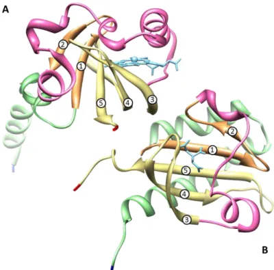
Figure 1.9 – Structure of the heme pocket region of G. sulfurreducens methyl-accepting chemotaxis protein GSU0935 sensor domain and schematic representation of heme group with its binding residues. ... 18
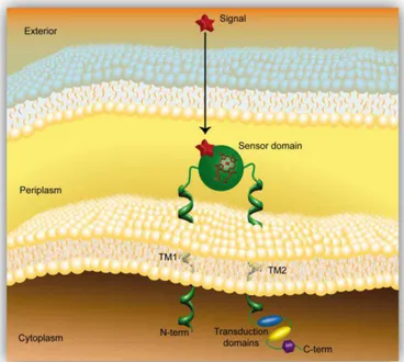
Figure 1.10 – Structure of the FixL and DosP PAS sensor domains. ... 21 Figure 1.11 – Model of a methyl-accepting chemotaxis protein containing a periplasmic heme
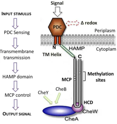
sensor domain that is anchored to the inner membrane by two transmembrane helices ... 25
Figure 1.12– Proposal sensing mechanism of G. sulfurreducens heme MCP sensors ... 26
Figure 1.13 – Sequence alignment of sensor domains GSU0935 and GSU0582 from G. sulfurreducens ... 27
Figure 1.14 – Structure of the sensor domains GSU0582 and GSU0935 in the oxidized form ... 27 Figure 1.15 – GSU0582 and GSU0935 predicted monomer model constructed using the program
3D-PSSM. ... 28
2.
E
XPERIMENTAL PROCEDURESFigure 2.1 – Schematic representation of the plasmid pA02 and pA91 regions carrying genes for
the antibiotic resistance, lac promoter, ELP tag and the cleavage site for TEV followed by residues required for cloning the gene of interest ... 33
xvi
3.
NMR
F
EATURES OF CHEMOTAXIS HEME SENSORSGSU0582
ANDGSU0935
FROMG.
SULFURREDUCENS
Figure 3.1 – Diagram of a heme c numbered according to the IUPAC-IUB nomenclature. ... 42
Figure 3.2 – 1D-1H NMR spectra of the reduced and oxidized unlabeled cytochrome PccH from
Geobacter sulfurreducens obtained at 25 ºC and pH 7.0.. ... 43
Figure 3.3 – 1D-1H-NMR of oxidized and reduced sensor domains prepared in D2O (pH 8.0 and
16 ºC). ... 48
Figure 3.4– Monitorization by 1D-1H-NMR of the GSU0935 sensor domain reduction with D. vulgaris (Hildenborough) hydrogenase.. ... 49
Figure 3.5 – 1D-1H-NMR spectra of 8%D2O/2H2O reduced sample of GSU0935 sensor domain
at pH 7.4. ... 50
Figure 3.6 – Connectivities observed between the heme proton signals in the 2D-1H NOESY and
2D-1H TOCSY ... 51
Figure 3.7 – 2D-1H-TOCSY of GSU0582 and GSU0935 sensor domains with 60 ms mixing-time
at pH 8.0 and 16 ºC ... 52
Figure 3.8– 2D-1H-NOESY pectrum of GSU0582 and GSU0935 sensor domains with 150 ms
mixing-time at pH 8.0 and 16 ºC ... 53
Figure 3.9 – Expansion of the high field region of 1D-1H NMR spectra acquired with different
concentration of GSU0935 sensor domain. ... 55
4.
F
OLDING MODULATES THE SWAPPED DIMERIZATION MECHANISM OF CHEMOTAXIS HEMESENSORS
GSU0582
ANDGSU0935
FROMG.
SULFURREDUCENSFigure 4.1 – Origin of the CD effect. The left and right circularly polarized components of
plane-polarized radiation. ... 64
Figure 4.2 – Far‐UV CD spectra associated with various types of secondary structure in
proteins ... .64
Figure 4.3 –Analytical gel filtration profiles of sensor GSU0935 on a calibrated Superdex-75
XK-16 column at different concentrations...68
Figure 4.4– Spectroscopic characterization of GSU0935 sensor domain conformers. ... 69 Figure 4.5 – Conformational stability of the protein sensors ... 71 Figure 4.6 – Disordered segments in the monomer and swapped dimers in PAS-like heme
sensor domains. ... 73
Figure 4.7 – Structural (far-UV CD) and conformational characterization of GSU0582 sensor
domain as a function of pH ... 75
Figure 3.10 – 1H-15N and 1H-13C NMR spectra of sensor domain GSU0935 acquired at pH
xvii
Figure 4.8 – Structural (far-UV CD) and conformational characterization of GSU0935 as a
function of pH ... 76
5.
T
HERMODYNAMIC AND KINETIC CHARACTERIZATION OF THE CHEMOTAXIS HEMESENSORS
GSU0582
ANDGSU0935
FROMG.
SULFURREDUCENSFigure 5.1 – Photo of the stopped-flow system implemented at the Instituto de Tecnologia
Química e Biológica (ITQB) ... 85
Figure 5.2– Schematic diagram of a stopped-flow analyzer ... 86 Figure 5.3 – Redox titrations followed by visible spectroscopy for GSU0935 and GSU0582
sensor domains at different pH and ionic strength values ... 90
Figure 5.4 – pH dependence of the reduction potential (Em) of sensors GSU0582 and GSU0935.. 92
Figure 5.5 – Dependence of the observed rate constants (kobs) on the square root of the sodium dithionite concentration at pH 7.0 for sensor domains GSU0935 and GSU0582. ... 94
Figure 5.6 – Kinetics of reduction by sodium dithionite of GSU0582 and GSU0935 obtained at
401, 414 and 551 nm (pH 7.0 and 200 mM ionic strength) ... 96
Figure 5.7– Dependence of the logarithm of the second order rate constant for the reduction of
the heme sensor domains by sodium dithionite, ln(k), on the square root of the ionic strength at different pH values for sensors GSU0582 and GSU0935. ... 97
Figure 5.8 – Ribbon diagram of swapped dimers of heme sensors GSU0582 and GSU0935.. .... 101
6. O
NGOING STUDIESFigure 6.1 – Far-UV CD and thermal denaturation of GSU0935 mutants ... 111 Figure 6.2– Redox titrations followed by visible spectroscopy for the mutants of the GSU0935
sensor domain at 100 mM Tris-maleate buffer at pH 8.0. ... 112
Figure 6.3 – Evolution of the UV-visible spectrum during NO binding experiments to the sensor
domain GSU0935. ... 114
Figure 6.4 – Dependence of kobs on the concentration of NO in GSU0935 sensor domain ... 115
Figure 6.5 – Proposed mechanism for NO binding to oxidized GSU0582 and GSU0935 sensor
domains. ... 115
Figure 6.6 – Proposed mechanism for NO binding to reduced GSU0582 and GSU0935 sensor
domains. ... 116
7.
C
ONCLUDING REMARKSFigure 7.1 – First derivative plot of cyclic voltammetry of Geobacter sulfurreducens biofilms at different stages of growth ... 120
Figure 5.9 – Surface electrostatic potential around the heme region calculated by the program
xviii
Figure 7.2 – Ilustration of the proposed signal transduction mechamism of the Geobacter sulfurreducens. ... 121
A.
A
PPENDIXFigure A.1 – Venn diagram showing the relationship of the 20 naturally occurring amino acids
to a selection of physio-chemical properties thought to be important in the determination of protein structure... 120
Figure A.2–NONOate peak decay………...139
xix
T
ABLES INDEX
1. I
NTRODUCTIONTable 1.1 – Selection of some two-component systems with their corresponding function and
signal molecule in some examples of different microorganism..………...5
Table 1.2 – Classification and function of the middle components of the chemotaxis mechanism. ... 9 Table 1.3 – Respiratory versatility of representative Geobacter species .. ………...23
Table 1.4– Number of chemotaxis and che genes obtained from the analysis of Geobacter species available genomes ... 24
3.
NMR
FEATURES OF CHEMOTAXIS HEME SENSORSGSU0582
ANDGSU0935
FROMG.
SULFURREDUCENS
Table 3.1 – Chemical shifts (ppm) of the heme protons of and GSU0582 and GSU0935 sensor
domains in the reduced state at pH 8.0 and 16 ºC. ... 54
4. F
OLDING MODULATES THE SWAPPED DIMERIZATION MECHANISM OF CHEMOTAXIS HEMESENSORS
GSU0582
ANDGSU0935
FROMG.
SULFURREDUCENSTable 4.1 – Thermodynamic parameters of denaturation of sensors GSU0935 and GSU0582 for
chemical denaturation by urea and GuHCl at pH 8.0... 72
Table 4.2 – Thermal stability and secondary structure content of sensor GSU0582 as a function
of pH. ... 75
Table 4.3 – Thermal stability and secondary structure content of sensor GSU0935 as a function
of pH. ... 77
5. T
HERMODYNAMIC AND KINETIC CHARACTERIZATION OF THE CHEMOTAXIS HEMESENSORS
GSU0582
ANDGSU0935
FROMG.
SULFURREDUCENSTable 5.1 – Midpoint reduction potentials in mV (versus SHE) of heme sensor domains GSU0582 and GSU0935 measured at different pH and ionic strength values. ... 91
Table 5.2 – Values of second order rate constants of the reduction of the two heme sensor
domains, at different pH values and ionic strengths. ... 94
Table 5.3 – Effective charge on the protein (Z1), second order rate constant extrapolated to infinite ionic strength (k∞) and reduction potential Em∞ extrapolated to infinite ionic strength, at
xx
Table 5.4 – Comparison between measured and predicted rate constants for each sensor
domain. ... 99
Table 5.5 – Comparison of the rate constants of the two sensor domains. ... 99
6. O
NGOINS STUDIESTable 6.1 – Summary of the expression trials for the different mutated heme sensor domains. ... 110 Table 6.2 – Midpoint redox potentials in mV (versus SHE) for heme sensor domain GSU0582 and GSU0935 mutants (pH 8.0) ... 113
A.
A
PPENDIXTable A.1 – Primers used for site-directed mutagenesis to prepare the sensor domains
xxi
A
BBREVIATIONS
,
SYMBOLS AND CONSTANTS
1D – one dimensional 2D – two dimensional
2xYT– 2x yeast extract-tryptone medium 5c– five-coordinated
6c – six-coordinated
AI-2 – quorum signal autoinducer-2 AMP – ampicillin
ANTAR – AmiR and NasR transcription
antitermination regulators
ATP – Adenosine TriPhosphate
BLAST– Basic Local Alignment Search Tool
B. subtilis–Bacillus subtilis
Cache – calcium channels and chemotaxis
receptor
CBD – Chromophore Binding Domain
C. crescenthus – Caulobacter crescentus
CCW – CounterClockWise CD – Circular Dichroism
CHASE– cyclases/histidine kinases
associated sensory extracellular
CLO – chloramphenicol
cNMP – cyclic Nucleotide-MonoPhosphate cGMP – cyclic Guanosine MonoPhosphate COSY – COrrelation SpectroscopY
CW – ClockWise
DBD– DNA Binding Domains DHp– Dimerization Histidine
phosphotransfer
di-GMP – cyclic diguanylate DNA – deoxyribonucleic acid
D. vulgaris – Desulfovibrio vulgaris
EAL – phosphodiesterase domain that
catalyzes the hydrolysis of di-GMP
E. coli – Escherichia coli
EDTA – EthyleneDiamine Tetraacetic Acid ELP– Elastin-Like-Polypeptide
EPR – Electron Paramagnetic Resonance FAD – Flavin Adenine Dinucleotide FID – Free Induction Decay
GAF – non-catalytic cGMP-binding domain GGDEF – guanylate cyclase domain that
catalyzes synthesis of di-GMP from two GTP molecules
G. sulfurreducens–Geobacter sulfurreducens
G. metallireducens – Geobacter metallireducens
G. uraniireducens – Geobacter uraniireducens
GTP – Guanosine-5'-triphosphate
HAMP – domain found in Histidine kinases,
Adenylate cyclases, Methyl binding proteins and Phosphatases
HD-GYP – conserved domain found in
response regulator modules of various signal transduction systems
HK – Histidine Kinase
HPLC – High-Performance Liquid
Chromatography
HPT– Histidine-containing PhosphoTransfer
protein
Hnr – regulators of stress sigma factor RpoS HS – High-Spin
HSQC – Heteronuclear Single-Quantum
xxii
HtrII-SrII – phototaxis sensory rhodopsin
II-transducer complex
IPTG – isopropyl β-D-1-thiogalactopyranoside IUPAC-IUB– International Union of Pure and
Applied Chemistry and International Union of Biochemistry
K. pneumoniae – Klebsiella pneumonia
LS – Low-Spin
MAP – Mitogen-Activated Protein MCP – Methyl-accepting Chemotaxis
Proteins
MFC–Microbial Fuel Cells
M. xanthus – Myxococcus xanthus
NIT – Nitrate/Nitrite sensor
NMR – Nuclear Magnetic Resonance NOE – Nuclear Overhauser Effect
NOESY – Nuclear Overhauser Enhancement
SpectroscopY
ox – oxidized
PAS– Period clock protein, Aryl hydrocarbon
receptor and Single-minded protein
PBP – Periplasmic solute-Binding Protein PCD – Photosensory Core Domain PDB – Protein Data Bank
PDC – PAS-like domain typified by PhoQ,
DcuS, CitA proteins
PHY – phytochrome pI– Isoelectric point ppm– parts per million
REC – CheY-like phosphoacceptor red - reduced
RNA – ribonucleic acid r.m.s. – root-mean-square
RpoS – RNA polymerase, sigma S RR – Respose Regulator
RRR– Response Regulator Receiver domain SAH– S-adenosylhomocysteine
SAM – S-adenosylmethionine
S. typhimurium – Salmonella enterica enterica sorovar Typhimurium
SDS-PAGE – Sodium Dodecyl Sulfate
PolyAcrylamide Gel Electrophoresis S. enterica – Salmonella enterica
SHE – Standard Hydrogen Electrode SMART – Simple, Modular, Architecture,
Research Tool
S. meliloti – Sinorhizobium meliloti
TCS – Two-Component Systems TEV – Tobacco Etch Virus TLP– Transducer-Like Proteins TM– TransMembrane
TOCSY – TOtal Correlation SpectroscopY Tris – tris(hydroxymethyl)aminomethane UV – Ultra-Violet
V. cholerae – Vibrio cholerae
δ – chemical shift
I– nuclear spin quantum number S– electron spin quantum number
π/2 – pulse with a flip angle of 90º
ωLarmor frequency or precession frequency
gyromagnetic ratio t1 – evolution period
t2 – detection period
B0 – static magnetic field
B1 – rotating magnetic field
𝑽𝒕 – total bed volume
𝑲𝒂𝒗 – partition coefficient
𝑽𝒆 – elution volume
𝑽𝟎 – column void volume
𝜽– ellipticity
𝑨 – absorbance
𝑵– native
xxiii
𝒇 – fraction
𝑲 – equilibrium constant
𝑻 – absolute temperature
∆𝑮𝟎 – standard free energy change
∆𝑮𝑼𝑯𝟐𝑶 – value of standard free enthalpy at zero concentration of denaturant
𝒎– denaturant effectiveness
Cm– lower midpoint denaturant concentration
Tm– midpoint thermal unfolding or melting temperature
kdiss – equilibrium dissociation constant
Em– midpoint redox potential
r – effective radius
Z – effective charges
k –kinetic rate constant
ε– molar absorption coefficient
– reorganisation energy
I - ionic strength
R– molar gas constant (8.314 J mol-1 K-1) F – Faraday constant (96500 C mol-1)
kobs– observed rate constant
A
MINO ACID ABBREVIATIONSAlanine Ala A
Arginine Arg R
Asparagine Asn N
Aspartate Asp D
Cysteine Cys C
Glutamate Glu E
Glutamine Gln Q
Glycine Gly G
Histidine His H
Isoleucine Ile I
Leucine Leu L
Lysine Lys K
Methionine Met M Phenylalanine Phe F
Proline Pro P
Serine Ser S
Threonine Thr T Tryptophan Trp W
Tyrosine Tyr Y
1
C
HAPTER
1
3
1. I
NTRODUCTION
Two-component signal transduction systems
1.1
Two-component systems (TCS) serve as a basic stimulus-response coupling mechanism to allow organisms to sense and respond to changes in many different environmental conditions including fundamental processes such as metabolism and chemotaxis [1].
The prototypical system involves a phosphotransfer reaction between a conserved histidine kinase (HK) domain, containing a kinase core, and a response regulator (RR) protein, containing a regulatory domain, which exerts a modulatory response depending upon its phosphorylation state [2, 3]. Typically, the RR is the output of the system that triggers the cellular response, and is regulated by the HK, which is the input component of the pathway. The activity of HK is modulated by the extracellular stimuli and transfers a phosphoryl group to the RR, in a reaction catalyzed by the RR. Phosphotransfer to the RR results in activation of a downstream effector domain that elicits the specific response [3].
These findings led to the propose of a phosphorylated two-component system concept for sensing a large variety of stimuli and perform a variety of functions (Figure 1.1A). The ATP-dependent phosphorylation of a conserved histidine residue of the first component and the subsequent transferring the phosphate to a conserved aspartate residue of the second component causes different responses like changing the direction of the rotation of flagella or controlling the expression of a particular gene.
The TCS are the most abundant multistep signaling pathways in Nature and despite of the inherent interest as a fundamental signaling strategy, the physiological signal that modulates their action are not precisely understood. TCS are sophisticated signaling systems marked by a highly modular design that has been adapted and integrated into a wide variety of cellular signaling circuits.
4
amoeba Dictyostelium and in plants, they are involved in important processes, such as osmoregulation, cell growth and differentiation [12, 13]. To date, TCS have not been identified in animals and are not encoded by the human genome [14, 15].
In prokaryotic and eukaryotic signaling systems apparently, there are significant changes in the way that the two-component proteins are used in different circumstances. Distinct arrangements of conserved domains and diverse integration of proteins into pathways provide adaptations of the basic scheme to meet the specific regulatory needs of many different signaling systems. Notably, in most prokaryotic systems, the output response is made directly by the RR, which functions as a transcription factor. In eukaryotic systems, two-component proteins are found at the beginning of signaling pathways where they interface with more conventional eukaryotic signaling strategies such as mitogen-activated protein (MAP) kinase and cyclic nucleotide cascades [16].
Similar to most signaling pathways, the architecture of the TCS is not always linear from the environment to transcription (Figure 1.1B).
5
Parallel sensory kinases affect the activity of a phosphotransferase domain within a more complex phosphorelay such as that seen for Vibrio cholerae (V. cholerae) quorum sensing [17] or for control of sporulation in Bacillus subtilis (B. subtilis) [6, 18]. Many eukaryotic signaling cascades involve protein kinases that phosphorylate both themselves and other protein substrates at specific serine, threonine or tyrosine residues thereby regulating protein activities. Several recent examples have identified response regulators containing the GGDEF domain, as PleD in Caulobacter crescentus (C. crescentus) [19] that synthesizes cyclic diguanylate (di-GMP) from GTP (guanosine-5'-triphosphate) to regulate targets that affect differentiation and biofilm formation.
The efforts to characterized systems such as bacterial chemotaxis [20], aerobic/anaerobic regulation in E. coli [21, 22], the sporulation system of B. subtilis [23, 24], differentiation in C. crescentus [25-27] and Myxococcus xanthus (M. xanthus) [28, 29] have provided a basic understanding of how these systems transduce extracellular signals and elicit appropriate cellular responses. In Table 1.1 is indicated a selection of some TCS with their corresponding function and signal molecule.
Table 1.1 – Selection of some two-component systems with their corresponding function and signal molecule in some examples of different microorganism. The unknown signal molecules are indicated by question marks. Adapted from [30] and [31].
System Microorganism Function Signal molecule
ArcB/ArcA Escherichia coli Sensing of oxygen and redox states Quinones reflecting the redox state
NarX/NarL Escherichia coli Nitrate and nitrite respiration Nitrate, nitrite
CitA/CitB Klebsiella pneumonia Transport and anaerobic metabolism of citrate Citrate
FixL/FixJ Bradyrhizobium japonicum Nitrogen fixation O2, CO, NO
LovK/LovR Caulobacter crescentus Bacterial cell attachment Blue light
TodS/TodT Pseudomonas putida Degradation of benzene derivatives Monoaromatic compounds
NtrB/NtrC Escherichia coli Nitrogen utilization 2-ketoglutarase, glutamine
KdpD/KdpE Escherichia coli K+ supply K+
VanS/VanR Streptomyces coelicolor Antibiotics resistance Vancomycin
EnvZ/Ompr Escherichia coli Osmosensing ?
KinB/Spo0F Bacillus subtilis Sporulation ?, ATP as cosignal?
BvgS/BvgA Bordetella pertussis Virulence Temperature, sulfate ions, nicotinic acid
6 (continued)
System Microorganism Function Signal molecule
DesR/DesK Bacillus subtilis Lipid modification Temperature
DosS/T/DosR Mycobacterium tuberculosis Redox sensor/ hypoxia sensor O2, CO, NO
PhoQ/PhoP Salmonella enterica Virulence Extracytoplasmic levels of Mg2+ and Ca2+
DcuS/DcusR Escherichia coli anaerobic fumarate Regulation of the
respiratory pathway
C4-dicarboxylates
NreB/NreC Staphylococcus carnosus Dissimilatory nitrate/nitrite reduction O2
MmoS/MmoQ Methylococcus capsulatus Controlling gene expression Copper
NifL/NifA Azotobacter vinelandii Nitrogen fixation O2
DctB/DctD Sinorhizobium meliloti Controlling gene expression C4-dicarboxylates
1.1.1 A specialized two-component system: chemotaxis
Bacterial chemotaxis is the reason for the movement towards regions that contain either higher concentrations of beneficial compounds or lower concentrations of toxic chemicals. The bacterial chemotaxis machinery was first identified in E. coli and Salmonella enterica subspecies I, serovar Typhimurium (S. typhimurium) for the regulation of their flagellar-based motility. The E. coli chemotaxis pathway is so sensitive that it is able to sense a change in similar molecules [32, 33].
Most sequenced bacterial genomes include homologues of genes that are known to encode components of flagella and chemosensory pathways indicating that motility is widespread and probably provides a selective advantage, especially in non-homogeneous, nutrient-limiting environments. However, all bacteria in which motility has been studied seem to be chemotaxis (although two sequenced Archaea, Aquifex aeolicus and Methanocaldococcus jannaschii, with putative flagellar genes have no obvious chemosensory genes [34]). So, the ability to move is not, by itself, essential and it must be predisposed by the growth of certain bacteria in specific environments.
7
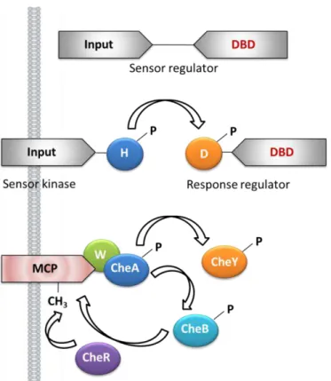
Figure 1.2 – One-component, two-component and chemosensory signal transduction in bacteria. The figure illustrates the prototypical one-component system (top), a two-component system (middle) and the specialized chemotaxis system (bottom). Represented are histidine kinase domains (H; CheA), aspartyl receiver domains (D), CheW coupling proteins (W), methyl-accepting chemotaxis protein (MCP), CheY response regulator and flagellar rotation controller, CheB methylesterase, the CheR methyltransferase and DNA binding domains (DBD). Adapted from [35].
Inputs for chemotaxis systems are transduced by the dedicated transmembrane chemoreceptor sensor proteins [methyl-accepting chemotaxis proteins (MCP) or transducer-like proteins (TLP)], which have a cytoplasmic domain, rather than by a fused sensory domain of a histidine protein kinase as is found for most systems that regulate transcription.
1.1.2 Methyl-accepting chemotaxis proteins
8
methylated site of the MCP. This MCP-like receptor mediates aerotactic responses by monitoring redox changes in the electron transport chain undergoing a sensory adaptation through a poorly-understood, methylation-independent mechanism [38-41].
MCP are the predominant chemoreceptors in bacteria and have been found in many Archaea. The best-studied is the Tar chemoreceptor from E. coli, which has four MCP (Tar, Tsr, Tap and Trg) and a fifth MCP-like protein (Aer). Roger Alexander and Igor Zhulin detected 2125 MCP sequences in 152 genomes of Bacteria and Archaea [42]. Barbara A. Methé et al. found multiple (~70) homologs of chemotaxis genes in the Geobacter sulfurreducens (G. sulfurreducens) genome, 34 MCP genes and six major che gene clusters [43].
The prototypical MCP are transmembrane receptors that contain a periplasmic ligand binding domain and a cytosolic signaling domain (Figure 1.3). In general, MCP has two transmembrane-spanning domains that create a periplasmic (or extracellular) loop functioning as a ligand binding domain and a cytoplasmic component comprising the broadly conserved helical signaling domain, methylation helices for adaptation and the highly conserved domain that regulates the kinase activity [44-46]. However, large subsets of receptors that are completely cytoplasmic and detect soluble cytoplasmic signals have been identified [47] including the well-characterized B. subtilis aerotactic sensor HemAT. The conformation of the cytoplasmic portion of the receptor is an extended α-helical coiled coil. For transmembrane receptors, a stimulus or ligand-binding event is transduced by a piston like motion across the cytoplasmic membrane and converted to helical rotation within the cytoplasmic portion of the receptor [48].
9
The middle components of the chemotaxis mechanism that bridge the stimulus and the flagella switch, are the Che proteins (Table 1.2). These are coded by the che genes, and carry out “excitation” and “adaptation” steps. The che genes have been found universally among bacteria. By responding to the MCP interaction with an attractant or repellent, CheA, CheW, CheY and CheZ transmit the flagella response. Then, CheB and CheR carry out adaptation to the attractant or repellent by methylating MCP or demethylating it.
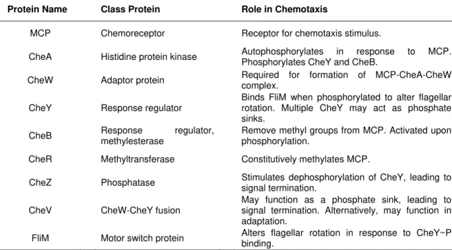
Table 1.2 – Classification and function of the middle components of the chemotaxis mechanism.
Protein Name Class Protein Role in Chemotaxis
MCP Chemoreceptor Receptor for chemotaxis stimulus.
CheA Histidine protein kinase Autophosphorylates in response to MCP. Phosphorylates CheY and CheB.
CheW Adaptor protein Required for formation of MCP-CheA-CheW complex.
CheY Response regulator Binds FliM when phosphorylated to alter flagellar rotation. Multiple CheY may act as phosphate sinks.
CheB Response methylesterase regulator, Remove methyl groups from MCP. Activated upon phosphorylation.
CheR Methyltransferase Constitutively methylates MCP.
CheZ Phosphatase Stimulates dephosphorylation of CheY, leading to signal termination.
CheV CheW-CheY fusion May function as a phosphate sink, leading to signal termination. Alternatively, may function in adaptation.
FliM Motor switch protein Alters flagellar rotation in response to CheY~P binding.
10
conformational change that counters the initial change induced by ligand binding and therefore brings about adaptation for a ligand-bound receptor complex. In E. coli chemotaxis, an auxiliary protein, the phosphatase CheZ, oligomerizes with the phosphorylated CheY (CheY∼P) and increase the spontaneous dephosphorylation rate of CheY∼P allowing the rapid signal termination [56].
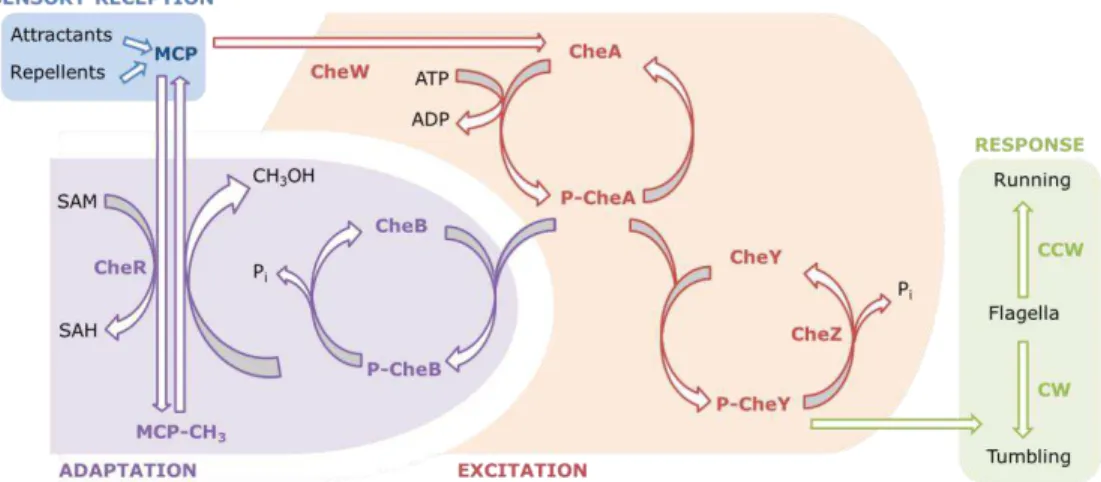
The ability of cells to respond to small changes in the concentration of a large number of chemical stimuli depends on the repertoire and specific affinities of the chemoreceptors. A decreased concentration of attractant results in decreased attractant binding to the MCP, which stimulates CheA trans-autophosphorylation. This results in an increase in the CheY∼P concentration. CheY∼P then binds to the flagellar motor and causes it to switch to clockwise rotation, which results in cell tumbling and direction change. CheB is also phosphorylated by CheA∼P, which results in an increased methylesterase activity and an increased demethylation of the MCP. Demethylated MCP has a reduced ability to induce CheA autophosphorylation (even in the presence of a low concentration of attractant), so the rate of CheA autophosphorylation and therefore the rate of rotation switch return to the pre-stimulus level. The system has now adapted and is primed to sense any subsequent increases or decreases in ligand binding (Figure 1.4). On the other hand, an increased concentration of attractant inhibits the autophosphorylation of CheA, which reduces the concentration of CheY∼P and therefore the frequency of motor switching. This causes the bacterium to swim in this positive direction for longer. CheB phosphorylation and therefore activity is also reduced, which allows the constitutive methyltransferase CheR to increase the methylation of the MCP. Highly methylated MCP are efficient in the stimulation of the CheA autophosphorylation, even in the continued presence of a chemoattractant, so this returns CheA autophosphorylation to the pre-stimulus level and the bacterium to a normal frequency of direction changing (Figure 1.4).
11
called excitation. Then adaptation follows: repellents cause demethylation of the methylated MCP and attractants cause its methylation. CCW, counterclockwise rotation; CW, clockwise rotation; SAH, S-adenosylhomocysteine; SAM, S-adenosylmethionine. Adapted from [57].
1.1.3 Effector domains: diversity of sensing domains
The great diversity of attached or independent effector domains creates an almost limitless variety of output responses that can be controlled through RR [58]. In prokaryotes, RR are usually the last components of signaling pathways, directly effecting the responses, while in eukaryotic TCS, RR are often intermediates, interfacing with proteins linked to common eukaryotic strategies such as MAP kinase cascades or cyclic nucleotide second messengers [59]. As noted above,
∼17% of prokaryotic RR exist as stand-alone CheY-like conserved receiver domains. Most of the remaining RR contain domains can be categorized into a relatively small set of nucleic acid binding, enzymatic and protein/ligand binding domains. The majority of RR contain DNA binding effector domains dominated by a small number of structural subfamilies. These include the OmpR/PhoB winged-helix domain (30%) [60], the NarL/FixJ four-helix helix-turn-helix domain (17%) [61], the NtrC/DctD AAA+ ATPase domain fused to a factor of inversion (Fis)-type helix-turn-helix domain (10%) [62] and the recently characterized LytTR domain with an unusual, predominantly β fold (3%) [63]. RNA binding domains are found in only ∼1% of RR and are mostly of the ANTAR (AmiR and NasR transcription antitermination regulators) family, functioning as antitermination factors [64]. Enzymatic domains are found in ∼13% of RRs. Most of these RR (6% of all RR) are involved in regulation of cyclic diguanylate (di-GMP) [65]. Enzymatic domains include GGDEF diguanylate cyclase domains (guanylate cyclase domain that catalyzes synthesis of di-GMP from two GTP molecules) and/or phosphodiesterase domains of the EAL (phosphodiesterase domain that catalyzes the hydrolysis of di-GMP) or HD-GYP, a conserved domain found in response regulator modules of various signal transduction systems. The remaining enzymatic subfamilies, in order of prevalence, are chemotaxis methylesterase CheB domains; HK domains, which are often coupled with additional PAS (Period clock protein, Aryl hydrocarbon receptor and Single-minded protein) and/or GAF (non-catalytic cGMP-binding domain) domains. Many additional enzymatic domains in RR occur infrequently. A small and diverse group of effector domains that mediate interactions with other proteins or ligands occur in 3% of RR. Approximately half of these RR correspond to chemotaxis CheV-like proteins containing CheW domains. Additional subfamilies include tetratricopeptide repeat, Hnr-like regulators of stress sigma factor RpoS (RNA polymerase, sigma S), HPT (histidine containing phosphotransfer), PAS, GAF, and cyclic nucleotide-monophosphate (cNMP) binding domains. Structures are available for representative members of most of these effector domains.
12
turgor pressure, cell envelope stress, redox potential and electrochemical gradients. The individual sensing domains are highly variable in sequence and this is particularly evident for domains that are flanked by transmembrane domains and localized outside the cytoplasm [6].
Stimuli are sensed by these domains either directly or indirectly through protein-protein interactions with auxiliary signal transduction proteins. How the individual domains that comprise the sensor complex function to augment or attenuate the signal output in response to ligand binding has not been determined even for relatively simple systems. It seems clear that the multiplicity of domains in the sensor complex allows multiple signal output and may permit metabolic, cell cycle and other cellular processes promoting the sensor histidine kinase activity and ultimately the cell response. It is reasonable to suppose that the diversity of signals detected by the sensing domains required the evolution of a large number of distinct folds. Few common topologies have been observed in sensory domains and these systems are classified into three major groups on the basis of these membrane topologies [66] (Figure 1.5).
13
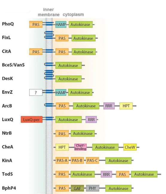
Figure 1.5 – Domain organization and topology of selected sensor kinases based on domain annotation by SMART complemented with structural information. Abbreviations: PAS, PER-ARNT-SIM; HAMP, domain found in histidine kinases, adenylyl cyclases, methyl binding proteins and phosphatases; RRR, response regulator receiver domain; HPT, histidine containing phosphotransfer; GAF domain (sensory domain first characterized in cGMP-dependent phosphodiesterase, adenylyl cyclases and E. coli FhlA); PHY,
phytochrome. The unknown domain is indicated by question mark. Reproduced from [30].
1.1.3.1 Cytoplasmic sensor domains
14
domains are typically between 100-120 amino acids in length [67-69]. The core of a PAS domain is a five stranded β-sheet and interspersed within this core are α-helices that provide ligand/signal specificity [67, 70]. The position of α-helices can vary depending on the cellular location; specifically, cytoplasmic PAS domains have α-2β-4α-3β topology (Figure 1.6A) and extracytoplasmic PAS domains (named PAS-like - see section 1.1.3.3) have 3α-2β-1/2α-3β-α topology (Figure 1.6B) [70-73].
Figure 1.6 – Representative three-dimensional structures of PAS (A) and PAS-like (B) domains. The first corresponds to Sinorhizobium meliloti (S. meliloti formely Rhizobium meliloti) oxygen sensor FixL protein (PDB:
1D06) and the second to the ligand-binding domain of the Klebsiella pneumoniae sensor kinase CitA protein
(PDB: 1P0Z). For both structures, the core β strands are labeled from 1 to 5. Schematic models were generated by CHIMERA [74]. Each region is colored as follows: the amino end with dark blue, the leading α-helix region with green, the first two β-strands with orange, the inter-domain α-helix region with pink, the last three β-strands with yellow, and the C-terminal with red. The heme and citrate molecules are shown in light blue stick models. Adapted from [75].
15
PAS sensor domain contains redox-active cysteine residues and is regulated by changes in redox states of quinone and menaquinone pools [88, 89].
Figure 1.7 – Structure and ligand binding in flavin adenine dinucleotide (FAD)-binding PAS redox sensor domains of the Methylococcus capsulatus MmoS (PDB: 3EWK). The domain architecture and ribbon of the MmoS PAS1 domain bound to FAD. β-strands are rendered in light green, α-helices in blue. Atoms of the flavin cofactor bound to the PAS1 domain are colored by element: carbon, green; nitrogen, blue; oxygen, red. Adapted from [31].
A second structural family of cytoplasmic sensor domains features a GAF fold [90]. GAF sensor domains consist of a six stranded anti-parallel β-sheet core, related in structure to PAS domains and are often found coupled in the cytoplasmic histidine kinase sensors. Both PAS and GAF domains have rather high sequence variability and structures plasticity, making them versatile signaling domains not only for stimuli recognition but also for signal transduction, owing their propensity for protein-protein interactions.
16
1.1.3.2 Membrane-embedded sensor domains
Histidine kinase receptors that contain multiple transmembrane segments often lack obvious extracellular domains and in subsets of such cases, the sensory region may lie within the transmembrane segments. Biochemical and structural studies of the DesK temperature sensor show that the transmembrane regions are required for a thermal response [96, 97]. In the membrane-embedded SenS redox sensor, regulation requires binding of the secreted octameric heme-binding protein HbpS to its N-terminal transmembrane region [98]. In addition, evidence suggests that the plant Etr1 histidine kinase detects ethylene within the hydrophobic N-terminal transmembrane region [99] and the AgrC quorum sensor detects an autoinducing peptide via two short extracellular loops in proximity to the transmembrane region [100, 101]. Although no structures exist for the transmembrane regions of histidine kinase receptors, the structure of the phototaxis sensory rhodopsin II-transducer complex (HtrII-SrII) [102], in which the transducer protein forms a four-helix bundle in the membrane, provides insight into the transmembrane helical arrangement of histidine kinases.
1.1.3.3 Extracytoplasmic sensor domains
Owing to the great sequence variability of extracytoplasmic sensing domains, only a limited number were recognized as a periplasmic sensor domains, such as the periplasmic solute-binding PBP domain, Cache (Calcium channels and Chemotaxis receptor), a series of CHASE (Cyclases/Histidine kinases Associated Sensory Extracellular) and NIT (Nitrate/Nitrite sensor) domains.
17
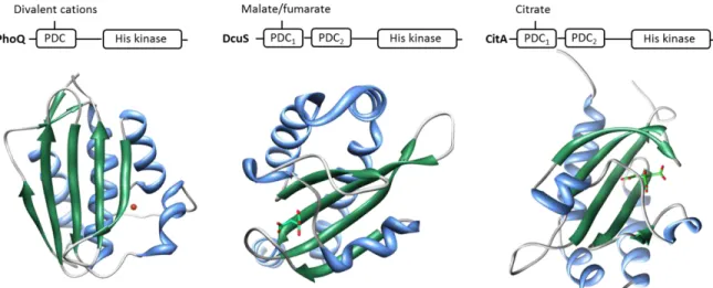
parallel evolution from PAS domains [109]. Structures of PDC domains are typified by the PhoQ, DcuS and CitA proteins in Figure 1.8.
Figure 1.8 – Structure and ligand binding in the PDC domains of the PhoQ (PDB: 1ID0), DcuS (PDB: 3BY8) and CitA (PDB: 1P0Z) sensor histidine kinases. The domain architecture and ribbon of Salmonella enterica PhoQ PDC domain, E. coli DcuS PDC1 domain and K. pneumoniae CitA PDC1 domain are shown as
well as the bound Mg2+, atoms of the malate cofactor ions and citrate ion, respectively. β-strands are represented in green and α-helices in blue. Magnesium ion is colored in red. Atoms of the malate cofactor and citrate bound to the PAS domain are colored by element: carbon, green; oxygen, red. Adapted from [31].
Additions to the collection of PDC sensor domain structures include the oligosaccharide sensor AbfS [110] and the PhoR periplasmic domain [71]. The activities of some sensors are regulated by auxiliary proteins, which can be the primary ligand-binding protein. Structures of proteins that regulate their associated sensor kinase have become available, including LuxP which regulates above-mentioned LuxQ, the periplasmic membrane-tethered proteins YycH and YycI of B. subtilis known to regulate the essential YycG sensor kinase [111, 112] and the cytoplasmic proteins pXO1-118 and pXO2–61 of Bacillus anthracis which regulates the sporulation [113]. The dicarboxylate transport system in the Rhizobiaceae (and other) species is regulated by the DctB sensor kinase which senses C4-dicarboxylic acids. DctB is membrane integrative sensor histidine kinase and shares the basic signal transduction scheme with other periplasmic sensor histidine kinases which detect periplasmic stimuli with N-terminal sensory domains transmitting the signals to cytoplasmic C-terminal kinase/phosphatase domains via transmembrane segments and the cytoplasmic helices [3].
PAS-18
like domains with c-type heme groups [115] (Figure 1.9). Interestingly, this places redox-active sensing domains in the periplasm. By contrast, the MCP with small periplasmic domains are more likely to detect signals via associations with other proteins, as in the case of DifA of M. Xanthus [116, 117], or detect intracellular signals when the MCP have no TM segments [118]. DifA is an MCP homolog with two putative transmembrane domains which lacks an apparent periplasmic domain and is therefore probably unable to promote direct ligand binding, as with classical bacterial chemoreceptors [119].
Figure 1.9 – (A) Structure of the heme pocket region of G. sulfurreducens methyl-accepting chemotaxis protein GSU0935 sensor domain and (B) schematic representation of heme group (red) with its binding residues (black). β-strands are rendered in light green, α-helices in blue and the heme-binding site of the monomer in purple . Atoms of the heme cofactor bound to the sensor domain are colored by element: carbon, green; nitrogen, blue; oxygen, red; iron, orange. Adapted from [31].
Signaling Mechanisms
1.2
19
kinase dimer surface and that these perturbations are relayed to the kinase core domains. The recent structural evidence suggests that communication between sensor and transmitter domains in histidine kinase signaling is mediated by subtle structural changes along the dimer interface and that there may be related aspects of symmetry and asymmetry.
In recent years, structural details for several systems with known ligands have been clarified, revealing diverse ligand binding modes and the overall folds of both extracytoplasmic and cytoplasmic sensory domains. A few classes of similar folds seem to be shared despite low sequence homology and common features in TM signaling mechanisms are beginning to emerge.
Functional chimeric proteins of Tar and various systems, as EnvZ and NarX, have been successfully constructed with interchanged periplasmic sensory domains to explore the potential shared TM signaling mechanism [124, 125]. Ligand binding at the dimer interface results in a symmetrical piston-like displacement of two N-terminal helices towards the TM region and presumably, these piston-like movements are transmitted through the TM helices and reach the cytoplasmic domains, although the exact conformational changes that occur there remain unknown. The quorum sensing LuxQ from Vibrio harveyi indirectly senses the quorum signal autoinducer-2 (AI-2) through interactions with the periplasmic AI-2 binding protein LuxP. Structures of (LuxPQp)2 complexes with or without the ligand AI-2 indicate little difference in the conformations of the tandem PAS-like folds of the LuxQ periplasmic domains (LuxQp) [106]. Instead, ligand binding causes the formation of an asymmetrical dimer, and the asymmetrical positioning of individual proteins is thought to be transduced into the cytoplasm, disrupting the symmetrical dimer of kinase core domains and thereby shutting off kinase activity. Unlike the indirect sensing mechanism of LuxQ, E. coli and Salmonella enterica (S. enterica) PhoQ proteins directly sense divalent cations, as Mg2+, and antimicrobial peptides while the sensory domains of
Klebsiella pneumonia (K. pneumoniae) CitA, S. meliloti DctB, V. cholerae DctB, and E. coli DcuS bind to small metabolic molecules of citrate or C4-dicarboxylates such as succinate, fumarate, and malate.
four-20
helical coiled-coil with each monomer contributing two parallel helices connected by a long loop [48]. Furthermore, the coiled-coils adopted unusual knobs-to-knobs packing, deviating from the conventional knobs-to-holes packing that can be achieved by helix rotation. Hence, a rotary mechanism has been suggested for the signaling of HAMP domains; however, it is not known whether this helical rotation mechanism is generally conserved for all proteins containing HAMP domains or how the TM signaling events, sometimes piston-like movements, are converted to helix rotation in HAMP domains. Structures of individual cytoplasmic sensory domains have also been determined for a few systems, such as the PAS domains from both B. subtilis KinA [128] and the heme based oxygen sensor FixL in S. meliloti [79, 81], the GAF domains of the O2/NO sensors DevS/DosT from Mycobacterium [129, 130] and the chromophore-binding bacteriophytochromes containing PAS, GAF and PHY domains [91, 93]. Binding of ligands and cofactors causes conformational changes in specific regions of these similar structures, yet it remains unknown how these changes are coupled to kinase activity until the spatial arrangements of the kinase and sensory domains are characterized. Certainly, without the barrier of the plasma membrane, signaling can be executed through signal-mediated direct contacts with the kinase as well as quaternary structural changes owing to the general plasticity of these domains. A structure of the full photosensory core domain from Pseudomonas aeruginosa bacteriophytochrome provides an interesting insight into the spatial organization of multiple sensory domains [91]. A long continuous helix directly connects the GAF and PHY domains and forms a parallel dimer with individual domains resembling gall-like extrusions from the central helical stem. Long coiled-coils immediately preceding the kinase domain typically extend into the four helix bundle of the DHp domain (dimerization histidine phosphotransfer). Such long helices are commonly found in various prokaryotic signaling proteins. Termed the signaling helix, this motif joins a wide variety of signaling domains such as PAS, GAF, HAMP, TM helices, DHp, REC (CheY-like phosphoacceptor), GGDEF, EAL and Ser/Thr/Tyr kinases [131].
Heme-based sensors
1.3
Heme proteins are one of the most versatile groups of proteins in Nature and they are involved in many different types of reactions such as electron transfer, oxygen transport and storage, reduction of peroxides or sensing. Though the basic structure of heme is identical among these heme proteins, the presence or absence and the nature of the axial ligands to the iron atom and the effect of the polypeptide chain of the protein on its surroundings, all contribute to this diversity of properties.
21
The currently known based sensors are grouped into four families accordingly to their heme-binding domains: heme-PAS, CooA, globin-coupled sensor and heme-NO-heme-binding [132-135].
The heme-PAS domain family members are extraordinarily versatile and relatively well studied. Members are already known that respond specifically to O2 or CO and whose heme-binding domains couple these dissimilar but widespread activities [132, 136-141]. These sensors demonstrably transduce signals and mediate adaptive responses by diverse strategies, including chemical modification of proteins, control of second-messenger levels and regulation of macromolecular interactions.
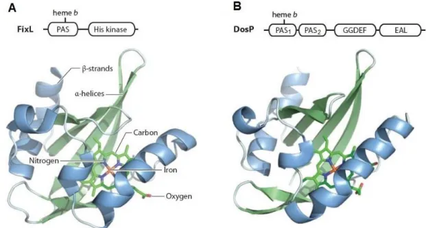
1.3.1 b-type heme sensors
As mentioned above, FixL is a heme-based oxygen sensor which controls the relative expression of proteins that function under microaerobic or anaerobic conditions [142-145] and contains one b-type heme group [137]. A related class of b-type heme-binding PAS domain is encoded by the E. coli direct oxygen sensor DosP (EcDOS) [138]. This sensor functions as an oxygen-regulated phosphodiesterase and catalyzes the cleavage of cyclic di-GMP into the linear dinucleotide pGpG [146, 147]. FixL-PAS and DosP-PAS1 share a common site of heme ligation via a conserved histidine residue (Figure 1.10). The conservation of this ligation site suggests a common evolutionary origin for this gas-sensing PAS domain.