Evaluation of
In-Labelled Exendin-4
Derivatives Containing Different Meprin
β-Specific Cleavable Linkers
Andreas Jodal1, Fabienne Pape1,2, Christoph Becker-Pauly3, Ole Maas4, Roger Schibli1,2, Martin Béhé1
*
1Center for Radiopharmaceutical Sciences ETH-PSI-USZ, Paul Scherrer Institute, Villigen, Switzerland, 2Department of Chemistry and Applied Biosciences, ETH Zurich, Zurich, Switzerland,3Institute of Biochemistry, University of Kiel, Kiel, Germany,4Department of Radiology and Nuclear Medicine, Division of Nuclear Medicine, University Hospital Basel, Basel, Switzerland
Abstract
Background
Cleavable linkers, which are specifically cleaved by defined conditions or enzymes, are powerful tools that can be used for various purposes. Amongst other things, they have been successfully used to deliver toxic payloads as prodrugs into target tissues. In this work novel linker sequences targeting meprinβ, a metalloprotease expressed in the kidney brush-border membrane, were designed and included in the sequence of three radiola-belled exendin-4 derivatives. As radiolaradiola-belled exendin-4 derivatives strongly accumulate in the kidneys, we hypothesised that specific cleavage of the radiolabelled moiety at the kid-ney brush-border membrane would allow easier excretion of the activity into the urine and therefore improve the pharmacological properties of the peptide.
Results
The insertion of a cleavable linker did not negatively influence thein vitroproperties of the peptides. They showed a good affinity to the GLP-1 receptor expressed in CHL cells, a high internalisation and sufficiently high stability in fresh human blood plasma.In vitro di-gestion with recombinant meprinβrapidly metabolised the corresponding linker se-quences. After 60 min the majority of the corresponding peptides were digested and at the same time the anticipated fragments were formed. The peptides were also quickly metabo-lised in CD1 nu/nu mouse kidney homogenates. Immunofluorescence staining of meprinβ
in kidney sections confirmed the expression of the protease in the kidney brush-border membrane. Biodistribution in GLP-1 receptor positive tumour-xenograft bearing mice re-vealed high specific uptake of the111In-labelled tracers in receptor positive tissue.
Accu-mulation in the kidneys, however, was still high and comparable to the lead compound
111In-Ex4NOD40.
OPEN ACCESS
Citation:Jodal A, Pape F, Becker-Pauly C, Maas O,
Schibli R, Béhé M (2015) Evaluation of¹¹¹In-Labelled
Exendin-4 Derivatives Containing Different Meprin β-Specific Cleavable Linkers. PLoS ONE 10(4): e0123443. doi:10.1371/journal.pone.0123443
Academic Editor:Marek Cebecauer, J. Heyrovsky Institute of Physical Chemistry, CZECH REPUBLIC
Received:January 13, 2015
Accepted:March 4, 2015
Published:April 9, 2015
Copyright:© 2015 Jodal et al. This is an open access article distributed under the terms of the
Creative Commons Attribution License, which permits unrestricted use, distribution, and reproduction in any medium, provided the original author and source are credited.
Data Availability Statement:All relevant data are within the paper and its Supporting Information files.
Funding:This work was supported by the following sources of funding: Seventh Framework Programme (http://ec.europa.eu/research/fp7/index_en.cfm) grant numbers 222980 and 602812; Juvenile Diabetes
Research Foundation International (http://jdrf.org/)
grant number 37-2012-4; and Deutsche
Forschungsgemeinschaft (http://www.dfg.de/en/index.
jsp) grant number SFB 877/2 2014; Projekt A9. The
Conclusion
In conclusion, we show that the concept of cleavable linkers specific for meprinβis feasible, as the peptides are rapidly cleaved by the enzyme while retaining their biological properties.
Introduction
Over recent years cleavable linkers targeting specific physiologic environments or enzymes have proven to be a versatile tool for various medical applications. Cleavable linkers are suc-cessfully used to reduce the side effects of toxic drugs, for example when camptothecin is con-jugated to the carrying molecule substance P via a cleavable linker, it acts as a prodrug and is not toxic. Upon reaching its target, however, the linker is specifically cleaved and releases its cy-totoxic payload into the desired tissue. This concept significantly improves the specificity of
the drug and thereby minimises off-target side-effects [1,2]. Cleavable linkers are also used
di-agnostically in a new approach to identify diseases such as cancer or inflammation. New near infrared (NIR) probes, highly sensitive tools for the diagnosis of such conditions, have two fluorescent, self-quenching dyes attached to the probe via a specific self-immolative linker. This linker is cleaved in the targeted tissue, thereby liberating the dye and resulting in a specific
fluorescent signal [3,4].
Nuclear medicine is another field that could benefit from the use of cleavable linkers. Accu-mulation of radioactivity in non-target tissues can both increase the background signal in diag-nostic purposes, as well as potentially damage sensitive tissues in therapeutic approaches. Tracer containing linker sequences that are degradable by enzymes could reduce the unwanted accumulation and increase the specificity of the signal.
Many hydrophilic radiolabelled compounds with a molecular weight below 60 kD are
excret-ed by the kidneys [5]. These radiopharmaceuticals are often reabsorbed in the proximal tubuli
in the kidneys, resulting in a strong accumulation that can hinder diagnostic imaging. Addition-ally, as they are organs sensitive to radiation, a high radiation dose to the kidneys can cause per-manent damage, potentially leading to future complications after radiotherapeutic interventions
[6]. Kidneys are the dose-limiting organs in several nuclear medicine therapies [7]. Cleavable
linkers located shortly before the radiolabelled moiety that are degradable by enzymes specifical-ly expressed in the kidney brush border membrane would allow the easy cleavage and excretion of the radioactive metabolites into the urine. This concept was previously demonstrated when the introduction of a Gly-Lys linker into radiolabelled antibody fragments significantly reduced
kidney accumulation [8,9]. The transfer to peptides, however, was not successful meaning that
targeting different enzymes is necessary [10]. In a recently published study a new cleavable
link-er was proposed.67NOTA-MI-Fab was shown to release67Ga-NOTA-Met upon proteolysis in
the lysosomes of the brush-border membrane, however, the structure of the radiometabolites
and the cleavable linkages are not yet completely understood [11].
Meprinαand meprinβ, two astacin metalloproteases strongly expressed on the brush
bor-der membranes of kidney proximal tubular cells, could be potentially utilised to cleave linkers
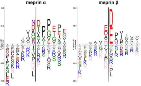
[12]. Meprinβ, in contrast to meprinα, has a stronger preference for negatively charged amino
acid residues around the scissile bond as determined by proteomics (Fig 1) [13]. The unique
specificity of meprinβmakes the design of specific linker sequences easier. Various substrates
of meprins have been identified, almost all of them confirming the cleavage specificity [14,15].
Among the substrates of meprinβare gastric peptides such as cholecystokinine 8 and gastrin
that share anionic amino acids around the cleavage site [16]. While most of those peptides are
degraded, meprinβcan also perform other functions like activating cytokines, as in the case of
the processing of interleukin-1βprecursor [10,17]. It has also been shown that meprins are
ex-pressed in a number of pathological conditions including cancer and fibrotic diseases, making
these interesting targets for prodrugs with meprin-specific cleavable linkers [18–20].
Introducing a linker into the amino acid sequence of a peptide, however, is challenging. The insertion of an additional sequence into a peptide might influence its secondary structure and potentially lead to reduced affinity and altered pharmacokinetic properties. Our model sub-stance, the 39 amino acid peptide exendin-4, is an agonist for the glucagon-like peptide 1
re-ceptor (GPL-1R) specifically expressed onβ-cells. These cells are located in the islets of
Langerhans in the pancreas, where their activation mainly affects insulin secretion, insulin
syn-thesis andβ-cell proliferation [21]. Based on known substrates we designed three derivatives of
exendin-4 to evaluate the feasibility of cleavable linkers as substrates for meprinβ.
Pathologically alteredβ-cells can lead to imbalanced glucose levels with potentially severe
complications. Both apoptosis ofβ-cells and their decreased sensitivity towards insulin can
lead to diabetes and result in hyperglycaemia, while an increased function caused by either
in-sulin-producing tumours (e.g. insulinoma) or by hyperplasticβ-cells (e.g. nesidioblastosis)
may lead to hypoglycaemia. In the latter cases particularly, accurate localisation is crucial to
surgically remove thosefoci, currently the only curative treatment option for these conditions.
111In-labelled exendin-4 derivatives have been successfully tested in clinical trials for detection
of insulinoma and to evaluate theβ-cell mass of type 1 diabetic patients and transplanted
β-cells. One remaining problem, however, is the high accumulation in the kidneys obstructing
the view on the pancreas [22–24].
In this work we describe the characterisation of three peptides based on exendin-4 that con-tain a cleavable linker between the binding moiety and the In-labelled chelator that were
de-signed as cleavable substrates for meprinβ.
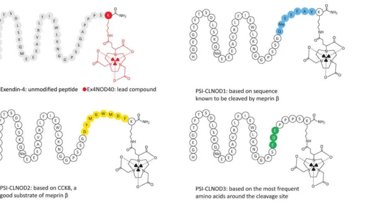
The linker of PSI-CLNOD1 is based on a sequence that can be also found within the binding sequence of exendin-4 spanning from Q13 to V19, which we have shown to be cleaved by Fig 1. The most common amino acids around the cleavage sites of both meprinαandβ.The size of the one letter code of the amino acid represents the frequency of that amino acid in that particular position. The figure was generated using icelogo, based on peptide cleavage assays described previously [13]. Peptide sequences were aligned on the scissile bond between P1 and P10indicated by a black line. Statistically
significant amino acid residue occurrences present (P<0.05) were plotted. Those amino acids that were completely absent are shown below in pink.
meprinβ. PSI-CLNOD2 contains a linker based on cholecystokinine-8 (CCK-8), a very good
natural substrate for meprinβ. Lastly, the cleavable linker of PSI-CLNOD3 was based on the
three most frequent amino acids around the cleavage site of the protease [13]. We determined
the binding properties of these peptides on Chinese hamster lung (CHL) cells expressing the GLP-1R as well as the internalisation kinetics. Further, we tested the stability of the probes. We assessed their metabolic stability in fresh human blood plasma as well as the feasibility of the
linkers in both kidney homogenates and with recombinant meprinβ. To verify the expression
of meprinβin mouse kidneys CD1 kidney sections were stained with meprinβ-specific
immu-noflourescent antibody. The biodistribution of the111In-labelled derivatives was performed in
CD1 nude mice bearing a CHL-GLP-1R positive tumour xenograft.
Material and Methods
Radiolabelling of the exendin-4 derivatives
Ex4NOD40, PSI-CLNOD1, PSI-CLNOD2 and PSI-CLNOD3, as seen inFig 2andS1 Table,
were synthesised by Peptide Specialty Laboratories (Heidelberg, Germany). The fragments of
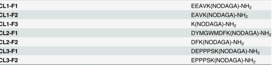
the peptides, seen inTable 1, were synthesised by piCHEM (Graz, Austria). The chelator
1-(1,3-carboxypropyl)-1,4,7-triazacyclononane-4,7-diacetic acid (NODAGA) was attached to
theε-amino group of the amidated lysine at the C-terminal end of the peptide. Unmodified
exendin-4 was purchased from Bachem (Bubendorf, Switzerland).
All peptides were labelled with111InCL3(Mallinckrodt, Netherlands) by adding 9.4 MBq
ac-tivity to 25μL 0.02 M HCl and 0.23 to 0.25 nmol of the respective compound and 5μL 0.5 M
ammonium acetate (pH 5.5). Following incubation at 95°C for 30 min the quality of the label-ling was assessed using reversed-phase high-performance liquid chromatography (RP-HPLC)
Fig 2. Graphical representation of exendin-4 and its derivatives.The linkers are highlighted in different colours.
on a C18 reversed-phase column (Dr. Maisch Reprospher 300 C18-TN, 4.6 mm x 150 mm;
5μm). Before injection the sample was diluted in 0.1 mM sodium DTPA and eluted with water
containing 0.1% TFA (trifluoroacetic acid) with a linear gradient from 15% to 95% acetonitrile for 10 minutes followed by an isocratic elution at 95% acetonitrile for an additional 5 min with
a flow rate of 1 mL/min. For thein vitromeprinβdigestion 10 nmol of the respective peptide
were labelled as described above. After the labelling the remaining unlabelled chelators were
saturated withnatInCl3(10 nmol, 1eq.) and once again incubated for 20 min at 95°C.
Cell culture
Chinese hamster lung cell line stably transfected with the GLP-1 receptor gene (CHL-GLP-1R
positive cells), a kind gift of Prof. Brigitte Lankat-Buttgereit, were cultured in Dulbecco’s
Modi-fied Eagle’s Medium (DMEM) with 4.5 g/L D-glucose and GlutaMax. In addition, the media
contained 10% fetal calf serum, 100 IU/mL penicillin G, 10 mg/mL streptomycine, 500μg/mL
geneticin sulfate, 1 mM sodium pyruvate and 0.1 mM non-essential amino acids. The cells
were maintained in a humidified 5% CO2atmosphere at 37°C and were harvested by
trypsini-sation with trypsin/EDTA [25].
Labelling of exendin-4 derivatives with
natInCl
3Peptides were labelled by adding 10 nmol of the respective peptide to 20 nmolnatInCl3
(Sigma-Aldrich, Switzerland) solution in 60μL 0.4 M ammonium acetate buffer (pH 5.5) and
subse-quent incubation for 15 min at 90°C. The labelling was verified by liquid chromatography–
mass spectrometry (LC/MS) on an Atlantis C18 (25 cm x 4.6 mm; 5μm) column.
IC
50binding assay
The experiments were conducted as described previously [26]. The half-maximal inhibitory
concentrations (IC50) ofnatIn-labelled PSI-CLNOD1, PSI-CLNOD2 and PSI-CLNOD3 were
determined using CHL-GLP-1R positive cells. 4 kBq (0.1–0.6 pmol)111In-labelled Ex4NOD40
was used for detection of the binding. The cells were treated with111In-Ex4NOD40 and various
concentrations of eithernatIn-labelled exendin-4 derivative or unmodified exendin-4 with the
final concentration ranging from 10-10to 10-6M. The total volume was adjusted to 1 mL with
medium (DMEM with 0.1% BSA) and the cells incubated on ice for 60 minutes. For the total
binding nonatIn-labelled peptide was added. After incubation the cells were washed twice with
phosphate buffered saline (PBS), solubilised with 1 mL 1 M sodium hydroxide (NaOH),
col-lected and the activity quantified using aγ-counter (Packard Cobra II Auto Gamma, Perkin
Elmer, Switzerland). The IC50values were calculated by fitting the data with non-linear
regres-sion using least squares fit with GraphPad Prism (GraphPad Software, La Jolla, CA). Experi-ments were performed on triplicate samples. The statistical significance was determined using Table 1. Amino acid sequences of the peptide fragments.
CL1-F1 EEAVK(NODAGA)-NH2
CL1-F2 EAVK(NODAGA)-NH2
CL1-F3 K(NODAGA)-NH2
CL2-F1 DYMGWMDFK(NODAGA)-NH2
CL2-F2 DFK(NODAGA)-NH2
CL3-F1 DEPPPSK(NODAGA)-NH2
CL3-F2 EPPPSK(NODAGA)-NH2
a one-way ANOVA test and corrected for multiple comparisons using Tukey’s honest significance test.
Internalisation assay
The internalisation kinetics of111In-PSI-CLNOD1,111In-PSI-CLNOD2 and111
In-PSI-CL-NOD3 were evaluated as described previously [26]. 4kBq (0.2–1 pmol) of each111In-labelled
peptide was used as a probe. After addition of the tracer the cells were incubated for certain time points (5 min, 15 min, 30 min, 60 min and 120 min, respectively) at 37°C; non-specific
binding was determined by the addition of the correspondingnatIn-labelled probe to a final
concentration of 1μM. After incubation, the supernatant was aspirated and the cells washed
with PBS. Both the supernatant and wash fractions were used to determine the non-bound fraction. In order to determine the peptide fraction bound on the surface the cells were incubat-ed with 1 mL glycine buffer pH 2.6 for 5 min at room temperature and collectincubat-ed separately. The internalised fraction was identified by adding 1 mL 1M NaOH to the cells with subsequent
collection of the lysates. The activity in all three fractions was measured in aγ-counter
(Pack-ard Cobra II Auto Gamma, Perkin Elmer, Switzerland). Experiments were performed on tripli-cate samples. The statistical significance was determined using the one-way ANOVA test and
corrected for multiple comparisons using Tukey’s honest significance test.
Plasma stability
Plasma stability was qualitatively determined as described previously [26]. 5 MBq (0.4 nmol)
labelled peptide was added to fresh human blood plasma and incubated at 37°C for 48 h. Sam-ples were taken before incubation as well as after 1 h, 4 h, 24 h and 48 h. Plasma proteins were precipitated using a solution containing 50% methanol and 50% acetonitrile with 0.1% TFA.
The sample was then filtered through a Thomson Single StEP Filter vial 0.45μm PVDF
(Thom-son Instrument Company, Oceanside, CA) and analysed using RP-HPLC on a Discovery
Bio-Wide Pore C18 (15cm x 2.1mm; 3μm) column. The column was eluted with 95% water
containing 0.1% TFA and 5% acetonitrile containing 0.1% TFA for 5 min followed with a linear gradient from 5 to 70% acetonitrile for 15 minutes succeeded by a second linear gradient from
70–90% acetonitrile for 5 min and an isocratic gradient at 95% acetonitrile for the final 5 min
with a flow rate of 1 mL/min.
Meprin
β
immunofluorescence staining
Immunofluorescence staining was performed on kidney cryosections obtained from CD1 nu/
nu mice. The 3μm thick sections were transferred onto a microscope slide, fixed in acetone for
10 min at 4°C and washed in PBS with 0.4% Triton X-100 (PBST) for 5 min. The sections were then treated with 1% donkey serum (Dako, Baar, Switzerland) in PBST, followed by blocking with 5% donkey serum in PBS for 1 h. Subsequently, the cells were incubated with polyclonal
antibody directed against meprinβ(sc-23491) (Santa Cruz Biotechnology, Santa Cruz, USA)
In vitro
meprin digestion
Recombinant meprinαand meprinβwere expressed in High Five Cells (BTI-TN-5B1-4)
de-rived from the parentalTrichopulsia nicell line, purified and activated as described previously
[27,28]. 220 ng of each recombinant protein was added to 35.5μL 0.1 mM In-labelled peptide
solution in 37.5μL meprin assay buffer (50 mM Tris-HCl pH 7.5 + 1 mM MgCl2+ 1 mM
CaCl2) and 30μL 1 M Tris pH 7.5 and incubated at 37°C. Samples were taken at specific time
points (0 min, 5 min, 10 min, 20 min and 60 min) and transferred into precipitation solution
(methanol/acetonitrile with 0.1% TFA 1:1). The samples were centrifuged at 11’000 g for 2
min and qualitatively analysed using both RP-HPLC and liquid chromatography-mass
spec-trometry (LC-MS) on a Discovery Bio Wide Pore C18 (25 cm x 4.6 mm; 5μm) column. The
LC-MS samples were ionised using electrospray ionisation (ESI+) and the charged molecules
detected using time of flight mode (TOF). The column was eluted with water (0–5 min)
fol-lowed by a linear gradient from 0–95% acetonitrile for 40 min and an isocratic elution with
95% acetonitrile for the final 5 min at a flow rate of 1 mL/min for the whole run. For the LC-MS samples 0.1% formic acid was added to the eluents whereas for the RP-HPLC 0.1% TFA was supplemented.
Ex vivo
stability in kidney homogenates
Kidneys were extracted from CD1 nu/nu mice and shock-frozen in liquid nitrogen. The frozen tissue was crushed using a Bessman Tissue Pulveriser (Spectrum Chemical Manufacturing Corp., New Brunswick, NJ), transferred into a tube with Krebs-Henseleit buffer pH 7.4 and ho-mogenised using a 20G syringe.
1.2 MBq (9.5–45.5 nmol) of either of the111In-labelled peptides were added to the
homoge-nate and incubated at 37°C. Samples were taken before incubation as well as after 30 min, 60 min and 180 min and mixed with precipitation solution (50% and 50% acetonitrile with
0.1% TFA). The samples were centrifuged twice at 11’000 g and qualitatively analysed via
RP-HPLC.
Biodistribution
Biodistribution of111In-PSI-CLNOD1,111In-PSI-CLNOD2,111In-PSI-CLNOD3 and
111In-Ex4NOD40 were performed in CD 1 nu/nu mice with tumour xenografts that were
pre-pared as previously described [26]. Six week-old female CD1 nu/nu mice were subcutaneously
inoculated in both shoulder regions with 8 x 10⁶CHL-GLP-1R positive cells suspended in PBS
pH 7.4. After 4 weeks, the mice were randomly divided into groups of four mice. For the
111In-labelled fragments mice without tumours were used. The mice were injected with
ap-proximately 100 kBq (1–5 pmol) of the respective111In-labelled peptide diluted in PBS pH
7.4 with 0.1% BSA via the tail vein and sacrificed after 4 h by CO2asphyxiation. In order to
determine the GLP-1R mediated uptake of111In-PSI-CLNOD1,111In-PSI-CLNOD2,
111In-PSI-CLNOD3 and111In-Ex4NOD40 another group of four mice each were pre-injected
with an excess of 100μg exendin-4 to block the receptor and sacrificed after 4 h. Blood, heart,
lungs, spleen, kidneys, pancreas, stomach, intestine, liver, muscle, bone and tumour were
re-moved, weighted and activity determined in aγ-counter (Packard Cobra II Auto Gamma,
Per-kin Elmer, Switzerland). The percentage of injected activity per gram tissue (%iA/g) was calculated for each sample. The statistical significance was determined using the two-way
ANOVA test and corrected for multiple comparisons using Tukey’s honest significance test.
Ethics Statement
All animal experiments were reviewed by the“Ethical Animal Committee of the Cantons
Aarau, Baselland and Baselstadt), approved by the Cantonal Veterinarian Department of the Canton Aarau (permit number: 75531) and conducted in accordance with the Swiss law of animal protection.
Results and Discussion
Radiolabelling
All peptides were successfully labelled with111In resulting in specific activities from 37.6 to
40.9 MBq/nmol. For thein vitromeprinα/βdigestion the peptides were labelled with lower
specific activities ranging from 0.07 to 0.25 MBq/nmol. In all cases the radiochemical yield was
>95% as determined by RP-HPLC.111In-DTPA eluted after 2 min while the111In-labelled
peptides were retained for approximately 8 to 9 min.
IC
50binding assay
In a competitive binding assay we showed that thenatIn-labelled peptides with the cleavable
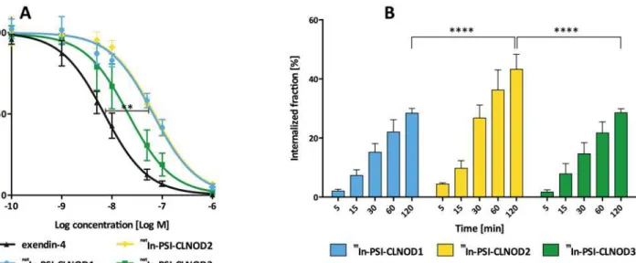
linkers retain a high affinity to the GLP-1R (Fig 3A). The binding ofnatIn-PSI-CLNOD3, the
peptide with the least modified amino acids, seems not to be impaired compared to unmodified
exendin-4 as the difference is not significant (IC50natIn-PSI-CLNOD3: 21 nM, 95% confidence
interval: 17 nM-26 nM; IC50exendin-4: 7.0 nM, 95% confidence interval: 5.9 nM-8.3 nM). The
other tested probes showed a significantly lower binding affinity (p<0.01) than exendin-4.
The IC50values of the other peptides were 66 nM (95% confidence interval 59–83 nM) for
natIn-PSI-CLNOD1 and 70 nM (95% confidence interval 60
–81 nM) fornatIn-PSI-CLNOD2,
respectively. Overall, all peptides have similar affinities as previously published peptides, which
indicates that the C-terminal modification seems not to impair the binding affinity [26,29].
Fig 3.In vitroassessment of the peptides containing cleavable linkers in CHL cells expressing GLP-1R.(A) IC50values ofnatIn labelled peptides. 0.1–
0.6 pmol111In-Ex4NOD40 was used as tracer. (B) Internalisation kinetics of the111In-labelled probes. 0.2–1 pmol of the respective111In-labelled peptide was used as tracer. Non-specific binding was determined by incubation with 1μM ofnatIn labelled peptide.
Internalisation
All the111In-labelled peptides internalise well, as seen inFig 3B. Both111In-PSI-CLNOD1 and
111In-PSI-CLNOD3 internalise over a similar timescale with approximately 28% uptake after 2 h,
which is in the same range as already published peptides [26].111In-PSI-CLNOD2 on the other
hand has a higher rate of internalisation with 43% of the peptide taken up into the cells after 2 h. The unspecific binding of PSI-CLNOD2, however, is also significantly higher (2.0 ± 0.8%) than
for the other two peptides (111In-PSI-CLNOD1: 1.0 ± 0.2%;111In- PSI-CLNOD3: 1.1 ± 0.7%)
(p<0.05).
Plasma stability
The plasma stability of the111In-labelled peptides is illustrated inFig 4. All peptides are>90%
stable after 4 h. After 48 h 71% of PSI-CLNOD1, 80% of PSI-CLNOD2 and 59% of PSI-CLNOD3 are still intact. While PSI-CLNOD3 is less stable than previously tested peptides, all probes are
>90% intact after 4 h, which is sufficient for SPECT imaging as, scans with111In labeled exendin
derivatives are usually performed after 4 h [22,26].
In vitro
meprin digestion
Figs5and6show the results of the digestion of the In-labelled probes with meprinαand
meprinβ, respectively. Both111In-PSI-CLNOD1 and111In-PSI-CLNOD3 are cleaved by
meprinβand quickly form metabolites with retention times of approximately 15 min. Closer
LC-MS analysis of the metabolites revealed four fragments for PSI-CLNOD1 and two
frag-ments for PSI-CLNOD3, respectively, all of which were in the predicted region as seen inFig 1.
After 60 min only 7% of full length PSI-CLNOD1 and 3% of PSI-CLNOD3 remain intact.
111In-PSI-CLNOD2, in contrast is not cleaved completely as after 60 min approximately 20%
of the full-length peptide remains in the sample.
All of the detected fragments were expected as the linkers were designed to be cleaved at
specific positions [12]. The quick metabolism of both PSI-CLNOD1 and PSI-CLNOD3 might
have been facilitated by a series of adjecent acidic amino acids that form a sequence recognised
by meprinβ, as seen inFig 1, while PSI-CLNOD2 on the other hand is not metabolised as
quickly as it contains only two, non-adjacent, aspartic acids. Furthermore, the localisation of one of the cleavage sites close to the chelator might sterically hinder the enzymatic digestion. The proximity to the chelator, however, is necessary as smaller fragmets are preferable since they are excreted more easily and rapidly.
Additionally, we tested the specificity of meprinαtowards the cleavable linkers, although
not as specific as meprinβbut also expressed in the kidney. Meprinαdigestion of the peptides
also led to rapid breakdown of PSI-CLNOD1 and PSI-CLNOD3 with only approximately 6% of each peptide remaining intact after 60 min, while approximately 25% of PSI-CLNOD2 was
intact 1 h after digestion. Cleavage sites for meprinαwithin the linker sequence were identified
for both PSI-CLNOD2 and PSI-CLNOD3, for PSI-CLNOD1 cleavage was only found within the binding sites.
As meprinαlacks the same high specifictiy for certain amino acid sequences it is not
sur-prising that those cleavage sites do not show a specific pattern [15]. Nevertheless, we were able
to identify several sequences that are cleaved by meprinα, some of which are also recognised
by meprinβ(Table 2).111In-Ex4NOD40 is also metabolised by both meprinαand meprinβ.
Cleavage by meprinβ, however, yields larger fragments that are more likely to be retained in
the kidneys. Multiple cleavage sites for meprinβwere identified between M14 and E17 while
Fig 4. Plasma stability of111In-labelled PSI-CLNOD1, PSI-CLNOD2 and PSI-CLNOD3 in fresh human
blood plasma.
Fig 5. Digestion of In-labelled probes with recombinant meprinβ.A, C, E, and G show the metabolism of the corresponding111In-labelled peptides.
Curves B, D, F and H show the degradation of the correspondingnatIn-labelled peptides and the formation of different fragments over time.
Fig 6. Digestion of In-labelled probes with recombinant meprinα.A, C, E, and G show the metabolism of the corresponding111In-labelled peptides.
Curves B, D, F and H show the degradation of the correspondingnatIn-labelled peptides and the formation of different fragments over time.
Meprin
β
immunofluorescence staining
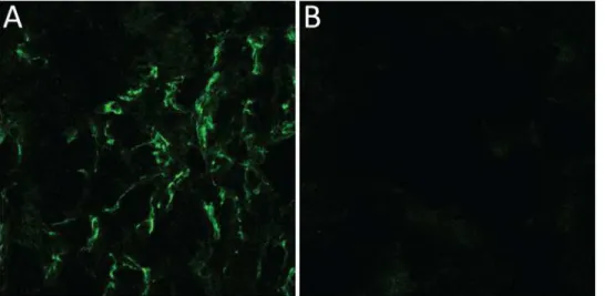
To verify the expression of meprinβin the kidneys we performed immunolfuorescence
stain-ing on kidney sections.Fig 7shows a clear flourescent signal originating from the brush-border
membrane confirming the viability of the target.
Ex vivo
stability in kidney homogenates
All the111In-labelled peptides with cleavable linkers are metabolised quickly after incubation
with CD1 kidney homogenates. Fragments with similar retention-times as after the meprinβ
digestion are formed within 30 min, as seen inFig 8. One metabolite not present in the meprin
βdigestion, however, is formed and elutes after 3 minutes, indicating a small, very polar
frag-ment. This fragment is formed in all peptides with cleavable linkers and is the only remaining
peak after 180 min for111In-PSI-CLNOD3 and the major peak for both111In-PSI-CLNOD1
and111In-PSI-CLNOD2. Radiolabelled Ex4NOD40 showed a similar pattern as within 30 min
the majority of the peptide was digested leaving the very polar fragment described above. These additional fragments are most likely a product of unspecific cleavage of the linkers by other proteases that are present in the homogenate and potentially the brush
border membrane.
Biodistribution
111In-labelled PSI-CLNOD1, PSI-CLNOD2 and PSI-CLNOD3 as well as Ex4NOD40 were
spe-cifically taken up in the receptor-positive tissues as tumour, pancreas and lungs of CD1 Table 2. Cleavage sites of In-labelled peptides digested with meprinαandβ.
natIn-PSI-CLNOD1 HGEGTFTSDLSKQNleEEEAVRLFIEWLKNGGPSSG"QNle#E#E#EAV#K(natIn-NODAGA)-NH 2 natIn-PSI-CLNOD2 HGEGTFTSDLSKQNleEEEAVRLFIEWLKNG"GPSSG#DYMGWM"#DFK(natIn-NODAGA)-NH
2 natIn-PSI-CLNOD3 HGEGTFTSDLSKQNleEEEAVRLFIEWLKNGGPSE#"D"#EPPPSK(nat
In-NODAGA)-NH2 nat
In-Ex4NOD40 HGEGTFTSDLSKQM#E#E#EAVRLF"IEWLKNGGPSS"GA"PPPSK(natIn-NODAGA)-NH 2
Cleavage sites for meprinαare illustrated with", for meprinβwith#. The linkers are highlighted in bold. In-labelled Ex4NOD40 was used as control.
doi:10.1371/journal.pone.0123443.t002
Fig 7. Immunofluorescence picture of murine kidney sections.(A) Anti-Meprinβantibody was used to detect the protease, visualised by fluorescence detection at 488 nm. (B) For the control goat igG was used.
Fig 8.Ex vivometabolism of the corresponding peptides in CD1 nu/nu kidney homogenates.
nude mice 4 h after administration. The uptake in those organs was blocked by the
pre-injection of 100μg exendin-4 (p<0.01). Out of the three peptides with the cleavable linkers
111In-PSI-CLNOD2 displayed the highest uptake in the tumour, its unspecific uptake in the
re-ceptor positive tissues, however, was also the highest (p<0.05), which correlates with the
re-sults from the internalisation assay (Fig 9andS2 Table). All of111In-labelled peptides with the
cleavable linkers had comparable uptake in GLP-1R positive tissue and were not significantly different from the uptake of the lead compound. Overall, the uptake in receptor positive tissue
is comparable to already published radiolabelled exendin-4 derivatives [26].
The most activity in all four cases, however, accumulates in the kidneys. It was shown previ-ously that this accumulation is mainly by non-GLP-1R mediated uptake and is primarily
facili-tated by various other mechanisms including the Megalin transporter [26,29–32]. None of the
111In-labelled peptides show a significant difference in kidney retention compared to the lead
compound111In-Ex4NOD40. A biodistribution of the111In-labelled fragments CL1F1, CL1F2,
CL1F3, CL2F1, CL2F2, CL3F1 and CL3F2 we identified after meprinβcleavage of the peptides
revealed that the fragments that are formed after digestion are not retained in the kidneys as less than 4% of the injected activity per gram tissue were detected at the end of the experiment (S3 Table). This leads to the assumption that the peptides are not cleavedin vivobefore being reabsorbed, either because the expression of meprin in the brush border membrane is too low to digest the linkers quickly enough or because the rate of the re-uptake mechanism of exen-din-4 exceeds the turnover rate of meprin.
Conclusions
In conclusion, we designed three new exendin-4 derivatives containing specific cleavage sites
for meprinβ.111In-labelled PSI-CLNOD1, PSI-CLNOD2 and PSI-CLNOD3 retain the
excel-lentin vitroproperties of previously tested exendin-4 derivatives as well as the high specific
Fig 9. Biodistribution of111In-labeled peptides in CD1 nu/nu mice with CHL-GLP1R positive
tumour-xenografts.Values are mean percentages injected dose per gram tissue (n= 4. Error bars SD). Blocking was performed by pre-injection of 100μg excess of unmodified exendin-4. Mice were sacrificed 4 h after injection. The significance was tested by a two-was ANOVA test corrected for multiple comparisons by Tukey’s honest significance test (**p<0.01;***p<0.001;****p>0.0001).
uptake into GLP-1R positive tissue. This suggests that the seven C-terminal amino acids may be altered without loss of affinity towards the GLP-1R receptor, allowing new strategies of modifying exendin-4 to be developed. Uptake of the peptides into the kidneys, however, was
not decreased even though we could show that meprinβis expressed in the brush border
mem-brane, leading to the assumption that the uptake mechanism of the peptides is faster than the
cleavage of the linker. The rapid and specific cleavage by meprinβ, and to a lesser extent by
meprinαin vitro, however, demonstrates the potential of the concept of specific linkers for
meprin targeting as prodrugs in conditions where meprinβexpression is upregulated.
Supporting Information
S1 Table. Amino acid sequences of the peptides used in this work.
(DOCX)
S2 Table. Biodistribution of111In-labelled peptides in CD1 nu/nu mice with CHL-GLP1R positive tumour xenografts.Values are mean percentages injected dose per gram tissue
(n= 4. Error bars SD). Blocking was performed by pre-injection of 100μg excess of unmodified
exendin-4. Mice were sacrificed 4 h after injection. (DOCX)
S3 Table. Biodistribution of the111Inlabelled fragments of the corresponding peptides in CD1 nu/nu mice.
(DOCX)
Acknowledgments
The authors wish to thank Alain Blanc and Olga Gasser for their technical support, Dr. Philipp Berger for support with the fluorescence microscopy, Prof. Erwin Sterchi for very fruitful dis-cussions and Dr. Laura Bailey for reviewing this manuscript.
Author Contributions
Conceived and designed the experiments: AJ MB. Performed the experiments: AJ FP OM CBP MB. Analyzed the data: AJ FP CBP RS MB. Contributed reagents/materials/analysis tools: CBP. Wrote the paper: AJ MB.
References
1. Zhang W, Song J, Mu L, Zhang B, Liu L, Xing Y, et al. Improving anticancer activity and selectivity of camptothecin through conjugation with releasable substance P. Bioorg. Med. Chem. Lett. Elsevier Ltd; 2011 Mar 1; 21(5):1452–5. doi:10.1016/j.bmcl.2011.01.013PMID:21282053
2. Leriche G, Chisholm L, Wagner A. Cleavable linkers in chemical biology. Bioorg. Med. Chem. Elsevier Ltd; 2012 Jan 15; 20(2):571–82. doi:10.1016/j.bmc.2011.07.048PMID:21880494
3. Sloniec J, Resch-Genger U, Hennig A. Photophysics and Release Kinetics of Enzyme-Activatable Opti-cal Probes Based on H-Dimerized Fluorophores on Self-Immolative Linkers. J. Phys. Chem. B. 2013 Nov 21; 117(46):14336–44. doi:10.1021/jp409388bPMID:24144206
4. Chen Y-J, Wu S-C, Chen C-Y, Tzou S-C, Cheng T-L, Huang Y-F, et al. Peptide-based MRI contrast agent and near-infrared fluorescent probe for intratumoral legumain detection. Biomaterials. 2014 Jan; 35(1):304–15. doi:10.1016/j.biomaterials.2013.09.100PMID:24120038
5. Trejtnar F, Laznicek M. Analysis of renal handling of radiopharmaceuticals. Q J Nucl Med. 2002 Sep; 46(3):181–94. PMID:12134135
7. Bodei L, Cremonesi M, Ferrari M, Pacifici M, Grana CM, Bartolomei M, et al. Long-term evaluation of renal toxicity after peptide receptor radionuclide therapy with 90Y-DOTATOC and 177Lu-DOTATATE: the role of associated risk factors. Eur J Nucl Med Mol Imaging. 2008 Apr 22; 35(10):1847–56. doi:10. 1007/s00259-008-0778-1PMID:18427807
8. Akizawa H, Imajima M, Hanaoka H, Uehara T, Satake S, Arano Y. Renal Brush Border Enzyme-Cleav-able Linkages for Low Renal Radioactivity Levels of Radiolabeled Antibody Fragments. Bioconjugate Chem. 2013 Feb 20; 24(2):291–9. doi:10.1021/bc300428bPMID:23330714
9. Fujioka Y, Satake S, Uehara T, Mukai T, Akizawa H, Ogawa K, et al. In Vitro System To Estimate Renal Brush Border Enzyme-Mediated Cleavage of Peptide Linkages for Designing Radiolabeled Antibody Fragments of Low Renal Radioactivity Levels. Bioconjugate Chem. 2005 Nov; 16(6):1610–6. PMID: 16287261
10. Yim C- B, Mikkola K, Fagerholm V, Elomaa V- V, Ishizu T, Rajander J, et al. Synthesis and preclinical characterization of [64Cu]NODAGA-MAL-exendin-4 with a Nε
-maleoyl-l-lysyl-glycine linkage. Nucl Med Biol. 2013 Nov; 40(8):1006–12. doi:10.1016/j.nucmedbio.2013.06.012PMID:23932646
11. Uehara T, Rokugawa T, Kinoshita M, Nemoto S, Fransisco Lazaro GG, Hanaoka H, et al.67/68Ga-Labeling
Agent That Liberates67/68Ga-NOTA-Methionine by Lysosomal Proteolysis of Parental Low Molecular
Weight Polypeptides to Reduce Renal Radioactivity Levels. Bioconjug Chem. 2014 Nov 19; 25 (11):2038–45. doi:10.1021/bc5004058PMID:25303645
12. Sterchi EE, Stöcker W, Bond JS. Meprins, membrane-bound and secreted astacin metalloproteinases. Mol Aspects Med. 2008 Oct; 29(5):309–28. doi:10.1016/j.mam.2008.08.002PMID:18783725 13. Becker-Pauly C, Barre O, Schilling O, auf dem Keller U, Ohler A, Broder C, et al. Proteomic Analyses
Reveal an Acidic Prime Side Specificity for the Astacin Metalloprotease Family Reflected by Physiologi-cal Substrates. Mol Cel Proteomics. 2011 Sep 8; 10(9):M111.009233–3.
14. Jefferson T, auf dem Keller U, Bellac C, Metz VV, Broder C, Hedrich J, et al. The substrate degradome of meprin metalloproteases reveals an unexpected proteolytic link between meprinβand ADAM10. Cell. Mol. Life Sci. 2012 Sep 1; 70(2):309–33. doi:10.1007/s00018-012-1106-2PMID:22940918 15. Broder C, Becker-Pauly C. The metalloproteases meprinαand meprinβ: unique enzymes in
inflamma-tion, neurodegenerainflamma-tion, cancer and fibrosis. Biochem. J. 2013 Feb 15; 450(2):253–64. doi:10.1042/ BJ20121751PMID:23410038
16. Bertenshaw GP, Turk BE, Hubbard SJ, Matters GL, Bylander JE, Crisman JM, et al. Marked differences between metalloproteases meprin A and B in substrate and peptide bond specificity. J. Biol. Chem. 2001 Apr 20; 276(16):13248–55. PMID:11278902
17. Herzog C, Kaushal GP, Haun RS. Generation of biologically active interleukin-1beta by meprin B. Cyto-kine. 2005 Sep 7; 31(5):394–403. PMID:16095909
18. Lottaz D, Maurer CA, Noël A, Blacher S, Huguenin M, Nievergelt A, et al. Enhanced Activity of Meprin-α, a Pro-Migratory and Pro-Angiogenic Protease, in Colorectal Cancer. Nguyen HTT, editor. PLoS ONE. 2011 Nov 11; 6(11):e26450. doi:10.1371/journal.pone.0026450PMID:22096485
19. Broder C, Arnold P, Vadon-Le Goff S, Konerding MA, Bahr K, Müller S, et al. Metalloproteases meprin αand meprinβare C- and N-procollagen proteinases important for collagen assembly and tensile strength. Proc. Natl. Acad. Sci. U.S.A. 2013 Aug 27; 110(35):14219–24. doi:10.1073/pnas. 1305464110PMID:23940311
20. Biasin V, Marsh LM, Egemnazarov B, Wilhelm J, Ghanim B, Klepetko W, et al. Meprinβ, a novel media-tor of vascular remodelling underlying pulmonary hypertension. J. Pathol. 2014 Jan 31; 233(1):7–17. doi:10.1002/path.4303PMID:24258247
21. Baggio LL, Drucker DJ. Biology of Incretins: GLP-1 and GIP. Gastroenterology. 2007 May; 132 (6):2131–57. PMID:17498508
22. Christ E, Wild D, Ederer S, Behe M, Nicolas G. Glucagon-like peptide-1 receptor imaging for the locali-sation of insulinomas: a prospective multicentre imaging study. Lancet Diabetes Endocrinol. 2013; 1 (2):115–22. doi:10.1016/S2213-8587(13)70049-4PMID:24622317
23. Brom M, Woliner-van der Weg W, Joosten L, Frielink C, Bouckenooghe T, Rijken P, et al. Non-invasive quantification of the beta cell mass by SPECT with111In-labelled exendin. Diabetologia. 2014 Feb 1; 57
(5):950–9. doi:10.1007/s00125-014-3166-3PMID:24488022
24. Pattou F, Kerr-Conte J, Wild D. GLP-1-receptor scanning for imaging of human beta cells transplanted in muscle. N. Engl. J. Med. 2010 Sep 23; 363(13):1289–90. doi:10.1056/NEJMc1004547PMID: 20860517
26. Jodal A, Lankat-Buttgereit B, Brom M, Schibli R, Béhé M. A comparison of three67/68Ga-labelled exendin-4 derivatives forβ-cell imaging on the GLP-1 receptor: the influence of the conjugation site of NODAGA as chelator. EJNMMI Research. 2014; 4:31. doi:10.1186/s13550-014-0031-9PMID:25006548 27. Becker-Pauly C, Höwel M, Walker T, Vlad A, Aufenvenne K, Oji V, et al. TheαandβSubunits of the
Metalloprotease Meprin Are Expressed in Separate Layers of Human Epidermis, Revealing Different Functions in Keratinocyte Proliferation and Differentiation. J Investig Dermatol. 2007 Jan 4; 127 (5):1115–25. PMID:17195012
28. Becker C, Kruse M- N, Slotty KA, Köhler D, Harris JR, Rösmann S, et al. Differences in the activation mechanism between the alpha and beta subunits of human meprin. Biol. Chem. 2003 May; 384 (5):825–31. PMID:12817480
29. Brom M, Joosten L, Oyen WJG, Gotthardt M, Boerman OC. Radiolabelled GLP-1 analogues for in vivo targeting of insulinomas. Contrast Media Mol. Imaging. 2012 Mar 6; 7(2):160–6. doi:10.1002/cmmi.475 PMID:22434628
30. Wild D, Béhé M, Wicki A, Storch D, Waser B, Gotthardt M, et al. [Lys40(Ahx-DTPA-111
In)NH2]exendin-4, a very promising ligand for glucagon-like peptide-1 (GLP-1) receptor targeting. J Nucl Med. 2006 Dec; 47(12):2025–33. PMID:17138746
31. Vegt E, Melis M, Eek A, de Visser M, Brom M, Oyen WJG, et al. Renal uptake of different radiolabelled peptides is mediated by megalin: SPECT and biodistribution studies in megalin-deficient mice. Eur J Nucl Med Mol Imaging. 2010 Dec 18; 38(4):623–32. doi:10.1007/s00259-010-1685-9PMID:21170526 32. Gotthardt M, van Eerd-Vismale J, Oyen WJG, De Jong M, Zhang H, Rolleman E, et al. Indication for