ABC- and SLC-Transporters in Murine and
Bovine Mammary Epithelium - Effects of
Prochloraz
Yagmur Yagdiran1, Agneta Oskarsson1, Christopher H. Knight2, Jonas Tallkvist1*
1Department of Biomedical Sciences and Veterinary Public Health, Swedish University of Agricultural Sciences, SE-750 07 Uppsala, Sweden,2Department of Veterinary Clinical and Animal Sciences, University of Copenhagen, 1870 Frederiksberg C, Denmark
*Jonas.Tallkvist@slu.se
Abstract
Some chemicals are ligands to efflux transporters which may result in high concentrations in milk. Limited knowledge is available on the influence of maternal exposure to chemicals on the expression and function of transporters in the lactating mammary gland. We deter-mined gene expression of ABC and SLC transporters in murine mammary tissue of different gestation and lactation stages, in murine mammary cells (HC11) featuring resting and secreting phenotypes and in bovine mammary tissue and cells (BME-UV). Effects on trans-porter expression and function of the imidazole fungicide prochloraz, previously reported to influenceBCRPin mammary cells, was investigated on transporter expression and function in the two cell lines. Transporters studied were BCRP, MDR1, MRP1, OATP1A5/OATP1A2, OCTN1 and OCT1. Gene expressions ofBCRPandOCT1in murine mammary glands were increased during gestation and lactation, whereasMDR1,MRP1,OATP1A5and
OCTN1were decreased, compared to expressions in virgins. All transporters measured in
mammary glands of mice were detected in bovine mammary tissue and in HC11 cells, while onlyMDR1andMRP1were detected in BME-UV cells. Prochloraz treatment induced MDR1 gene and protein expression in both differentiated HC11 and BME-UV cells and increased protein function in HC11 cells, resulting in decreased accumulation of the MDR1 substrate digoxin. In conclusion, our results demonstrate that murine (HC11) and bovine (BME-UV) mammary epithelial cells can be applied to characterize expression and function of transporters as well as effects of contaminants on the mammary transporters. An altered expression, induced by a drug or toxic chemical, on any of the transporters expressed in the mammary epithelial cells during lactation may modulate the well-balanced composition of nutrients and/or secretion of contaminants in milk with potential adverse effects on breast-fed infants and dairy consumers.
OPEN ACCESS
Citation:Yagdiran Y, Oskarsson A, Knight CH, Tallkvist J (2016) ABC- and SLC-Transporters in Murine and Bovine Mammary Epithelium - Effects of Prochloraz. PLoS ONE 11(3): e0151904. doi:10.1371/journal.pone.0151904
Editor:Cecilia Williams, University of Houston, USA, UNITED STATES
Received:February 11, 2016
Accepted:February 15, 2016
Published:March 30, 2016
Copyright:© 2016 Yagdiran et al. This is an open access article distributed under the terms of the
Creative Commons Attribution License, which permits unrestricted use, distribution, and reproduction in any medium, provided the original author and source are credited.
Data Availability Statement:All relevant data are within the paper and its Supporting Information file.
Funding:This study was funded by a grant from the research program TvärLivs, which is co-founded by the Swedish Research Council for Environment, Agricultural Sciences and Spatial Planning (Formas), Sweden's Innovation Agency (Vinnova), Swedish Farmer's Foundation for Agricultural Research, the Swedish Food Federation and the Swedish Food Retailer's Federation.
Introduction
Milk and dairy products are valuable foods for humans constituting an important nutrient source [1] but may also contain hazardous compounds [2–4]. Chemicals are secreted from blood to milk either via passive diffusion or active transport. Passive diffusion of chemicals can be predicted by taking physical and chemical properties into account [5] and result in levels of hydrophilic compounds in milk equal to or lower than in plasma. However, in cases where active transport mechanisms are operative chemical compounds may be concentrated in milk to a high extent which may pose a health threat to both breast-fed infants and dairy consumers [6,7].
Transporters are transmembrane proteins involved in cellular in- and efflux of essential and non-essential chemicals. Because these transporters are highly expressed in epithelia of tissues they serve an important role in absorption, tissue distribution and excretion of drugs and other chemicals [8]. Although a vast knowledge has been gained during the last decades concerning transport protein function, localization and expression in many tissues of the body, there is limited data on transporters in the mammary gland and their role for secretion of contami-nants into milk [7–12]. In addition, very little attention has been paid to the influence of mater-nal exposure to chemicals on the expression and function of transporters in the lactating mammary gland.
Expressions of transport proteins belonging to both the ATP-binding cassette (ABC-) and Solute Carrier (SLC-) superfamilies vary with lactation stage of the mammary gland [7,9,10]. The transporters play pivotal roles in the delivery of e.g. vitamins, fatty acids, sterols, porphy-rins, thyroid hormones, carnitine, and amino acids to the breast-fed offspring [6,13,14]. How-ever, some of these transporters feature broad substrate specificities and have been
demonstrated to mediate active transport also of drugs and toxic chemicals into milk [7,9,14– 16]. Transporters with broad substrate specificities and altered expressions in lactating as com-pared to resting mammary epithelial cells include breast cancer resistance protein (BCRP/ ABCG2) [6,7,13,17,18], multidrug resistance protein 1(MDR1/ABCB1) [7,18], multidrug resistance-associated protein 1 (MRP1/ABCC1) [7,10], solute carrier organic anion trans-porter family member 1A2 (OATP1A2/SLCO1A2), organic cation transtrans-porter novel protein type 1 (OCTN1/SLC22A4) [10] and organic cation transporter 1 (OCT1/SLC22A1) [16]
Prochloraz (N-propyl-N-[2-(2,4,6-trichlorophenoxy)ethyl]-1H-imidazole-1-carboxamide), an imidazole fungicide used in agriculture and horticulture [19] is an endocrine disruptor with various modes of action [20,21]. It has been reported that prochloraz treatment increases BCRPexpression and efflux activity through AhR activation in primary bovine mammary epi-thelial cells and in Madin-Darby canine kidney II (MDCK II) cells transfected with caprine BCRPcDNA [22,23].
The aims of the present study were to (i) investigate gene expressions of endogenousBCRP, MDR1,MRP1,OATP1A5/OATP1A2,OCTN1andOCT1in mammary glands of mice in virgins and at various gestation and lactation stages as well as in mammary glands of lactating cows (ii) characterize gene expressions of the transporters in HC11 and BME-UV cells and (iii) assess the effect of prochloraz on expression and function of transporters in the two cell lines.
Materials and Methods
Reagents and chemicals
Roswell Park Memorial Institute (RPMI) 1640 basal medium, gentamicin, heat–inactivated fetal bovine serum (FBS), Ham’s F12, NCTC 135 and 0.05% Trypsin-EDTA, Hank’s Balanced Salt Solution with CaCl2and MgCl2were from Gibco (Invitrogen). Human insulin, epidermal
growth factor (EGF), prolactin, hydrocortisone, lactose, lactalbumin hydrolysate, GSH, L-ascorbic acid, prochloraz (N-propyl-N-[2-(2,4,6-trichlorophenoxy)ethyl]-1H-imidazole-1-car-boxamide) and N-(2-hydroxyehtyl) piperazine-N’-(2-ethanesulfonic) acid (HEPES) were obtained from Sigma-Aldrich. Nucleospin RNA purification kit was purchased from Macherey-Nagel and Quant-iT™RiboGreen1RNA Assay Kit from Life Technologies. One-tube QuantiTect™SYBR1Green RT-PCR Kit was obtained from Qiagen and Cell TagTM 700 Stain In-Cell WesternTM Assay Kit was purchased from Li-Cor. Digoxin was kindly provided by Dr. Per Artursson, Department of Pharmacy, Uppsala University, Sweden and3H-digoxin purchased from PerkinElmer.
Mammary gland isolation from mouse and cow
NMRI-mice were given a standard pellet diet and tap water ad libitum under standard condi-tions of temperature and light. Animals were killed by cervical dislocation and mammary glands from virgin, pregnant (gestation days 13 and 18), lactating (lactation days 2 and 9) and weaning (weaning day 2) mice were rapidly excised, placed in RNAlater (Invitrogen), snap-fro-zen in liquid nitrogen and stored at -70°C pending isolation of total RNA. Animal experiments were reviewed and approved by the Council for Animal Experimentation of the Danish Minis-try of Food, Agriculture and Fisheries at the Danish Veterinary and Food Administration (per-mit no. 2012-15-2934-00587) and conducted at the Danish Technical University, Copenhagen, Denmark.
Mammary gland tissues of lactating cows were collected fresh at the Funbo-Lövsta abattoir located at the Swedish Livestock Research Centre, Uppsala, Sweden, placed in RNAlater and stored at -70°C pending isolation of total RNA.
Cell culture
The HC11 murine mammary epithelial cell line was a generous gift from Dr. Nancy Hynes (Friedrich Miescher Institute for Biomedical Research, Basel, Switzerland) [29] and used with the permission of Dr. Bernd Groner (Institute for Biomedical Research, Frankfurt, Germany). Cells were cultured in sterile filtered RPMI 1640 medium containing 10% heat-inactivated FBS, 5 mg/L insulin and 10μg/L EGF and 50μg/ml gentamycin in polycarbonate flasks at 37°C
in 5% CO2. Medium was changed routinely every 2 or 3 days and cells subcultured by
and 1μM hydrocortisone. Differentiation of the cells was assessed by measuring induction ofβ
-casein(CSN2)gene expression as well as examination of cellular morphology as described pre-viously [18].
The BME-UV bovine mammary epithelial cell line deriving from lactating and pregnant Holstein cow was kindly provided by Dr. Bruce Schultz, College of Veterinary Medicine, Kan-sas State University, USA and cultured as described by [30]. Briefly, BME-UV cells were cul-tured in sterile filtered RPMI 1640 medium containing 10% heat-inactivated FBS, 40% Ham’s F12, 20% NCTC 135, 0.1% lactose, 0.1% lactalbumin hydrolysate, 1.2 mM GSH, 10μg/ml L
ascorbic acid, 1μg/ml hydrocortisone, 1μg/ml insulin and 50μg/ml gentamycin in
polycar-bonate flasks at 37°C in 5% CO2. In some instances, BME-UV cells were prolactin treated for
72 h as previously described [31].
Cell viability
MTS reduction test was applied to measure mitochondrial activity in viable cells where the tet-razolium compound is transformed to colored formazan. A total of 17,000 cells/well were seeded into 96-well plates in a volume of 100μl, cultured and differentiated as described above.
Stock solutions of prochloraz were dissolved in dimethyl sulfoxide (DMSO) and added to the basal medium, 1:1000, to achieve the decided concentrations of the chemical in the presence of 0.1% DMSO. Cells were treated for 24 h with prochloraz (vehicle control, 0.1, 1, 10, 30 and 100μM) in serum-free RPMI 1640 medium. DMSO at 10% was used as a positive control in
the MTS test. The cells were exposed to test substances for 24 h and 20μl CellTiter 961AQueos
One Solution Reagent (Promega Corporation) was added to each well, according to manufac-turer’s instructions. After 1 hour incubation at 37°C absorbance was measured at 490 nm using a Wallac Victor21420 microplate reader (Perkin-Elmer). Cell viability was assessed by compar-ing mean absorbance values from prochloraz treated cells and vehicle controls based on six replicates.
Cell exposure
HC11 and BME-UV cells were seeded in 6-well culture plates and HC11 cells differentiated as described above. After differentiation of the HC11 cells and accomplished confluency of the BME-UV cells an additional 24 h incubation with exposure-medium (basal medium supple-mented with prochloraz to a final concentration of 0.1, 1, 10, 30μM and vehicle control) was
performed. After prochloraz exposure, cells were washed with 2.5 mL 1 x PBS and then lysed with RA1 buffer (Macherey-Nagel) for RNA isolation. RA1 lysates were stored at -70°C freezer prior to isolation of RNA and gene expression analyzes as described below.
Isolation of total RNA and RT-qPCR analysis
Total RNA from tissues and cells was isolated by using the NucleoSpin RNA kit containing DNAse I (Macherey-Nagel) as recommended by the manufacturer. To check the integrity of the RNA the 28S and 18S ribosomal RNA bands were examined by UV-visualization following agarose gel electrophoresis. Quantification of the RNA was performed with the RNA specific Quant-iT RiboGreen protocol (Molecular Probes) as described by the manufacturer.
Quantitative gene expression was examined by RT-qPCR using a Rotor-Gene 3000 (Corbett Research) by applying the One-tube QuantiTect™SYBR1Green RT-PCR Kit (Qiagen), accord-ing to the manufacturer’s recommendations. Murine and bovine lactating mammary tissue, respectively, was used as positive amplification controls and all primer-pairs tested on RNA isolated from these tissues generated specific RT-PCR products with anticipated amplicon sizes and single melting curve peaks. Final primer concentration for all target genes was 0.4μM and
75 or 150 ng total RNA was used as template in 12.5μl RT-qPCR reactions. Non-template
con-trols served as blanks and melt curve analysis was performed for each sample to check the spec-ificity of the obtained PCR products. Expressions of target genes were normalized to the geometric average expression of three appropriate reference genes [32]. Murine reference genes were hypoxanthine-guanine phosphoribosyltransferase (Hprt), ribosomal protein L13A (Rpl13a) and glyceraldehyde 3-phosphate dehydrogenase (Gapdh) [33] and bovine reference genes were ubiquitously expressed transcript isoform 2 (UXT), ribosomal protein S9 (RPS9) and ribosomal protein S15 (RPS15) [34]. Relative quantification of mRNA expressions was per-formed by comparing the quantification cycle (Cq) between the tissues and treatment groups of cells according to the 2-(deltadeltaCq)–method [35]. Cq cycle 35 was used as cut-off for limit of detection of gene expression. Fold differences were calculated setting virgins or untreated con-trol cells to one.
In-Cell Western Assay
HC11 and BME-UV cells were seeded at a density of 17,000 cells/well in 96-well plates. The cells were cultured and differentiated as described above prior to treatment with 30μM
pro-chloraz. Control cells were treated with only vehicle. After 24 h of treatment cells were washed once with 1X PBS and fixed with 4% formaldehyde (1X PBS) for 20 min. Cells were then treated with permeabilization solution (1X PBS containing 0.1% Triton X-100) 5 times for 5 min. After permeabilization cells were incubated in blocking buffer (Li-Cor) for 90 min at RT. Primary Mdr1 antibody (JSB-1, Abcam) diluted 1:200 in blocking buffer, was added to the wells and incubation was performed for 2.5 h under gentle shaking. Cells were then washed five times with 1X PBS containing 0.1% Tween-20 (PBS-T) and then hybridized for 1 h with a polyclonal secondary antibody (IRDye 800CW anti-mouse IgG (H+L) (Li-Cor) diluted 1:1000 in blocking buffer. Cell Tag 700 (Li-Cor) was used for normalization and added (1:500) to the
Table 1. Primer sequences used for the RT-qPCR analyses.Abbreviations, m: murine; b: bovine.
Genes and accession numbers Primer sequences
mCSN2NM_009972 CTTAACCCCACCGTCCAAT (forward) AGCATGATCCAAAGGTGAAA (reverse) mBCRPNM_011920.3 CGCAGAAGGAGATGTGTT (forward) TTGGATCTTTCCTTGCTGCT (reverse) mMDR1NM_011076.2 ATTTGGCAAAGCTGGAGAGA (forward) ACCCTGTAGCCCCTTTCACT (reverse) mMRP1NM_008576.3 CTGTGCTCACGATTGCTCAT (forward) CAGAGACCAGCTCACACCAA (reverse) mOAPT1A5NM_001267707.1 GCACAGAGAAAAAGCCAAGG (forward) CTCCAGGTATTTGGGCAAGA (reverse) mOCTN1NM_019687.3 CCTGTTCTGTGTTCCCCTGT (forward) GGTTATGGTGGCAATGTTCC (reverse) mOCT1NM_009202.5 CAGGTTTGGCCGTAAGCTCT (forward) GCAACATGGATGTATAGTCTGGG (reverse) bCSN2M16645.1 GTGAGGAACAGCAGCAAACA (forward) AGGGAAGGGCATTTCTTTGT (reverse) bBCRPEU570105.1 AACGGCATTCCAGAGACAAC (forward) ATGTGGATCCTTCCTTGCAG (reverse) bMDR1XM_590317.6 GCAACATTCTTCACCGGTTT (forward) TTGTCCTCCAAATGCAATCA (reverse) bMRP1AB082124.1 CCGTCCCTGTTCAAAGTGTT (forward) TGACGAAGCAGATGTGGAAG (reverse) bOATP1A2NM_174654.2 GCTTGTCTTGCTGGTTGTGA (forward) CAGGGATGGCAGATAAGGAA (reverse) bOCTN1NM_001206989.1 TTCTCGGCTCCTTTGTGTCT (forward) GCCACCACGTAGTTGGAGAT (reverse)
diluted secondary antibody hybridization solution. For background subtraction some wells were used as controls and only incubated with secondary antibody. Cells were washed five times with PBS-T and plates were scanned with detection at both 700 and 800 nm using an Odyssey instrument (Li-Cor Biosciences) and data were analyzed by applying Odyssey soft-ware. In each well, secondary antibody signal from the 800 nm channel was normalized to Cell Tag 700 signal from the 700 nm channel. Fold differences were calculated setting vehicle-treated controls to one. The whole procedure was performed at room temperature.
The JSB-1 antibody is reported to cross react with pyruvate carboxylase (PC) with a MW of 130 kDa [36]. However, cross reactivity was not apparent in the HC11 cells under the condi-tions used in our experiments (S1 Fig). Furthermore, data from human mammary glands dem-onstrates that the abundance of PC is very low [37].
Accumulation studies
HC11 and BME-UV cells were seeded in 12 well plates and cultured and treated as described above. Digoxin (Sigma) was chosen as a marker to examine the function of MDR1 in the cells using3H-digoxin (Perkin-Elmer Life Sciences), with a specific radioactivity of 5 Ci/mmol, as tracer. Cells were rinsed with 2 x 1.5 ml of 37°C Hank’s Balanced Salt Solution with CaCl2and
MgCl2(Invitrogen), pH 7.4 containing 25 mM HEPES (HBSS) and then pre-incubated for 30
min at 37° in 1.5 ml HBSS. After pre-incubation the cells were incubated at 37°C for 60 min with 37°C HBSS containing 1–5μM digoxin and 3000 Bq3H-digoxin/ml as tracer. At the end
of the experiment the cells were rinsed with 3 x 1.5 ml ice-cold HBSS and thereafter lysed by adding 1 ml 0.5 M NaOH to each well. The digoxin concentration in the cells was calculated from the radioactivity measurement byβ-spectrometry using a 2810 TR Tri-Carb1Liquid Scintillation Analyzer (PerkinElmer) and normalized to total protein concentration in each sample measured by applying the BCA-method.
Statistical analysis
Statistical analyses were performed by using Minitab 16 software. The results were analyzed by Kruskal-Wallis to detect any significant differences among the various treatment groups, fol-lowed by Mann-Whitney to examine statistically significant differences between two groups. The level of significance was set at p0.05.
Results
Gene expressions in mouse mammary gland
A limited number of cows and without specific data on lactation stages was analyzed. In the bovine mammary gland tissueCSN2,BCRP,MDR1,MRP1,OATP1A2andOCTN1gene expressions were detected (data not shown).
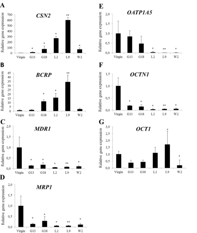
Fig 1. Relative gene expression ofCSN2and transporters (A-G) in the mammary gland of mice.Mammary glands were taken from virgin, pregnant (gestation day 13 and 18), lactating (lactation day 2 and 9) and weaning (weaning day 2) mice. Normalized gene expressions (A-G) are shown relative to virgin and the data is presented as means±SD; n = 3–4. Statistically significant differences as compared to virgins*p0.05;**p0.01.
Gene expressions in cultured mammary epithelial cells
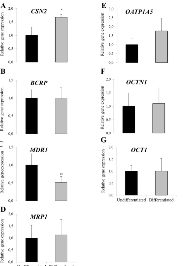
Statistically significant up-regulation inCSN2gene expressions was observed in the differenti-ated HC11 cells as compared to the undifferentidifferenti-ated controls (Fig 2A).MDR1gene expression was statistically significantly reduced in the differentiated HC11 cells as compared to the undif-ferentiated controls (Fig 2C). No statistically significant difference due to differentiation was observed onBCRP,MRP1,OATP1A5,OCTN1andOCT1gene expressions (Fig 2B, 2D, 2E, 2F and 2G, respectively).
Among the bovine genes examined onlyMDR1andMRP1were detected in the BME-UV cells (Fig 3). No difference was observed inMDR1orMRP1gene expressions after prolactin treatment of the cells (Fig 3A and 3B).
Cell viability in prochloraz-treated cells
Cell viability after prochloraz treatment of cells was assessed by MTS. For both cell lines cell viability remained>80% after incubation with prochloraz up to 30μM compared to vehicle
control and declined dramatically after treatment with 100μM prochloraz (Fig 4).
Gene expression in prochloraz-treated cells
The effect of prochloraz on gene expression ofCSN2and transporters was studied in mammary epithelial cells from both cell lines. Prochloraz treatment at non-cytotoxic concentrations resulted in a down regulation of gene expressions ofBCRPin differentiated HC11 cells (Fig 5B). In contrast,MDR1gene expression was significantly up-regulated at the two highest con-centrations of prochloraz (10 and 30μM) (Fig 5C) and OCT1 was significantly up-regulated at
the highest concentration (Fig 5G). No statistically significant differences were observed for gene expressions ofCSN2,MRP1,OATP1A5andOCT1after prochloraz treatment (Fig 5A, 5D, 5E and 5G).
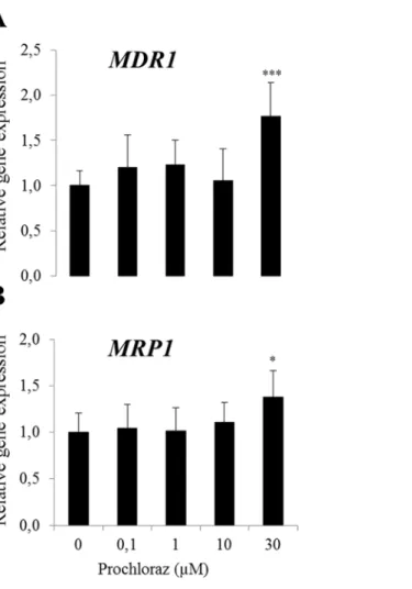
In BME-UV cells, gene expressions ofMDR1andMRP1were induced at the highest con-centration of prochloraz (30μM) (Fig 6A and 6B).
Protein expression in prochloraz-treated cells
Differentiated HC11 and confluent BME-UV cells were exposed to 30μM prochloraz for 24 h
and MDR1 protein expression was examined by an In-Cell Western Assay. MDR1 protein expression was significantly upregulated following 30μM prochloraz treatment in both HC11
and BME-UV cells (Fig 7).
Accumulation assay in prochloraz-treated cells
Differentiated HC11 and confluent BME-UV cells were exposed to 30μM prochloraz for 24 h
and the function of MDR1 was studied using digoxin as a substrate. Accumulation of digoxin was statistically significantly reduced in prochloraz treated HC11 cells compared to controls cells (Fig 8A). Also in BME-UV cells a lower, although not statistically significant, accumula-tion of digoxin was observed after prochloraz treatment (Fig 8B).
Discussion
Fig 3. Relative gene expression of transporters in BME-UV cells without (control) and with lactogenic hormone stimulation (prolactin treated).Normalized gene expressions are presented as means±SD; n = 6). Experiment repeated twice and data shown from one representative experiment.
doi:10.1371/journal.pone.0151904.g003
Fig 4. Cell viability in HC11 and BME-UV cells treated with prochloraz for 24h.The data represent means±SD; n = 6. Statistically significant differences as compared to vehicle controls*p0.01. Experiment repeated twice and data shown from one representative experiment.
substrates for transporters in mammary gland and study the function of transporters, cell mod-els are valuable tools.
We have previously demonstrated gene expressions ofBCRPandMDR1in HC11 cells [18]. In the present investigation we identified gene expressions ofMRP1,OATP1A5,OCTN1and
Fig 5. Relative gene expression ofCSN2and transporters (A-G) following prochloraz treatment in differentiated HC11 cells.Normalized gene expressions are presented as means±SD; n = 6 pooled from 2 separate experiments. Statistically significant differences as compared to vehicle controls *p0.05;**p0.01.
OCT1in HC11 cells. In the bovine mammary epithelial BME-UV cells onlyMDR1andMRP1 gene expressions were detected. The lack of expressions ofBCRP,OATP1A2andOCTN1in the BME-UV cells may be due to a number of reasons such as loss of transcription factors and selection of cells during passages and experimental conditions [38–40].
In line with previous reports our results demonstrate that both BCRP and OCT1 were most abundant during peak lactation as assessed by the peak inCSN2(β-casein) expression, whereas MDR1andMRP1expressions were reduced at this stage [7,9,10,16,41,42]. Stage dependent expression profiles ofBCRPandCSN2were similar, as were the profiles ofMDR1,MRP1and OCTN1.
In the present investigation the imidazole fungicide prochloraz was chosen as a test com-pound to examine the effect on the transporters expressed in the HC11 and BME-UV cell lines. Interestingly, the results showed that prochloraz induced gene and protein expression of MDR1 in both differentiated HC11 and in BME-UV cells. In addition,MRP1expression was upregulated by prochloraz in BME-UV cells. Based on predominant expressions ofMDR1and MRP1in excretory organs which have roles in elimination of drugs and xenobiotics, one possi-ble explanation can be that both MDR1 and MRP1 undertake protective roles as efflux pumps in order to prevent accumulation of chemicals and toxic effects in the cells. The apical
Fig 6. Relative gene expression ofMDR1(A) andMRP1(B) following prochloraz treatment in BME-UV cells.Normalized gene expressions are presented as means±SD; n = 8–12 pooled from 2 separate experiments. Statistically significant differences as compared to vehicle controls,**p0.01;***p0.001.
Fig 7. Relative expression of MDR1 following 30μM prochloraz treatment for 24 h in HC11 and
BME-UV cells.The data is presented as means±SD; n = 4–10. Statistically significant differences as compared to vehicle controls*p0.05;**p0.01.
doi:10.1371/journal.pone.0151904.g007
Fig 8. Accumulation of3H-digoxin in differentiated HC11 (A) and confluent BME-UV (B) cells, treated with 30μM prochloraz for 24 h.The data is presented as means±SD of 3–4 samples and expressed as pmol mitoxantrone/mg cellular protein. Statistically significant differences between groups,*p0.05.
localization and induction of MDR1 in the apical membranes of mammary epithelial cells may result in an active transport of toxic compounds into milk.
Both gene and protein expressions of MDR1 were increased after treatment of HC11 and BME-UV cells with prochloraz. Our accumulation studies with digoxin, a MDR1 substrate, showed a reduced accumulation in HC11 cells after treatment with prochloraz, indicating an increased function of the efflux transporter MDR1. Also in BME-UV cells treated with pro-chloraz the accumulation of digoxin tended to be reduced although not statistically significant. The increased MDR1 expression and reduced digoxin accumulation in prochloraz-treated murine mammary epithelial cells emphasizes the risk that chemicals may affect the expression and function of transporters and increase secretion of hazardous chemicals in milk.
The endogenous role of the ABC transporter BCRP has not been clarified, but has been sug-gested to involve secretion of vitamins including vitamin K3, riboflavin and folic acid into milk [6,43,44]. Thus, the induced BCRP expression during lactation in mammary glands may be important for the milk composition in terms of vitamin content. Prochloraz has been reported to increaseBCRPexpression in primary bovine mammary epithelial cells [22] and is trans-ported to milk [45]. However, in the differentiated HC11 cells prochloraz did not induce, but rather reduceBCRPexpression. Causes for this discrepancy can only be speculated upon but may depend on differences in experimental conditions such as prochloraz concentrations used, exposure times, or species differences in the display of transcription factors and regulation in BCRPgene expression. It can be noted that 2,3,7,8-tetrachlorodibenso-p-dioxin (TCDD) treat-ment in the human intestinal C2bbe1 (a subclone of Caco2) cells as well as in other human sec-ondary carcinoma cells of the colon, liver, and mammary glands resulted in induction ofBCRP transcripts but failed in mouse-derived hepatic (hepa1c1c7), mammary (EMT 6), and intestinal (CMT93) cell lines [46].
In summary, the results demonstrate that murine mammary epithelial HC11 cells featuring a secreting phenotype express endogenousBCRP,MDR1,MRP1,OATP1A5,OCT1and OCTN1. Gene and protein expression of the efflux protein MDR1 is upregulated by prochloraz treatment in both murine HC11 and in bovine mammary epithelial BME-UV cells, which in the HC11 correlates to a decreased accumulation of the MDR1 substrate digoxin. Our findings demonstrate that murine (HC11) mammary epithelial cells can be applied to characterize effects of toxic compounds on expression or function ofBCRP,MDR1,MRP1,OATP1A5, OCTN1andOCT1, whereasMDR1andMRP1can be assessed in bovine (BME-UV) mammary epithelial cells. An altered expression on any of the transporters expressed in the mammary epithelial cells during lactation may modulate the composition of nutrients and/or secretion of contaminants in milk with potential adverse effects on breastfed infants and dairy consumers.
Supporting Information
S1 Fig. Western blot of HC11cell lysates showing distinct MDR1 bands (170 kDa) and only weak bands at other MWs, including 130 kDa, which is the MW of pyruvate carboxylase.
Cells were cultured in T75 tissue culture flasks at 37°C in 15 ml sterile filtered RPMI 1640 medium containing L-glutamine and 25 mM HEPES supplemented with 50μg/ml bovine
insu-lin, 10 ng/ml epidermal growth factor, 7.5% NaHCO3and 10% heat-inactivated fetal bovine
serum in an atmosphere of 95% air and 5% CO2 in 95% relative humidity. Cells were harvested by trypzination and homogenized with 5 volumes of RIPA lysis buffer in 1.5 ml Eppendorf tubes by pipetting. Homogenates were incubated on ice for 30 min and then centrifuged at 16,000xg for 30 min at 4°C and supernatant protein concentrations determined by the BCA method. Triplicate samples of 20, 50 and 100μg of cellular protein were separated on a 10%
Blocking was performed with 5% non-fat dry milk powder in Tris-buffered saline containing 0.05% Tween-20 (TBS-T) overnight at 4°C. Hybridization was then performed with primary MDR1 antibody (JSB1, Abcam) diluted 1:200 in TBS-T. Primary MDR1 antibodies were detected by HRP-conjugated secondary antibodies (ab6728, Abcam) diluted 1:7.500 in TBS-T. HRP was detected by ECL Advance (GE Healthcare) and ChemiDoc instrument (Bio-Rad). (TIF)
Author Contributions
Conceived and designed the experiments: YY AO JT. Performed the experiments: YY JT. Ana-lyzed the data: YY AO JT. Contributed reagents/materials/analysis tools: AO CK. Wrote the paper: YY AO JT.
References
1. Haug A, Hostmark AT, Harstad OM. Bovine milk in human nutrition—a review. Lipids in health and dis-ease. 2007; 6:25. Epub 2007/09/27. doi:10.1186/1476-511X-6-25PMID:17894873; PubMed Central PMCID: PMC2039733.
2. Oskarsson A, Palminger Hallen I, Sundberg J, Petersson Grawe K. Risk assessment in relation to neo-natal metal exposure. The Analyst. 1998; 123(1):19–23. Epub 1998/05/15. PMID:9581014.
3. Oskarsson A, Palminger Hallen I, Sundberg J. Exposure to toxic elements via breast milk. The Analyst. 1995; 120(3):765–70. Epub 1995/03/01. PMID:7741226.
4. Hallen IP, Breitholtz-Emanuelsson A, Hult K, Olsen M, Oskarsson A. Placental and lactational transfer of ochratoxin A in rats. Natural toxins. 1998; 6(1):43–9. Epub 1998/12/16. PMID:9851511.
5. Breitzka RL, Sandritter TL, Hatzopoulos FK. Principles of drug transfer into breast milk and drug dispo-sition in the nursing infant. Journal of human lactation: official journal of International Lactation Consul-tant Association. 1997; 13(2):155–8. Epub 1997/06/01. PMID:9233209.
6. van Herwaarden AE, Wagenaar E, Merino G, Jonker JW, Rosing H, Beijnen JH, et al. Multidrug trans-porter ABCG2/breast cancer resistance protein secretes riboflavin (vitamin B2) into milk. Molecular and cellular biology. 2007; 27(4):1247–53. Epub 2006/12/06. doi:10.1128/MCB.01621-06PMID:
17145775; PubMed Central PMCID: PMC1800714.
7. Jonker JW, Merino G, Musters S, van Herwaarden AE, Bolscher E, Wagenaar E, et al. The breast can-cer resistance protein BCRP (ABCG2) concentrates drugs and carcinogenic xenotoxins into milk. Nature medicine. 2005; 11(2):127–9. Epub 2005/02/03. doi:10.1038/nm1186PMID:15685169. 8. Klaassen CD, Aleksunes LM. Xenobiotic, bile acid, and cholesterol transporters: function and
regula-tion. Pharmacological reviews. 2010; 62(1):1–96. Epub 2010/01/28. doi:10.1124/pr.109.002014PMID:
20103563; PubMed Central PMCID: PMC2835398.
9. Gilchrist SE, Alcorn J. Lactation stage-dependent expression of transporters in rat whole mammary gland and primary mammary epithelial organoids. Fundamental & clinical pharmacology. 2010; 24 (2):205–14. Epub 2009/08/26. doi:10.1111/j.1472-8206.2009.00760.xPMID:19702690.
10. Alcorn J, Lu X, Moscow JA, McNamara PJ. Transporter gene expression in lactating and nonlactating human mammary epithelial cells using real-time reverse transcription-polymerase chain reaction. The Journal of pharmacology and experimental therapeutics. 2002; 303(2):487–96. Epub 2002/10/22. doi:
10.1124/jpet.102.038315PMID:12388627.
11. Al-Bataineh MM, van der Merwe D, Schultz BD, Gehring R. Cultured mammary epithelial monolayers (BME-UV) express functional organic anion and cation transporters. Journal of veterinary pharmacol-ogy and therapeutics. 2009; 32(5):422–8. Epub 2009/09/17. doi:10.1111/j.1365-2885.2009.01057.x PMID:19754907; PubMed Central PMCID: PMC2747106.
12. Obaidat A, Roth M, Hagenbuch B. The expression and function of organic anion transporting polypep-tides in normal tissues and in cancer. Annual review of pharmacology and toxicology. 2012; 52:135– 51. Epub 2011/08/23. doi:10.1146/annurev-pharmtox-010510-100556PMID:21854228; PubMed Central PMCID: PMC3257355.
13. Vlaming ML, Lagas JS, Schinkel AH. Physiological and pharmacological roles of ABCG2 (BCRP): recent findings in Abcg2 knockout mice. Advanced drug delivery reviews. 2009; 61(1):14–25. Epub 2009/01/03. doi:10.1016/j.addr.2008.08.007PMID:19118589.
and comparative physiology. 2006; 290(3):R793–802. Epub 2005/10/01. doi:10.1152/ajpregu.00087.
2005PMID:16195500.
15. Kindla J, Rau TT, Jung R, Fasching PA, Strick R, Stoehr R, et al. Expression and localization of the uptake transporters OATP2B1, OATP3A1 and OATP5A1 in non-malignant and malignant breast tis-sue. Cancer biology & therapy. 2011; 11(6):584–91. Epub 2011/02/01. PMID:21278488.
16. Ito N, Ito K, Ikebuchi Y, Kito T, Miyata H, Toyoda Y, et al. Organic cation transporter/solute carrier family 22a is involved in drug transfer into milk in mice. Journal of pharmaceutical sciences. 2014; 103 (10):3342–8. Epub 2014/09/02. doi:10.1002/jps.24138PMID:25175747.
17. Maliepaard M, Scheffer GL, Faneyte IF, van Gastelen MA, Pijnenborg AC, Schinkel AH, et al. Subcellu-lar localization and distribution of the breast cancer resistance protein transporter in normal human tis-sues. Cancer research. 2001; 61(8):3458–64. Epub 2001/04/20. PMID:11309308.
18. Tallkvist J, Yagdiran Y, Danielsson L, Oskarsson A. A model of secreting murine mammary epithelial HC11 cells comprising endogenous Bcrp/Abcg2 expression and function. Cell Biol Toxicol. 2015; 31 (2):111–20. Epub 2015/03/21. doi:10.1007/s10565-015-9298-5PMID:25791223.
19. EFSA. Conclusion on the peer review of the pesticide risk assessment of the active substance pro-chloraz. EFSA Journal. 2011; 9(7):2323.
20. Vinggaard AM, Hass U, Dalgaard M, Andersen HR, Bonefeld-Jorgensen E, Christiansen S, et al. Pro-chloraz: an imidazole fungicide with multiple mechanisms of action. International journal of andrology. 2006; 29(1):186–92. Epub 2006/02/10. doi:10.1111/j.1365-2605.2005.00604.xPMID:16466539. 21. Ohlsson A, Ulleras E, Oskarsson A. A biphasic effect of the fungicide prochloraz on aldosterone, but
not cortisol, secretion in human adrenal H295R cells—underlying mechanisms. Toxicology letters. 2009; 191(2–3):174–80. Epub 2009/09/08. doi:10.1016/j.toxlet.2009.08.020PMID:19733639. 22. Halwachs S, Wassermann L, Lindner S, Zizzadoro C, Honscha W. Fungicide prochloraz and
environ-mental pollutant dioxin induce the ABCG2 transporter in bovine mammary epithelial cells by the arylhy-drocarbon receptor signaling pathway. Toxicological sciences: an official journal of the Society of Toxicology. 2013; 131(2):491–501. Epub 2012/10/20. doi:10.1093/toxsci/kfs304PMID:23081912. 23. Halwachs S, Wassermann L, Honscha W. A novel MDCKII in vitro model for assessing ABCG2-drug
interactions and regulation of ABCG2 transport activity in the caprine mammary gland by environmental pollutants and pesticides. Toxicology in vitro: an international journal published in association with BIBRA. 2014; 28(3):432–41. Epub 2014/01/07. doi:10.1016/j.tiv.2013.12.015PMID:24389113. 24. Graber-Maier A, Gutmann H, Drewe J. A new intestinal cell culture model to discriminate the relative
contribution of P-gp and BCRP on transport of substrates such as imatinib. Molecular pharmaceutics. 2010; 7(5):1618–28. Epub 2010/08/13. doi:10.1021/mp100040fPMID:20701289.
25. Marte BM, Meyer T, Stabel S, Standke GJ, Jaken S, Fabbro D, et al. Protein kinase C and mammary cell differentiation: involvement of protein kinase C alpha in the induction of beta-casein expression. Cell growth & differentiation: the molecular biology journal of the American Association for Cancer Research. 1994; 5(3):239–47. Epub 1994/03/01. PMID:8018556.
26. Desrivieres S, Prinz T, Castro-Palomino Laria N, Meyer M, Boehm G, Bauer U, et al. Comparative proteomic analysis of proliferating and functionally differentiated mammary epithelial cells. Molecular & cellular proteomics: MCP. 2003; 2(10):1039–54. Epub 2003/07/30. doi:
10.1074/mcp.M300032-MCP200PMID:12885951.
27. Ball RK, Friis RR, Schoenenberger CA, Doppler W, Groner B. Prolactin regulation of beta-casein gene expression and of a cytosolic 120-kd protein in a cloned mouse mammary epithelial cell line. The EMBO journal. 1988; 7(7):2089–95. Epub 1988/07/01. PMID:3416834; PubMed Central PMCID: PMC454494.
28. Al-Bataineh MM, van der Merwe D, Schultz BD, Gehring R. Tumor necrosis factor alpha increases P-glycoprotein expression in a BME-UV in vitro model of mammary epithelial cells. Biopharmaceutics & drug disposition. 2010; 31(8–9):506–15. Epub 2010/11/26. doi:10.1002/bdd.731PMID:21104926; PubMed Central PMCID: PMC3034978.
29. Hynes NE, Taverna D, Harwerth IM, Ciardiello F, Salomon DS, Yamamoto T, et al. Epidermal growth factor receptor, but not c-erbB-2, activation prevents lactogenic hormone induction of the beta-casein gene in mouse mammary epithelial cells. Mol Cell Biol. 1990; 10(8):4027–34. Epub 1990/08/01. PMID:
2196443; PubMed Central PMCID: PMC360913.
30. Zavizion B, van Duffelen M, Schaeffer W, Politis I. Establishment and characterization of a bovine mam-mary epithelial cell line with unique properties. In vitro cellular & developmental biology Animal. 1996; 32(3):138–48. Epub 1996/03/01. PMID:8925136.
32. Bustin SA, Benes V, Garson JA, Hellemans J, Huggett J, Kubista M, et al. The MIQE guidelines: mini-mum information for publication of quantitative real-time PCR experiments. Clin Chem. 2009; 55 (4):611–22. Epub 2009/02/28. doi:10.1373/clinchem.2008.112797PMID:19246619.
33. Han LQ, Yang GY, Zhu HS, Wang YY, Wang LF, Guo YJ, et al. Selection and use of reference genes in mouse mammary glands. Genet Mol Res. 2010; 9(1):449–56. Epub 2010/04/15. doi:
10.4238/vol9-1gmr724PMID:20391330.
34. Bionaz M, Loor JJ. Identification of reference genes for quantitative real-time PCR in the bovine mam-mary gland during the lactation cycle. Physiol Genomics. 2007; 29(3):312–9. Epub 2007/02/08. doi:10.
1152/physiolgenomics.00223.2006PMID:17284669.
35. Livak KJ, Schmittgen TD. Analysis of relative gene expression data using real-time quantitative PCR and the 2(-Delta Delta C(T)) Method. Methods. 2001; 25(4):402–8. Epub 2002/02/16. doi:10.1006/
meth.2001.1262PMID:11846609.
36. Abcam. Anti-P Glycoprotein antibody [JSB-1]. Available from: http://www.abcam.com/p-glycoprotein-antibody-jsb-1-prediluted-ab51893.html.
37. Atlas THP. Pyruvate carboxylase. Available from:http://www.proteinatlas.org/ENSG00000173599-PC/ tissue.
38. Monzani PS, Bressan FF, Mesquita LG, Sangalli JR, Meirelles FV. beta-casein gene expression by in vitro cultured bovine mammary epithelial cells derived from developing mammary glands. Genetics and molecular research: GMR. 2011; 10(2):604–14. Epub 2011/04/15. doi:10.4238/vol10-2gmr1034 PMID:21491370.
39. Larson BL. Comparative production of beta-lactoglobulin and orotic acid with lactose in bovine mam-mary cell cultures: effects of cell density and constituent inhibition. Journal of dairy science. 1976; 59 (11):1881–9. Epub 1976/11/01. doi:10.3168/jds.S0022-0302(76)84457-8PMID:993409.
40. Blum JL, Zeigler ME, Wicha MS. Regulation of mammary differentiation by the extracellular matrix. Environmental health perspectives. 1989; 80:71–83. Epub 1989/03/01. PMID:2647486; PubMed Cen-tral PMCID: PMC1567626.
41. Shi H, Zhu J, Luo J, Cao W, Yao D, Li J, et al. Genes regulating lipid and protein metabolism are highly expressed in mammary gland of lactating dairy goats. Functional & integrative genomics. 2014. Epub 2014/12/01. doi:10.1007/s10142-014-0420-1PMID:25433708.
42. Robinson GW, McKnight RA, Smith GH, Hennighausen L. Mammary epithelial cells undergo secretory differentiation in cycling virgins but require pregnancy for the establishment of terminal differentiation. Development. 1995; 121(7):2079–90. Epub 1995/07/01. PMID:7635053.
43. Shukla S, Wu CP, Nandigama K, Ambudkar SV. The naphthoquinones, vitamin K3 and its structural analogue plumbagin, are substrates of the multidrug resistance linked ATP binding cassette drug trans-porter ABCG2. Molecular cancer therapeutics. 2007; 6(12 Pt 1):3279–86. Epub 2007/12/11. doi:10.
1158/1535-7163.MCT-07-0564PMID:18065489; PubMed Central PMCID: PMC2398729.
44. Assaraf YG. The role of multidrug resistance efflux transporters in antifolate resistance and folate homeostasis. Drug resistance updates: reviews and commentaries in antimicrobial and anticancer che-motherapy. 2006; 9(4–5):227–46. Epub 2006/11/10. doi:10.1016/j.drup.2006.09.001PMID:
17092765.
45. Vinggaard AM, Christiansen S, Laier P, Poulsen ME, Breinholt V, Jarfelt K, et al. Perinatal exposure to the fungicide prochloraz feminizes the male rat offspring. Toxicological sciences: an official journal of the Society of Toxicology. 2005; 85(2):886–97. Epub 2005/03/25. doi:10.1093/toxsci/kfi150PMID:
15788727.