In Vitro
Reconstruction of Neuronal Networks
Derived from Human iPS Cells Using
Microfabricated Devices
Yuzo Takayama*, Yasuyuki S. Kida*
Biotechnology Research Institute for Drug Discovery, National Institute of Advanced Industrial Science and Technology (AIST), Tsukuba, Ibaraki, Japan
*[email protected](YT);[email protected](YSK)
Abstract
Morphology and function of the nervous system is maintained via well-coordinated processes both in central and peripheral nervous tissues, which govern the homeostasis of organs/tis-sues. Impairments of the nervous system induce neuronal disorders such as peripheral neu-ropathy or cardiac arrhythmia. Although further investigation is warranted to reveal the molecular mechanisms of progression in such diseases, appropriate model systems mimick-ing the patient-specific communication between neurons and organs are not established yet. In this study, we reconstructed the neuronal networkin vitroeither between neurons of the human induced pluripotent stem (iPS) cell derived peripheral nervous system (PNS) and cen-tral nervous system (CNS), or between PNS neurons and cardiac cells in a morphologically and functionally compartmentalized manner. Networks were constructed in photolithographi-cally microfabricated devices with two culture compartments connected by 20 microtunnels. We confirmed that PNS and CNS neurons connected via synapses and formed a network. Additionally, calcium-imaging experiments showed that the bundles originating from the PNS neurons were functionally active and responded reproducibly to external stimuli. Next, we confirmed that CNS neurons showed an increase in calcium activity during electrical
stimulation of networked bundles from PNS neurons in order to demonstrate the formation of functional cell-cell interactions. We also confirmed the formation of synapses between PNS neurons and mature cardiac cells. These results indicate that compartmentalized culture devices are promising tools for reconstructing network-wide connections between PNS neu-rons and various organs, and might help to understand patient-specific molecular and func-tional mechanisms under normal and pathological conditions.
Introduction
The nervous system consists of the central and peripheral systems that are connected with each other, and thus form an electrical signaling network throughout the body. Although each neu-ron type is differentiated from different stem/progenitor cell pools, interactions between vari-ous cell types are well-coordinated both morphologically and functionally. The peripheral nervous system (PNS) is connected to the central nervous system (CNS), and this functional
OPEN ACCESS
Citation:Takayama Y, Kida YS (2016)In Vitro Reconstruction of Neuronal Networks Derived from Human iPS Cells Using Microfabricated Devices. PLoS ONE 11(2): e0148559. doi:10.1371/journal. pone.0148559
Editor:Simone Di Giovanni, Hertie Institute for Clinical Brain Research, University of Tuebingen., GERMANY
Received:July 28, 2015
Accepted:January 19, 2016
Published:February 5, 2016
Copyright:© 2016 Takayama, Kida. This is an open access article distributed under the terms of the
Creative Commons Attribution License, which permits unrestricted use, distribution, and reproduction in any medium, provided the original author and source are credited.
Data Availability Statement:All relevant data are within the paper and its Supporting Information files.
sion or paracrine interactions, non-contacting co-culture conditions are required. In these cases, permeable co-culture systems have been used [5,6]. However, these conventional co-cul-ture methods are not suitable for neuronal co-culco-cul-tures, because neurons extend their neurites over a long distance, and establish connections with other types of cells. Furthermore, neuronal functions by the neurite connections are based on the parallel and distributed computations of network-wide activities [7,8]. Thus, cultured neurons and target organs have to be arranged correctly for reconstructing inter-organ neuronal networksin vitro.
Microfabrication techniques for preparing specific dishes are a promising way to overcome these problems in neuronal cultures. In 1977, R. B. Campenot developed a Teflon-based culture chamber to separate neurites from cell bodies [9]. The Campenot chamber, which enables a compartmentalized culturing technique, was primarily used for studying the local effects of growth factors in neurites. However, due to its size, the Campenot chamber is not suitable for culturing any neurons, and the preparation of the chamber requires practice [10]. Compared to Teflon, polydimethylsiloxane (PDMS) is a more suitable material for engineering cell culture devices, because it is biocompatible, optically transparent, and even submicron features are easy to fabricate [11]. We and others have used microfabricated PDMS chambers for the network-wide integration of rodent CNS neurons, sympathetic neurons, and cardiomyocytes [12–14]. However, these studies largely relied on animal cells because of difficulties in obtain-ing and handlobtain-ing patient-derived cells. Therefore, the construction of co-culture networks using PNS neurons, CNS neurons, or other cell types derived from human pluripotent stem cells (PSCs) could become a promisingin vitromodel system for studying peripheral neuron-related diseases.
In this study, we created co-culture networks using human PNS and CNS neurons. First, we fabricated a PDMS-based co-culture chamber, which consisted of two culture compartments connected with 20 microtunnels, and we cultured induced PNS and CNS neurons differenti-ated from human iPS cells. Development of their connections was evaludifferenti-ated with microscopic observations, immunochemical analysis, and calcium imaging. Furthermore, we prepared a co-culture system using PNS neurons and cardiomyocytes, both derived from the same human iPS cells, to confirm that our microfabricated device can be used with various cell types.
Materials and Methods
Ethnic statement
Device fabrication
The co-culture device was fabricated from PDMS using soft lithography and replica molding technique. For producing the master mold, SU-8 3005 (Microchem) was spin-coated on aφ76 silicon wafer (Matsuzaki Seisakusyo., Ltd.) at 4000 rpm for 60 s to reach a height of 5μm. The coated wafer was pre-baked at 95°C for 3 min. Then, the wafer was exposed to ultraviolet (UV) light with a UV crosslinker (CL-1000L; UVP) through a custom-made photomask. The photo-mask was designed to fabricate 20 microtunnels with a width of 50μm and a length of 3 mm. After UV exposure, the wafer was developed with the SU-8 developer (Microchem), and then it was rinsed with 2-propanol (Wako Pure Chemical Industries). After its development, the wafer was placed in a conventional culture dish (φ100 mm; Corning).
Mixture of the PDMS-prepolymer and curing catalyst (10:1 weight ratio; Silpot 184, Dow Corning) was poured over the fabricated wafer to achieve a thickness of 5 mm. Then, PDMS was cured in an oven at 70°C for 1h. After curing, the PDMS sheet was trimmed using a surgi-cal knife and was released from the master. To prepare the two culture compartments, which were connected by the microtunnel structures, holes were opened with a punch (φ8 mm; Har-ris Uni-Core; Ted Pella). We verified that each microtunnel was at least 1 mm in length; lengths450μm have been reported to allow only axons to pass through microtunnels [15]. The PDMS chamber was sealed with a cell culture dish (φ35 mm; Corning), which was previ-ously coated with 20μg/ml poly-L-ornithine (PLO; Aldrich). Then 5μg/ml laminin (Sigma-Aldrich) was poured into the PDMS chamber and was incubated in the chamber until cell plating.
Cell culture
Human iPS cells (201B7 line) were provided by the RIKEN Bioresource Center in Japan. iPS cells were plated on culture plates coated with Laminin-511-E8 (iMatrix511, Nippi) and were maintained in mTeSR1 WO 2ME/MV medium (Stemcell Technologies) at 37°C in a 5% CO2
incubator. The medium was changed every day. When the iPS cells were nearly confluent, iPS colonies were digested into single cells with accutase (Life Technologies), and then cells were passaged or induced as described below.
factor (BDNF; Wako Pure Chemical Industries), and 10 ng/ml glial cell line-derived neuro-trophic factor (GDNF; Wako Pure Chemical Industries). The medium was changed twice a week.
Differentiation of iPS cells into PNS neurons was performed according to the following pro-tocol. Similar to above, the EBs of iPS cells were cultured in KSR medium containing 2μM DM and 10μM SB on day 0. On day 2, the medium was changed to KSR medium containing 3μM CHIR99021 (CHIR; Cayman Chemical) and 20μM SB. The KSR medium containing 3μM CHIR and 20μM SB was changed on every second day until day 12. On day 12, EBs were disso-ciated with TrypLE, were plated in PLO/laminin-coated dishes at a density of 1 × 10⁵cells/cm2, and were maintained in NDM2, which consisted of DMEM-F12, 1% N2 supplement, 1% NEAA, and 1% P/S. NDM2 was also supplemented with 10μM forskolin, 50μg/ml ascorbic acid, 10 ng/ml BDNF, 10 ng/ml GDNF, 10 ng/ml nerve growth factor (NGF; Wako Pure Chemical Industries), and 10 ng/ml neurotrophin 3 (NT-3; Wako Pure Chemical Industries). The medium was changed twice a week.
For co-culture experiments in the PDMS chamber, PNS neurons were first plated into one chamber and were incubated for 30 min. After the adhesion of PNS neurons was confirmed, CNS neurons were plated into the other chamber. Each neuron type was plated at a density of 2–5 × 10⁵cells/cm2. The medium in each compartment was changed on every second day.
Differentiation of iPS cells into cardiomyocytes was performed as previously described [16]. Briefly, the EBs of iPS cells were transferred into RPMI-1640 medium (Wako Pure Chemical Industries) containing 2% B27 supplement without insulin (Life Technologies) and 7.5μM CHIR on day 0. 24 hours later, the medium was changed to fresh medium without CHIR. On day 3, the medium was changed to RPMI-1640 medium containing 2% B27 supplement with-out insulin and 10μM IWR-1 (Sigma-Aldrich). On day 5, the medium was changed to fresh medium without IWR-1. From day 7, EBs were maintained in RPMI-1640 containing B27 sup-plement (Life Technologies), and the medium was changed on every second day. For co-cul-tures with PNS neurons, contracting EBs after two weeks in culture were used.
PKH labeling
In order to differentiate neurons in the co-culture chamber, each neuron type was labeled with a PKH permanent fluorescent marker [17] before plating into the co-culture chamber. PNS
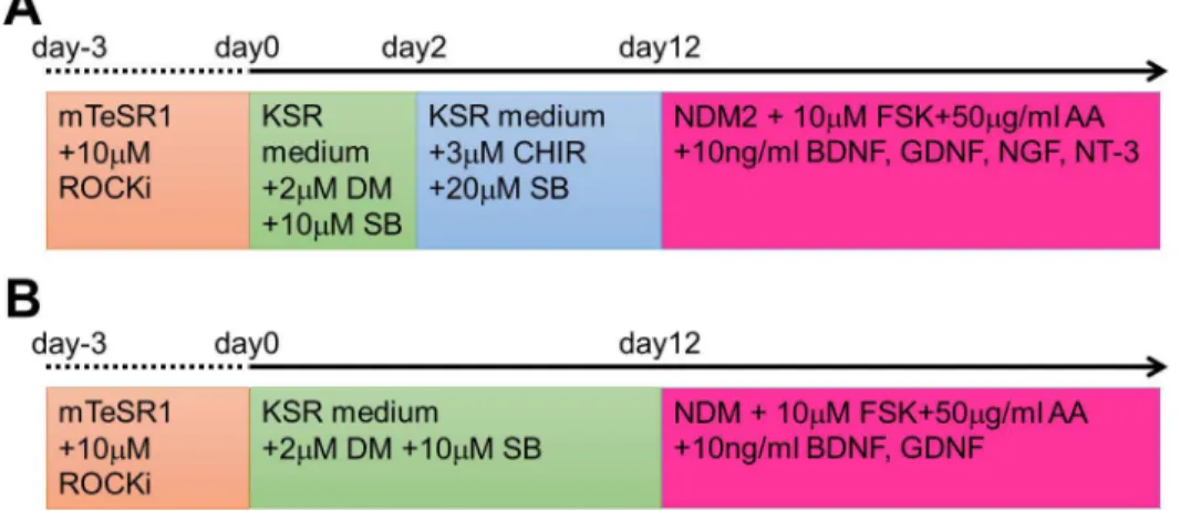
Fig 1. Schematic diagram showing the differentiation protocols from human iPS cells to mature neurons.Neurons of the peripheral nervous system (PNS; A) and the central nervous system (CNS; B) were induced from human induced pluripotent stem (iPS) cells with several compounds dissolved in serum-free media. Each medium and compound was added at the time indicated in (A) or (B).
and CNS neurons were labeled with 1 × 10⁻⁶M PKH26 (red fluorescence; Sigma-Aldrich) and 1 × 10⁻⁶M PKH67 (green fluorescence; Sigma-Aldrich), respectively. An electro multiplying (EM) charge coupled device (CCD) camera (iXon+; Andor) mounted on an inverted micro-scope (IX-81; Olympus) was used for detecting the fluorescence signal of PKH dyes. PKH-labeled neurons were also used for the calculation of axon growth rates. Only axonal regions that passed through microtunnels were used for the growth rate calculation in order to exclude non-linear lengths resultant from the circular shape of the culture wells. Images were visualized and analyzed with the ImageJ software (National Institutes of Health; available athttp:// imagej.nih.gov/ij/).
Immunochemical staining
The cultures were rinsed with phosphate-buffered saline (PBS) and then were fixed with 3.7% formaldehyde (Wako) in PBS for 10 min at room temperature (RT). After fixation, the cells were permeabilized with PBS containing 0.2% Tween-20 (Wako Pure Chemical Industries) for 10 min, and then were blocked with PBS containing 4% Block Ace (DS Pharma Biomedical) and 0.2% Tween-20 for 1 h at RT. Primary antibodies diluted in the Can Get Signal Solution 1 (Toyobo) were incubated with the cells overnight at 4°C. Next day, the cells were washed three times with PBS containing 0.2% Tween-20, and then were incubated for 2 h at RT with the sec-ondary antibodies (anti-mouse Alexa Fluor-488 and anti-rabbit Alexa Fluor-555; 1:1000; Life Technologies) diluted in the Can Get Signal 2 solution (Toyobo). The primary antibodies used in this study were as follows: mouse anti-class III beta-tubulin (TUJ1; 1:1000; Covance), mouse P75NTR (neurotrophin receptor) (1:200; Advanced Targeting Systems), mouse anti-HNK-1 (1:500; Sigma-Aldrich), mouse anti-cTnT (cardiac Trophonin T) (1:500; Abcam), rab-bit anti-Synapsin-1 (1:1000; Millipore), and rabrab-bit anti-Peripherin (1:1000; Millipore). To stain the nuclei, 0.2μg/ml Hoechst 33342 (Dojindo Molecular Technologies) was added to the Can Get Signal 2 solution.
Calcium Imaging
Calcium dynamics were visualized with a calcium imaging technique. The cells were labeled with 5μg/ml Fluo-4/AM (Invitrogen), a calcium indicator dye, for 30 min. After labeling, the medium was replaced with Ringer’s solution (148 mM NaCl, 2.8 mM KCl, 2 mM CaCl2, 1 mM
MgCl2, 10 mM HEPES, and 10 mM glucose; pH 7.4). Then, the cells were placed on the stage
of the inverted microscope, and fluorescence was detected with the EM CCD camera. In PNS-CNS co-culture experiments, electrical stimulation was used to evoke cellular responses. For stimulation, a pair of platinum electrodes (0.5 mm diameter; Nilaco) was immersed into the solution, and electrical pulses with a constant voltage were delivered to the cells using an electrical stimulator (SEN-3401; Nihon Kohden). Negative phase pulses with 3 ms duration and 10 V intensity were used. A frame rate of 2 frame/s was used for the PNS-CNS co-cultures. For cardiomyocytes, a frame rate of 14 frame/s was used. The recorded signals were analyzed with the ImageJ software.
Results
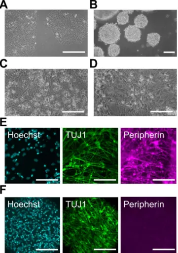
formation of EBs (Fig 2B). The induced cells were re-plated in PLO/laminin-coated dishes, and we continued the differentiationin vitrofor at least 2 weeks (Fig 2C). Most differentiated cells expressed the type III intermediate filament Peripherin, which is a marker of peripheral neu-rons, and TUJ1 in their neurites (Fig 2E). We also confirmed that Peripherin-positive neurons expressed the neural crest markers P75NTR and HNK-1 (S1 Fig). These data indicate that dif-ferentiated PNS neurons have characteristics of neural crest-lineage cells. On the other hand, we used a dual SMAD inhibition protocol under non-adherent conditions for the induction of CNS neurons. After 2 weeks of induction, the EBs were re-plated in PLO/laminin-coated dishes, and the cells started to extend neurites almost immediately (Fig 2D). Immunochemical staining revealed that the neurites of most cells expressed TUJ1 at a high level on day 15 (Fig 2F). However, Peripherin and P75NTR were not expressed, suggesting that these TUJ1-positive neurons were CNS neurons [20]. Thus, we prepared human PNS and CNS neurons in a large quantityin vitro.
Fig 2. Differentiation of human PNS and CNS neurons.(A) Phase-contrast image of pre-differentiated human induced pluripotent stem (iPS) cells under a feeder-free condition. (B) Phase-contrast image of embryonic bodies (EBs) before chemical induction on day 0. (C, D) Phase-contrast image of differentiated peripheral nervous system (PNS) neurons (C) and central nervous system (CNS) neurons (D). Induced neurites were detected on day 23 (C) and day 15 (D) in PNS and CNS neurons, respectively. (E, F) Immunofluorescent labeling with antibodies specific for class III beta-tubulin (TUJ1) and Peripherin in PNS (E) and CNS neurons (F) on day 22. Cell nuclei were counterstained with Hoechst 33342. Scale bar: 100μm.
Co-culture of PNS and CNS neurons in the fabricated culture device
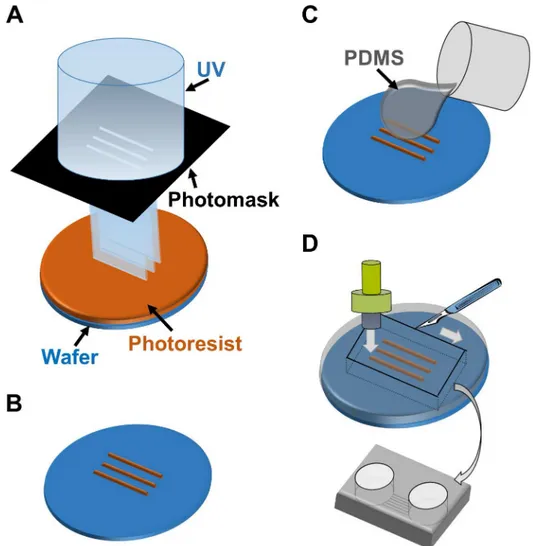
PNS neurons re-extend their neurites into peripheral tissues, including internal organs and limbs, in response to neuronal injury. The neurite re-innervates target organs and the CNS to re-establish bidirectional signals. However, the process of re-connection between the PNS and CNS is not fully understood yet. Therefore, we developed anin vitrocell culture device to image and understand the underlying mechanisms of neuronal wiring. Using PDMS, we fabri-cated a culture chamber with 2 separate wells and tunnels as outlined inFig 3. Schematic image of the fabricated culture chambers is also shown inFig 4A. Importantly, no cross-contamina-tion must occur between the wells, and the tunnels need to allow only the growth of extending neurites to the other side. Therefore, connecting tunnels were designed with a height of 5μm to exclude neuronal cell bodies from the microtunnels [12,15]. After PNS and CNS neurons were fully differentiated, each type of neuron was collected and re-plated in separate wells of the culture chamber. In order to monitor the cell bodies and neurites of each neuron type, cells were labeled with different dyes before re-plating. During co-culture experiments, neurites entered the microtunnels, but the cell bodies of neurons were excluded (Fig 4B). After 2 weeksFig 3. Schematic outline of the microfabrication process to manufacture the co-culturing device.(A) UV exposure of the photoresist through the photomask. (B) Development of micropatterns to fabricate the master mold. (C) Casting and curing of polydimethylsiloxane (PDMS) onto the maser mold. (D) Cutting, punching, and releasing of the PDMS chamber from the master mold.
of co-culture, neurites reached the opposing well and made neuronal connections. Interest-ingly, mostly the neurites of PNS neurons crossed the microtunnels (Fig 4C). In contrast, only a few CNS neurites passed through the microtunnels. To confirm these observations, we performed TUJ1 and Peripherin immunostaing, which showed that neurites were double-posi-tive for TUJ1 and Peripherin (Fig 4D). PNS neurites that passed through the microtunnels extended at a rate of approximately 0.2 mm/day during the 2 weeks of co-culture. This rate of growth was lower than thatin vivo[21,22]. Furthermore, the morphology of PNS neurites sug-gested that neurites gathered together and made bundles before and during entering the tun-nels (Fig 4E), and these bundles passed through the microtuntun-nels together.
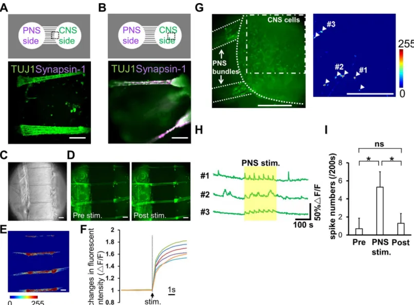
Additionally, we examined whether the connected neurons constructed a neuronal network. To reveal the structure of connection more closely, we evaluated Synapsin-1 expression at the locations of neurite arrival in the CNS well. Synapsin-1 is localized in synaptic vesicles, thus its presence is suggesting axon/neurite interactions. In the well containing CNS neurons, TUJ1-positive PNS bundles integrated with aggregated CNS neurons, as Synapsin-1 was detected in the terminal of the bundles, not in the microtunnels (Fig 5A and 5B). These data indicate that iPS cell-derived PNS and CNS neurons are able to connect with each other and make a neuro-nal network in our culture device. In addition, a functioneuro-nal assay was carried out using calcium imaging. Preliminarily studies demonstrated that differentiated CNS neurons exhibit sponta-neous activity with kinetics typical of neuronal calcium transients (a rapid onset and decay occurring within 10 s,Fig 5H). Alternatively, PNS neurons rarely exhibited spontaneous
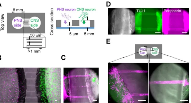
Fig 4. Co-culturing of PNS and CNS neurons in the PDMS chamber device.(A) Schematic diagram of the polydimethylsiloxane (PDMS) co-culture chamber. Diameter of the chambers was 8 mm. Width of the microtunnels was 50μm. Although the length of each microtunnel varied depending on its location, the length of each microtunnel was at least 1 mm. The neurites were able to pass through the 5-μm-high microtunnels. (B) Co-cultured neurons close to the microtunnels one day after cell plating. Peripheral nervous system (PNS) neurons were labeled with PKH26 (magenta), and central nervous system (CNS) neurons were labeled with PKH67 (green). (C) Most neurites of PNS neurons passed through the microtunnels 12 days after cell plating. Bundles (magenta) originating from PNS neurons reached the aggregated CNS neurons (green). (D) Phase-contrast images and immunofluorescent staining for class III beta-tubulin (TUJ1) and Peripherin. Neurites that passed through the microchannel originated from PNS neurons, which was confirmed by the expression of TUJ1 and Peripherin (arrowheads) 48 days after cell plating. (E) PNS neurites around the microtunnels 30 days after cell plating. Two different regions, indicated in the diagram, are shown at a high magnification. PNS neurites gathered together and made bundles before and during entering the tunnels. Scale bar: 100μm.
activity but did respond to the TRPM8 agonist menthol and the TRPV1 agonist capsaicin (data not shown, [23]), which classically activate peripheral afferent neurons. To monitor the responsiveness of PNS bundles to external stimuli, electrical stimulation was applied to the cells with an electric stimulator.Fig 5C and 5Dshow phase-contrast and fluorescent images around the microtunnel region on day 39. Fluorescence images show the amount of intracellu-lar calcium before and 1 s after applying the electrical pulse.Fig 5Eshows normalized
Fig 5. Structural and functional analysis of co-cultured neuronal networks.(A, B) Immunofluorescence images of co-cultured neurons 48 days after cell plating (bottom panel). Different regions indicated in the diagram are shown (top panel). Synapsin-1 staining was detected in the chamber with central nervous system (CNS) neurons, but not in the microtunnels. (C, D) Phase-contrast images of cells and the fluorescence images of a calcium probe on day 39. Fluorescence images before (pre stim) and 1 s after applying the electrical stimulation (post stim) are shown. Fluorescence was increased after the stimulus (arrowheads). (E) Calcium response after the stimulation normalized to the signal before the stimulation. The color bar shows fluorescence intensity. Neurites in the microtunnels were induced with electrical stimulation. (F) Kinetics of calcium transient onset during applying the electrical stimulation. Data of 6 peripheral nervous system (PNS) bundles were considered. (G) Identification of CNS neurons that responded to PNS bundle stimulation. Left, a co-culture sample at day 29 labelled with fluo-4. Right, normalized calcium response in CNS neurons following PNS bundle stimulation (dot-dashed line). The color bar indicates fluorescence intensity. White arrowheads identify CNS neurons that exhibited a calcium response to PNS bundle stimulation. (H) Kinetic plots of calcium transients in CNS neurons represented in panel G. The region highlighted by the yellow rectangle indicates the period of electrical stimulation of PNS bundles. (I) The effect of PNS bundle stimulation on the activity of CNS neurons. Calcium spiking in CNS neurons was averaged pre-, during and post-PNS bundle stimulation. Values are reported as mean±S.D. (n= 20 neurons from 3 samples). Statistical analyses using an unpaired t-test are shown here (ns not significant,*p<0.001; Scale bar: 100μm).
junction with the statistical data presented inFig 5I, our data confirm the presence of func-tional cell-cell interaction between differentiated PNS and CNS neurons. Thus, our culture device could serve as a promising tool to culture different types of neurons and to analyze their interactions.
Co-culture of PNS neurons and cardiomyocytes in the fabricated culture
device
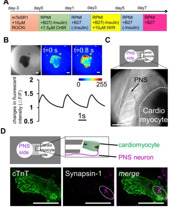
Next, we constructed another type of co-culture system using PNS neurons and cardiomyocytes derived from iPS cells. The heart beats autonomously throughout life, but it is also regulated by neuronal stimuli [24,25]. Cardiomyocytes are innervated by PNS neurons, which regulate the beating rate of the heart. Thus, preparing anin vitroco-culture system of PNS neurons and cardi-omyocytes, both differentiated from same human iPS cells, would be a promising model system for investigating the mechanisms that regulate heart beating, and for drug discovery in cardiovas-cular diseases associated with peripheral neuropathy. Therefore, we attempted to establish a co-culture using PNS neurons and cardiomyocytes derived from human iPS cells. Following the cell induction method shown inFig 6A, the EBs of human iPS cells were differentiated into cardio-myocytes. After 2 weeksin vitro, the cell aggregates started to contract autonomously (S1 Movie). We also checked the cardiomyocyte beats with calcium imaging, which revealed that almost all cells exhibited calcium oscillations (Fig 6BandS2 Movie).
To establish the compartmentalized co-culture system quickly, PNS neurons were first re-plated in the left side of the chamber and were cultured for two weeks. After we confirmed that the PNS bundles passed through the microchannels, differentiated contracting cardiomyocyte aggregates were isolated and transferred in the right side of the chamber. On the right side of the chamber, the bundles innervated the contracting aggregates (Fig 6CandS3 Movie). To confirm the formation of an apparent connection between PNS neurons and cardiomyocytes, we probed co-culture samples for expression of cTnT and syanpsin-1 in co-culture samples. cTnT expression indicated the presence of mature cardiomyocytes in the well, and PNS-derived Synapsin-1 was co-localized on these cells (Fig 6D). These data indicate the formation of a neuromascular connection between mature cardiomyocytes and PNS neurons in the PDMS devices. These results suggest that our PDMS chamber device is also useful for establish-ing a connection between PNS neurons and cardiomyocytes and for examinestablish-ing how PNS neu-rons control cardiomyocyte functions in response to various neuronal stimuli.
Discussion
Here, we reported the manufacturing of a co-culturing device, which was used for
compartmentalized device made from PDMS could be a useful experimental platform for mon-itoring neurite/cell interactions in various cell types, for screening chemicals, or to investigate the effectiveness or safety of drugs.
Fig 6. Reconstruction of neuronal networks innervating the heart using iPS cells co-cultured in a microfabricated device.(A) Schematic diagram of the differentiation protocol for iPS cell-derived
cardiomyocytes. (B) Calcium imaging of cardiomyocyte-aggregates on day 16. Contracting embryonic bodies (EBs) showed sustained calcium dynamics. The color bar shows fluorescence intensity, and the change in fluorescence signals confirms that these cell-aggregates contained cardiomyocytes. The graph is showing the kinetics of calcium transients during the recording. (C) Co-culture of peripheral nervous system (PNS) neurons and cardiomyocytes on day 44 after plating PNS neurons. PNS-derived bundles extended from left chamber (arrow) and reached a cardiomyocyte-aggregate, which was in the right chamber (within white dash line). (D) Immunostaining for the cardiomyocyte marker cTnT and the synaptic vesicle marker Synapsin-1 on day 24 after plating PNS neurons. Top, the positional relationship of the axon from PNS neurons and cardiomyocyte. Bottom, localization of Synapsin-1 on a cardiomyocyte (dot line region). Scale bar: 100μm.
mature astrocytes and oligodendrocytes in CNS require an induction for 2–3 months [30–32] compared with neurons that require one month from human PSCs. Thus, the cellular environ-ment providing support for neurons might not be present in the CNS chamber. Optimizing co-culture conditions to enhance neurite growth should be considered in further investigations. Furthermore, our device might be useful for identifying chemical compounds and growth fac-tors that modulate neurite growth, especially if we can reconstruct the outgrowth of defected neurites, such as those derived of patients with neuropathy.
Our microtunnel structures only allow the isolation axons, but not dendrites, from their cell bodies [12,15]. Hence, PNS bundles originating from PNS neurons, which enter the CNS chamber, are thought to be axons. Accordingly, we did not find synapses within the bundles themselves (Fig 5A). Furthermore, PNS bundles exhibited reproducible evoked responses to electrical stimulations. These functions of PNS cellsin vitroare consistent with those described in a previous study [14]. As these dynamics are easy to regulate, the system might be suitable for investigating neuronal signal-based inter-organ communicationin vitro.
Currently, dopaminergic neurons [33], motor neurons [34], and/or other specific types of cells from various organs [35] can be differentiated from human iPS or ES cells. In addition, direct cell conversion techniques are useful for obtaining a purified population of cells [36,37]. Ourin vitroco-culture model system is suitable for examining morphological and molecular mechanisms underlying tissue development, homeostasis, and diseases using specific types of neurons.
Conclusions
We co-cultured PNS and CNS neurons derived from human iPS cells in a microfabricated device. We validate methods and protocols for future research on the integration of PNS and CNS neurons to form network structures. In addition, we showed that the co-culture device can be applied for the integration of PNS neurons and other types of cells, such as cardiomyo-cytes. These data suggest that the compartmentalized PDMS co-culture device is a promising tool for investigating the network-wide integration of PNS neurons, and might help to under-stand the details of neuronal network mechanisms and the underlying functional mechanisms in normal and pathological conditions.
Supporting Information
crest lineage. Scale bar: 100μm. (TIF)
S1 Movie. Contracting cardiomyocytes derived from human iPS cells on day 16.Regions (1.78 mm × 1 mm) were captured in real time.
(WMV)
S2 Movie. Calcium imaging of the contracting cardiomyocytes on day 16.Regions (1 mm × 1 mm) were captured in real time.
(WMV)
S3 Movie. Calcium imaging of co-cultured PNS neurons and cardiomyocytes in the device 44 days after the plating of PNS neurons.1 mm × 1 mm regions were captured. Speed of the movie is 3 times faster than the real time.
(WMV)
Acknowledgments
We thank Drs. Hiroko Kushige, Yoichiro Shibuya, and Yasuhiko Jimbo for useful discussion and reagents, and Asako Nakasu and Kumiko Ishihama for administrative assistance. The human iPS cell line 201B7 was provided from the RIKEN Bioresource Center.
Author Contributions
Conceived and designed the experiments: YT YSK. Performed the experiments: YT. Analyzed the data: YT YSK. Wrote the paper: YT YSK.
References
1. Slaugenhaupt SA, Gusella JF. Familial dysautonmia. Curr Opin Genet Dev 2002; 12: 307–311. PMID: 12076674
2. Calvo M, Dawes JM, Bennett DLH. The role of the immune system in the generation of neuropathic pain. Lancet Neurol 2012; 11: 629–642. doi:10.1016/S1474-4422(12)70134-5PMID:22710756 3. Callaghan BC, Cheng HT, Stables CL, Smith AL, Feldman EL. Diabetic neuropathy: clinical
manifesta-tions and current treatments. Lancet Neurol 2012; 11: 521–534. doi:10.1016/S1474-4422(12)70065-0 PMID:22608666
4. Van Dam PS, Cotter MA, Bravenboer B, Cameron NE. Pathogenesis of diabetic neuropathy: focus on neurovascular mechanisms. Eur J Pharmacol 2013; 719: 180–186. doi:10.1016/j.ejphar.2013.07.017 PMID:23872412
5. Kim J-Y, Kim DH, Kim JH, Yang YS, Oh W, Lee EH et al. Umbilical cord blood mesenchymal stem cell protect amyloid-β42 neurotoxicity via paracrine. World J Stem Cells 2012; 4: 110–116. doi:10.4252/ wjsc.v4.i11.110PMID:23293711
6. Mooren OL, Li J, Nawas J, Cooper JA. Endothelial cells use dynamic actin to facilitate lymphocyte transendothelial migration and maintain the monolayer barrier. Mol Biol Cell 2014; 25: 4115–4129. doi: 10.1091/mbc.E14-05-0976PMID:25355948
7. Downes JH, Hammond MW, Xydas D, Spencer MC, Becerra VM, Warwick K et al. Emergence of a small-world functional network in cultured neurons. PLoS Comput Biol 2012; 8: e1002522. doi:10. 1371/journal.pcbi.1002522PMID:22615555
8. Buckner RL, Krienen FM. The evolution of distributed association networks in the human brain. Trends Cogn Sci 2013; 17: 648–665. doi:10.1016/j.tics.2013.09.017PMID:24210963
9. Campenot RB. Local control of neurite development by nerve growth factor. Proc Natl Acad Sci 1977; 74: 4516–4519. PMID:270699
10. Millet LJ, Gillette MU. Over a century of neuron culture: from the hanging drop to microfluidic devices. Yale J Biol Med 2012; 85: 501–521. PMID:23239951
17. Horan PK, Slezak SE. Stable cell membrane labeling. Nature 1989; 340: 167–168. PMID:2662017 18. Menendez L, Kulik MJ, Page AT, Park SS, Lauderdale JD, Cunningham ML et al. Directed
differentia-tion of human pluripotent cells to neural crest stem cells. Nat Protoc 2013; 8: 203–212. doi:10.1038/ nprot.2012.156PMID:23288320
19. Mica Y, Lee G, Chambers SM, Tomishima MJ, Studer L. Modeling neural crest induction, melanocyte specification, and disease-related pigmentation defects in hESCs and patient-specific iPSCs. Cell Rep 2013; 3: 1140–1152. doi:10.1016/j.celrep.2013.03.025PMID:23583175
20. Lee G, Kim H, Elkabetz Y, Shamy GA, Panagiotakos G, Barberi T et al. Isolation and directed differenti-ation of neural crest stem cells derived from human embryonic stem cells. Nat Biotechnol 2007; 25: 1468–1475. PMID:18037878
21. Burnett MG, Zager EL. Pathophysiology of peripheral nerve injury: a brief review. Neurosurg Focus 2004; 16: 1–7.
22. Pfister BJ, Gordon T, Loverde JR, Kochar AS, Mackinnon SE, Cullen DK. Biomedical engineering strat-egies for peripheral nerve repair: surgical applications, state of the art, and future challenges. Crit Rev Biomed Eng 2011; 39: 81–124. PMID:21488817
23. Blanchard JW, Eade KT, Szűcs A, Lo Sardo V, Tsunemoto RK, Williams D et al. Selective conversion of fibroblasts into peripheral sensory neurons. Nat Neurosci 2015; 18: 25–35. doi:10.1038/nn.3887 PMID:25420069
24. Parati G, Esler M. The human sympathetic nervous system: its relevance in hypertension and heart fail-ure. Eur Heart J 2012. doi:10.1093/eurheartj/ehs041
25. Florea VG, Cohn JN. The autonomic nervous system and heart failure. Circ Res 2014; 114: 1815–
1826. doi:10.1161/CIRCRESAHA.114.302589PMID:24855204
26. Bloch J, Fine EG, Bouche N, Zurn AD, Aebischer P. Nerve growth factor- and neurotrophin-3-releasing guidance channels promote regeneration of the transected rat dorsal root. Exp Neurol 2001; 172: 425–
432. PMID:11716566
27. Lee AC, Yu VM, Lowe JB III, Brenner MJ, Hunter DA, Mackinnon SE et al. Controlled release of nerve growth factor enhances sciatic nerve regeneration. Exp Neurol 2003; 184: 295–303. PMID:14637100 28. Kreitzer FR, Salomnis N, Sheehan A, Huang M, Park JS, Spindler MJ et al. A robust method to derive
functional neural crest cells from human pluripotent stem cells. Am J Stem Cell 2013; 2: 119–131. 29. Liu Q, Spusta SC, Mi R, Lassiter RNT, Stark MR, Höke A et al. Human neural crest stem cells derived
from human ESCs and induced pluripotent stem cells: induction, maintenance, and differentiation into functional schwann cells. Stem Cells Transl Med 2012; 1: 266–278. doi:10.5966/sctm.2011-0042 PMID:23197806
30. Hu B-Y, Du Z-W, Li X-J, Ayala M, Zhang S-C. Human oligodendrocytes from embryonic stem cells: con-served SHH signaling networks and divergent FGF effects. Development 2009; 136: 1443–1452. doi: 10.1242/dev.029447PMID:19363151
31. Hu B-Y, Weick JP, Yu J, Ma L-X, Zhang X-Q, Thomson JA et al. Neural differentiation of human induced pluripotent stem cells follows developmental principles but with variable potency. Proc Natl Acad Sci 2010; 107: 4335–4340. doi:10.1073/pnas.0910012107PMID:20160098
32. Wang S, Bates J, Li X, Schanz S, Chandler-Militello D, Levine C et al. Human iPSC-derived oligoden-drocytes progenitor cells can myelinate and rescue a mouse model of congenital hypomyelination. Cell Stem Cell 2013; 12: 252–264. doi:10.1016/j.stem.2012.12.002PMID:23395447
33. Kriks S, Shim J-W, Piao J, Ganat YM, Wakeman DR, Xie Z et al. Dopamine neurons derived from human ES cells efficiently engraft in animal models of Parkinson’s disease. Nature 2011; 480: 547–
34. Qu Q, Li D, Louis KR, Li X, Yang H, Sun Q et al. High-efficiency motor neuron differentiation from human pluripotent stem cells and the function of Islet-1. Nat Commun 2014; 5. doi:10.1038/ ncomms4449
35. Pagliuca FW, Millman JR, Gürtler M, Segel M, Dervort AV, Ryu JH et al. Generation of functional human pancreaticβcells in vitro. Cell 2014; 159: 428–439. doi:10.1016/j.cell.2014.09.040PMID: 25303535
36. Pfisterer U, Kirkeby A, Torper O, Wood J, Nelander J, Dufour A et al. Direct conversion of human fibro-blasts to dopaminergic neurons. Proc Natl Acad Sci 2011; 108: 10343–10348. doi:10.1073/pnas. 1105135108PMID:21646515