of Human Embryonic Stem Cell (hESC)-Derived Male
Germ Cells
Jung Jin Lim1, Myung Sun Shim1, Jeoung Eun Lee1, Dong Ryul Lee1,2*
1Fertility Center of CHA Gangnam Medical Center, College of Medicine, CHA University, Seoul, Korea,2Department of Biomedical Science, College of Life Science, CHA University, Seoul, Korea
Abstract
The low efficiency of differentiation into male germ cell (GC)-like cells and haploid germ cells from human embryonic stem cells (hESCs) reflects the culture method employed in the two-dimensional (2D)-microenvironment. In this study, we applied a three-step media and calcium alginate-based 3D-culture system for enhancing the differentiation of hESCs into male germ stem cell (GSC)-like cells and haploid germ cells. In the first step, embryoid bodies (EBs) were derived from hESCs cultured in EB medium for 3 days and re-cultured for 4 additional days in EB medium with BMP4 and RA to specify GSC-like cells. In the second step, the resultant cells were cultured in GC-proliferation medium for 7 days. The GSC-like cells were then propagated after selection using GFR-a1 and were further cultured in GC-proliferation medium for 3 weeks. In the final step, a 3D-co-culture system using calcium alginate encapsulation and testicular somatic cells was applied to induce differentiation into haploid germ cells, and a culture containing approximately 3% male haploid germ cells was obtained after 2 weeks of culture. These results demonstrated that this culture system could be used to efficiently induce GSC-like cells in an EB population and to promote the differentiation of ESCs into haploid male germ cells.
Citation:Lim JJ, Shim MS, Lee JE, Lee DR (2014) Three-Step Method for Proliferation and Differentiation of Human Embryonic Stem Cell (hESC)-Derived Male Germ Cells. PLoS ONE 9(4): e90454. doi:10.1371/journal.pone.0090454
Editor:Hyunjung Jade Lim, Konkuk University, Korea, Republic of
ReceivedSeptember 25, 2013;AcceptedFebruary 3, 2014;PublishedApril 1, 2014
Copyright:ß2014 Lim et al. This is an open-access article distributed under the terms of the Creative Commons Attribution License, which permits unrestricted use, distribution, and reproduction in any medium, provided the original author and source are credited.
Funding:This research was supported by the Bio & Medical Technology Development Program (2012M3A9C6049723; PI: D.R.L) of the National Research Foundation (NRF) funded by the Ministry of Education, Science and Technology, South Korea. No additional external funding received for this study. The funders had no role in study design, data collection and analysis, decision to publish, or preparation of the manuscript.
Competing Interests:The authors have declared that no competing interests exist. * E-mail: [email protected]
Introduction
Mouse and human embryonic stem cells (ESCs), which are derived from the inner cell mass of the blastocyst, have the capacity to self-renew and differentiate into all three germ layers [1–2]. ESCs can also spontaneously differentiate into primordial germ cell (PGC)-like cells and advanced germ cells
in vitro[3–5]. Thein vitrogeneration of sperm cells and oocytes from ESCs is beneficial for the basic and clinical study of reproduction.
Millions of mature sperm are produced from spermatogonia during spermatogenesis. These spermatogonia originate from PGCs in the genital ridge [6]. Genetic analysis using targeted mutations and co-culture has revealed that bone morphogenic protein (BMP) signaling is required for the generation of PGCs from early embryonic stage cells [7–10]. In addition, retinoic acid (RA), which regulates the transcriptional activity of various target genes, has been shown to induce the differentiation of PGCs into germ (spermatogonial) stem cells (GSCs, self-renewed spermatogonia) or differentiated spermatogonia [11–12]. Puta-tive PGCs, derived from mouse ESCs through BMP and/or RA induction, have been found to produce sperm with morpholog-ical characteristics after transplantation into busulfan-treated adult testis or haploid male gametes with fertilizing abilityin vitro
[4,8]. In humans, germ cell differentiation from ESCs via spontaneous or BMP-induced embryoid body (EB) formation
has been reported [9,13], and these studies revealed that human ESCs are capable of forming germ cellsin vitro. However, the efficiency of this differentiation is low, and a specific protocol using BMPs and RA for differentiation into germ cells has not been well-established.
Three-dimensional (3D)-culture systems based on biomate-rials are important tools for studying cell proliferation, differentiation and regeneration. Alginate is one of the biomaterials used in 3D-culture systems. Alginate is a natural polysaccharide obtained from brown seaweed, which forms a physical hydrogel in the presence of divalent cations, such as calcium [14]. Alginate is stable against mammalian enzymatic digestion but can be eliminated by in vivo mechanisms [15]. Owing to the biocompatibility, the mildness of gelation conditions and the low immunogenicity of purified alginate, this compound has been widely used in biomedical, biomate-rial and therapeutic applications [16].
Materials and Methods
Culture of Human ESCs
This study was undertaken under approval of the Institutional Review Board for Human research of CHA Gangnam Medical Center, Seoul, Korea and the National IRB board regarding the research using human ESCs. Protocols for the use of animals in these experiments were approved by the Institutional Animal Care and Use Committee of CHA University (IACUC). Human ESCs (CHA-hES15: hES12010028, 40,50 passages, Korea Stem Cell Registry, KNIH, Osong, South Korea; and H1: WiCell, 70,80 passages, Madison, WI; both normal 46XY) were maintained according to a previously described method [17]. Briefly, undifferentiated human ESCs were maintained on mitomycin-C treated mouse embryonic fibroblast (MEF) cells in ES medium [DMEM/F12 (Invitrogen, Grand Island, NY) supplemented with 20% (v/v) knockout serum replacement (KSR, Invitrogen), penicillin (100 IU/mL, Welgene, Daegu, South Korea), strepto-mycin (100mg/mL, Welgene), 0.1 mM non-essential amino acids (Invitrogen), 0.1 mM b-mercaptoethanol (Invitrogen) and 4 ng/ mL basic fibroblast growth factor (bFGF, Invitrogen)]. The medium was changed daily. For the maintenance of undifferen-tiated human ESCs, the ESCs cultures were mechanically passaged weekly and were subsequently transferred onto freshly prepared MEF cells. The cell lines were cultured at 37uC in 5% CO2.
The first step (Induction of GSC-like cells from ESCs) ESCs were differentiated in EB medium (DMEM/F12 medium) containing 10% fetal bovine serum (FBS), 0.5-fold N2 and B27 (Invitrogen), 0.05% bovine serum albumin (BSA, Sigma), 2 mM GlutaMAX (Invitrogen), 0.5 mM ascorbic acid (Sigma) and 4.561025 M 1-thioglycerol (Sigma) [18]. After trypsinization, the disassociated ESCs were seeded onto gelatin-treated plates for 40 min to remove the feeder cells. The unattached cells were subsequently cultured in EB medium for 72 hr [19]. After 72 hr, the specification into GSC-like cells from EBs was induced using either treatment with 100 ng/ml BMP4 and/or 0.1mM RA. Those four groups (EB medium, EB medium+BMP4, EB medium+RA or EB medium +BMP4/RA group) were cultured for 2–8 days at 37uC in 5% CO2.
The second step (Expansion of GSC-like cells)
After 4 days culture, GSC-like cells from EB medium+BMP4/ RA group were cultured in EB culture medium or still EB medium+BMP4/RA or germ cell (GC)-proliferation medium [StemPro-34 SFM (Invitrogen)] supplemented with 10mg/ml insulin-transferrin-selenium solution (ITS; Gibco), 6 mg/ml D-(+)-glucose, 30mg/ml pyruvic acid, 1ml/ml DL-lactic acid (Sigma), 5 mg/ml BSA (ICN Biomedicals, Costa Mesa, CA), 2 mM L-glutamine, 561025M 2-mercaptoethanol, MEM vitamin solution (Invitrogen), MEM non-essential amino acids solution (Invitrogen), 1024M ascorbic acid, 10mg/ml d-biotin, 30 ng/ml b-estradiol, 60 ng/ml progesterone (Sigma), 20 ng/ml mouse epidermal growth factor (EGF; Becton Dickinson, Bedford, MA), 10 ng/ml human basic fibroblast growth factor (bFGF; Becton Dickinson), 103 units/ml murine leukemia inhibitory factor (LIF; ESGRO, Invitrogen), 10 ng/ml Recombinant rat glial-derived neurotrophic factor (GDNF; R&D Systems, Minneapolis, MN) and 1% fetal calf serum (JRH Biosciences, Lenexa, KS) [20]. The cultures were maintained under the same conditions for an additional week. The medium was changed every 2 days. To collect and propagate GSC-like cells, the EBs were dissociated and sorted using a
GFR-a1 antibody (Chemicon, Billerica, MA) through a magnetic
activated cell sorting system (MACS; Dynal Flowcomp, Invitro-gen). MACS-sorted GSC-like cells were additionally cultured in GC-proliferation medium for 3 weeks. Cultured cells were divided into two groups. One group was used for differentiation into advanced germ cells, and the other group was used for global gene expression analysis.
The third step (In vitrodifferentiation of GSC-like cells into haploid germ cells using modified 3D-co-culture)
After culture, GSC-like cells from GC-proliferation medium group were harvested. Enzyme-dissociated cells were resuspended at 16109 cells/ml in DMEM/F12 medium containing 0.5% bovine calf serum (Hyclone) and 50mg/ml of phytohemagglutinin (Sigma). After incubation for 10 min at 37uC, the cells were centrifuged, and the supernatant was removed. The aggregated cells were extruded into 1 ml sodium alginate solution (0.01 mg/ ml in saline, custom-made RGD-coupled alginate with high glucouronic acid content, NovaMatrix FMC Biopolymer) in a Petri dish using a fire-polished Pasteur pipette [200mm outside diameter (o.d.)]. The extruded strands of alginate-treated cells were drawn with a column of alginate solution into the tip of a 90 Pasteur pipette (1 mm o.d.) and transferred into a calcium chloride solution (0.015 mg/ml in saline, Sigma) [21–22]. The alginate-encapsulated cell aggregates were co-cultured with and without testicular somatic cell (104/ml) in differentiation medium com-promising HEPES-buffered DMEM/F-12 medium supplemented with 10mg/ml ITS solution, 1024mol/l vitamin C (Sigma), 10mg/ml vitamin E (Sigma), 3.361027mol/l retinoic acid (Sigma), 3.361027mol/l retinol (Sigma), 1 mmol/l pyruvate (Sigma), 2.561025IU recombinant human FSH (Gonal-F; Merck-Serono, Modugno Bari, Italy), 1027mol/l testosterone (Sigma), 16antibiotic-antimycotic solution (ABAM, containing penicillin, streptomycin and amphotericin B; Gibco/Invitrogen) and 10% bovine calf serum (Hyclone) in a 24-well dish and cultured for up to 6 weeks at 32uC in a humidified atmosphere of 5% CO2. The medium was replaced every other day. After 2
weeks ofin vitrodifferentiation, the differentiated germ cells were analyzed through immunocytochemistry, RT-PCR, FISH and flow cytometric analysis. These 3-step methods were summarized in supporting information (Fig. S1).
Testis tissue samples and cell preparation
Reverse transcriptase-polymerase chain reaction (RT– PCR)
RT–PCR was performed to assess the expression of stage-specific marker genes (OCT4 and NANOGfor ESCs;VASA,Integrin
a6andIntegrinb1for GSC-like cells or spermatogonial cells; c-Kit for spermatogonia and spermatocytes; testis-specific histone protein 2B (TH2B) for spermatocytes; transition protein (TP-1) for spermatids) in 3D-co cultured cells induced from ESCs (CHA-hES15 and H1 cell lines). Total RNA was extracted from the cultured cells using the TRIzol method (Gibco). Reverse transcription was performed using 1mg of total RNA, 5 mmol/l MgCl2, and 1 IU of DNase I at 37uC for 30 min followed by the
addition of 1 mmol/l dNTP, 2.5mmol/l oligo-dT, and 2.5 IU reverse transcriptase (Superscript, Invitrogen) and incubation at 42uC for 1 hr [24]. After the reaction was complete, the cDNA were either directly used for PCR or stored at 220uC. The following targets were amplified from cDNA using the primers indicated in parentheses: OCT4 (F: 59-GAA AGG CTT CCC CCT CAG GGA A-39R: 59-AAG AAC ATG TGT AAG CTG CGG-39; 460 bp; GenBank accession number NM 002701),
NANOG (F: 59-ATG CAG GCA ACT CAC TTT AT-39 R: 59 -TTC AGG ATG TTG GAG AGT TC-39; 548 bp; GenBank accession number NM024865), VASA (F: 59-AAG AGA GGC GGC TAT CGA GAT GGA-39 R: 59-CGT TCA CTT CCA CTG CCA CTT CTG-39; 238 bp; GenBank accession number NM024415),Integrina6(F: 59-GGG AGC CTC TTC GGC TTC TC-39R: 59-CAC ATG TCA CGA CCT TGC CC-39; 286 bp; GenBank accession number NM000210), Integrinb1(F: 59-CTG CAA GAA CGG GGT GAA TG-39R: 59-CAC AAT GTC TAC CAA CAC GCC C-39; 301 bp; GenBank accession number BC020057) and 18S ribosomal RNA (F: 59-TAC CTA CCT GGT TGA TCC TG-39R: 59-GGG TTG GTT TTG ATC TGA TA-39; 255 bp; GenBank accession number K03432). The amplifica-tion was performed in a 20-ml reaction mixture containing 10 mmol/l Tris–HCl (pH 8.3), 2 mmol/l MgCl2, 50 mmol/l KCl, 0.25 mmol/l dNTP, 3–5 pmol of each primer, and 1.25 IU Taq polymerase (Gibco). The PCR was initiated with denatur-ation at 94uC for 5 min followed by 35–40 cycles of 30 sec at 94uC, 30 sec at 55–60uC, and 30 sec at 72uC with a final extension step for 10 min at 72uC. The PCR products were confirmed on a 1.5% agarose gel and visualized under the UV light after ethidium bromide staining. The negative controls included mock transcription without mRNA.
Immunocytochemical analysis
To investigate the localization and expression of ESC and GSC markers in cultured colonies or MACS-sorted cells, we performed immunocytochemistry using antibodies [OCT4 (Santa Cruz Biotechnology, Santa Cruz, CA), SSEA-4 (Chemicon), TRA-1-60 (Chemicon) and SSEA-3 (Chemicon) were used for ESC; integrin a6 (Santa Cruz Biotechnology) and integrin b1 (Santa Cruz Biotechnology) were used for ESC or GSC; VASA (R&D Systems) and GFR-a1 (Chemicon) were used for GSC or GSC-like cells; intra-acrosomal protein (IAP, Abcam) was used for spermatid [25]. The samples were washed three times in DPBS containing 5% FBS and subsequently fixed in paraformaldehyde (4% v/v in DPBS) for 24 hr. For permeabilization, the cells were incubated in 0.1% Triton X-100 in DPBS for 10 min. After washing three times with DPBS, non-specific antibody binding was suppressed through incubation in blocking solution (4% normal goat serum in DPBS) for 30 min at room temperature. After additional washing three times with PBS, the fixed samples were subjected to immunocytochemical staining through incubation with a primary antibody diluted to 1:200–1:500 with DPBS containing 1% BSA
for 60 min at room temperature or overnight at 4uC. The immunoreactive protein was subsequently detected using CY3 or FITC-conjugated secondary antibodies diluted to 1:500 with DPBS for 60 min at room temperature. The samples were counterstained using 1mg/ml 49,6-diamidino 2-phenyindiol (DAPI; Sigma). Following multiple washes, the cells were mounted in Vectashield mounting medium (Vector Laboratories, Burlin-game, CA). The stained cells were viewed on an inverted confocal laser scanning microscope (LSM 510; Carl Zeiss, Oberkochen, Germany) with fluorescence at 4006magnification. The micro-graphs were stored in LSM (Zeiss LSM Image Browser version 2.30.011; Carl Zeiss Jena GmbH, Jena, Germany). Negative control was performed by using the isotype IgG instead of primary antibody or protein Block (DAKO Corporation, Carpinteria, CA) without primary antibody. Percentage of VASA-positive cells in EBs derived from CHA-hES15 and H1 hESC lines were evaluated with a counting of 200 cells per each slide.
Fluorescence in situ hybridization (FISH)
FISH was performed to confirm the haploidy of presumptive spermatids derived from 3D co-cultured GSC-like cells. After 2 weeks ofin vitrodifferentiation, the differentiated germ cells were dispersed and placed in hypotonic solution [6 mg/ml bovine serum albumin and 0.5% sodium citrate (Sigma)] for 10 min, and fixed in Carnoy’s solution (methanol:acetic acid = 3:1) for 10 min. Next, fixed cells were spread onto cleaned glass slides, dehydrated, and examined with directly labeled DNA probes (Vysis Inc., Framingham, MA, USA), Chromosome 9 (spectrum red) and Chromosome X (spectrum green). The probe mixture was applied to the slide and covered with a cover glass. Probe and target cells were co-denatured by heating the slide to 73uC for 10 min. Hybridization was carried out in a moist chamber at 37uC for overnight. Following hybridization, the slides were washed using 0.46SSC/0.3% NP40 solution at 73uC for 3 min. The slides were air-dried, counterstained using DAPI and mounted. Following FISH, the nuclei and fluorescence signals were viewed using a fluorescence microscope.
Cell proliferation assay
Proliferations of PGC-like cell were assessed using PrestoBlue assay kit (Invitrogen) according to the manufacturer’s protocol. After 1 or 4 weeks culture, the cells were dissociated and seeded at 16104 cells/well in a 96-well plate for 24 hr. The cells were washed and incubated with PB reagent for 2 hr. The proliferation activities were detected using fluorescence spectroscopy. The fluorescence was read (excitation 560 nm; emission 590 nm). The cell proliferation was expressed as a relative fold change.
Flow cytometric analysis
Flow cytometric analyses were performed using a standard protocol. Induced GSC-like cells, dissociated in trypsin-EDTA, and aliquots of 106 cells were resuspended in 0.1 ml of PBS containing 2% fetal bovine serum (PBS/FBS) and subsequently incubated with primary antibodies. To quantify GSC-like cells, the cells were incubated with 10 mg/ml of GFR-a1 antibody (Chemicon). The primary antibodies were detected using 5 mg/ ml of a FITC-conjugated secondary antibody. The control cells were not treated with primary antibodies. The cells were maintained in the dark on ice until analysis on a Becton Dickinson FACS IV Calibur (Becton Dickinson, San Jose, CA). At least 5000 to 10000 events were acquired for each sample.
nylon mesh, the cells were fixed in cold 70% ethanol and maintained at 4uC until further analysis. For the DNA content assay, 16106cells were washed twice with DPBS and incubated in 500mL of 0.2% pepsin for 10 min at 37uC. After centrifugation, the cells were stained in a solution containing 25mg/ml propidium iodide (PI) (Sigma), 40mg/ml RNase (Gibco/Invitrogen), and 0.3% Tween-20 in PBS at room temperature for 20 min. The stained cells were analyzed using a FACS IV system.
Transplantation of ESC-derived GSC-like cells into seminiferous tubule of W/WVmice
Cultured ESC-derived GSC-like cells were transplanted into ten dominant-white spotting (W) locus mutant recipient mice (W/Wv; The Jackson Laboratory, Bar Harbor, ME) in the first (3 mice), second (3 mice) and third (4 mice) experiments. To transfer the cells into the seminiferous tubules of a recipient testis, we used a transfer technique via the efferent duct [26]. GSC-like cells derived from genetically modified human ESCs (CHA-hES3-GFP, hES12010001: Korea Stem Cell Registry, KNIH, Osong, South Korea, normal 46XY, [27]) were dissociated and suspended in DPBS medium containing 0.4% trypan blue (Sigma). The W/Wv recipient mouse was anesthetized, and the testis was exteriorized through a midline abdominal incision and immobilized. The injection pipette was constructed from a three-inch length of borosilicate glass with an internal diameter of 0.75 mm and an external diameter of 1 mm (World Precision Instruments #
TW100-3). The glass was drawn on a Kopf pipette puller (Model 750) creating two potential injection pipettes [40–60mm outside diameter (o.d.)]. The glass pipette was inserted into the efferent duct under a dissecting microscope, and the pressure in the injection tubing was raised until the cell suspension flowed into the seminiferous tubule. The flow was monitored based on the color change. A cell suspension of approximately 3–5ml (less than 16105 cells) was injected into a W/Wv recipient testis, filling ,50% of the tubules in each testis. One to three weeks after transplantation, the recipient testes were collected, detunicated and analyzed for GFP expression using a fluorescent microscope.
Microarray and data analysis
Each total RNA sample (200 ng) obtained from the undiffer-entiated hESC, hESC-derived GSC-like cells and testis-derived spermatogonia were labeled and amplified using the Low Input Quick Amp Labeling kit (Agilent Technologies, Palo Alto, CA). The Cy3-labeled RNAs were resuspended in 50ml of hybridiza-tion soluhybridiza-tion (Agilent Technologies). After the labeled RNAs were placed in an Agilent SurePrint G3 Human Gene Expression 8660K Microarray (Agilent Technologies) and covered with a Gasket 8-plex slide (Agilent Technologies), the slides were hybridized for 17 hr at 65uC. The hybridized slides were washed in 26SSC, 0.1% SDS for 2 min, 16SSC for 3 min, and 0.26 SSC for 2 min at room temperature. The slides were centrifuged at 3000 rpm for 20 sec to dry. The arrays were analyzed on an Agilent scanner using the associated software. The gene expression levels were calculated using Feature Extraction v10.7.3.1 (Agilent Technologies). The relative signal intensities for each gene were generated using the Robust Multi-Array Average algorithm. The data were processed based on the median polish normalization method using GeneSpring GX 7.3.1 (Agilent Technologies). This normalization method mediates the distribution of intensities for each array in a set of similar arrays. The normalized and log-transformed intensity values were subsequently analyzed using GeneSpring GX 7.3.1 (Agilent Technologies). The fold-change filters included a requirement that the genes are present in at least 200% of the controls for up-regulated genes and lower than 50%
of the controls for down-regulated genes. The hierarchical clustering data comprised clustered groups with similar charac-teristics across experiments using GeneSpring GX 7.3.1 (Agilent Technologies). The clustering algorithm represented the Euclidean distance and average linkage. Microarray data have been deposited in Gene Expression Omnibus (GEO) database, www. ncbi.nlm.nih.gov/geo/ (accession no. GSE52302).
Validation of microarray results (Quantitative RT-PCR) Quantitative RT-PCR was performed for the undifferentiated hESCs, GSC-like cells induced from hESCs and testicular spermatogonia using SYBRHgreen I reagents and the following probe sets:TENC1(F: 59-CCT CTT TGC AGA GCT TGA CC-39 R: 59-CTG GCC CAG TAG AAC TTT GG-39, 76 bp),
PTPRM(F: 59-GTG GCC CTG GAA TAC TTG AA-39R: 59 -GAA CGT CTT GGC TTT GTG GT-39; 84 bp),ENC1(F: 59 -CTC GAA CTG CTT TCG TCC AT-39R: 59-CCC AAC TAC AGC CCA CTC AT-39; 198 bp),NR0B1(F: 59-CTG TTC TTC AGG CCC ATC AT-39R: 59-CTT CCC TCAT GG TGA ACT GC-39; 124 bp), EPB41L3 (F: 59-AAA GAG CAG CAC CCT GAC AT-39 R: 59-CTA ACG GTT TGC ATG ACT GC-39; 134 bp),DUSP6(F: 59-TTG CTT GTG TTG TCG CAA A-39R: 59-TGC ATT TGA GGT GAC ACT CC-39; 150 bp),NID2(F: 59-CCT ACT GCC CAA CAG GAA GA-39 R: 59-TGC CTT TGC AGT CAC TGT TC-39; 100 bp), STARD13 (F: 59-CTG TAT GCC AGC ACA GGA GA-39R: 59-CTC TTG GAG CCC ATT GAC AT-39; 96 bp).
Statistical analysis
All experiments were replicated at least 3 times, and the data are presented as the means6SEMs. The statistical significance of the differences among treated groups was evaluated using one-way analysis of variance (ANOVA) with a log-linear model in the Statistical Analysis System (SAS, Cary, NC, USA). Values of P,0.05 were considered to be statistically significant.
Results
Characterization of hESCs and spermatogonia from human testis
Prior to the experiments, the expression of the pluripotent stem cell markers OCT4, NANOG, SSEA-3, SSEA-4, and TRA1-60 was reconfirmed in both of the undifferentiated human ESC lines: CHA-hES15 cells (Fig. S2) and H1 cells (data not shown). The expression of the GC-specific markers VASA and GFR-a1 was not observed in undifferentiated human ESC lines (Fig. S2). However, these markers were detected in the primordial germ cell and spermatogonia from the testes, respectively (Fig. S3A). In the fetal gonad, immunohistochemical staining revealed that the VASA protein was present in the primordial germ cell only. But, in the adult testis, VASA protein was present in the spermatogonia, spermatocytes and some mature spermatozoa (data not shown). The GFR-a1 protein was detected only in the spermatogonia located on the basement of a tubule (Fig. S3A), suggesting that this marker could be a more specific marker of spermatogonia. In addition, integrin a6 and b1 were expressed in both isolated testicular spermatogonia and human ESCs (Fig. S3B,C).
The first step: Induction of GSC-like cells from hESC-derived EBs
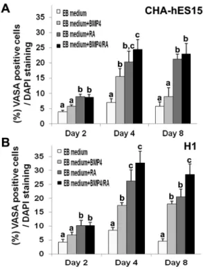
medium for 4 days. The EBs were attached and spread onto a culture dish in non-treated or BMP4-only treated groups. However, in the RA only- or BMP4/RA-treated groups, the EBs were clustered together at the periphery of the cells and were swollen (Fig. 1A). For effective induction of EBs into GSC-like cells, the specification rate into the GC lineage was compared based on the expression of VASA for 2–8 days after treatment of the cells with BMP4 and/or RA. The VASA protein was expressed on the membrane and in the cytoplasm of developing PGCs at the genital ridge [28]. Although there was no VASA expression in undifferentiated hESCs (Fig. S2), the VASA-positive cells were most frequently localized on the surface of EBs (Fig. 1B). Figure 2 shows the percentages of VASA-positive putative GSC-like cells. The results also indicated that the specification in CHA-hES15 and H1 cells could potentially be enhanced through treatment with BMP4 or RA, which are important for differen-tiation into GCs in vitro. On day 2, VASA was detected in EBs cultured with and without BMP4/RA. The percentage of VASA-positive cells in EBs cultured with RA (9.0%60.7, 10.3%61.8 in CHA-hES15 and H1) and BMP4/RA (8.8%60.9, 10.3%61.1) was significantly greater than that in EBs cultured without factors (4.0%60.4, 4.3%61.1) and that in the BMP4 (5.8%60.5, 6.8%61.1)-treated group (p,0.05). After 4 days, a significant increase in percentage of VASA positive cells was detected in EBs cultured with BMP4 (15.5%62.6 and 17.5%62.9 in CHA-hES15 and H1), RA (20.4%63.6 and 26.3%64.0) or BMP4/RA (24.5%63.2 and 32.8%64.2) compared with the EB medium group (7.0%61.3 and 8.5%61.7)(p,0.05). In particular, on day 4, the EBs treated with BMP4/RA showed the highest expression of VASA. The VASA expression on day 8 (EB medium group: 5.8%61.4 and 4.5%61.6 in CHA-hES15 and H1, BMP-treated: 9.0%62.9 and 18.0%63.4, RA-treated: 21.3%61.7 and 20.5%62.8, BMP4/RA-treated: 23.0%63.5 and 28.5%63.8) was not increased compared with that of the 4-day culture groups. Indeed, the VASA expression was reduced in certain groups, and there were slightly more dead cells in the EBs (data not shown). The number of putative GSC-like cells was higher in BMP4/RA-treated EBs than in the other groups. Thus, we decided to use BMP4/RA in further experiments.
The second step: Proliferation and isolation of PGC-like cells from hESC-derived EBs
GC-proliferation medium was used to propagate GSC-like cells for 1 to 3 weeks. The population of GSC-like cells (induced spermatogonia) was analyzed based on the expression of GFR-a1, which is a more reliable marker of spermatogonia detection than VASA. Before the flow cytometric analysis, we have counted total Figure 1. Differentiation of embryoid bodies (EBs) into male germ stem cell (GSC)-like cells.(A) Effects of treatment (EB medium; Non-treated, EB medium+BMP4, EB medium+RA or EB medium+BMP4/RA group) of EBs on the morphological features and induction of GSC-like cells after culture for 4 days. Scale bars: 50mm. a; undifferentiated CHA hES 15, b: embryoid body from CHA hES 15, c; EB medium, d; EB medium+BMP4, e; EB medium+RA, f; EB medium+BMP4/RA group (B) Immunocytochemical staining using anti-VASA antibody on 4-day-old EBs (the left panel) and dissociated cells (the right panel). The yellow box indicates the magnified area. The red arrows indicate the signals for VASA fluorescence.Note:Scale bars are 20mm.
doi:10.1371/journal.pone.0090454.g001
Figure 2. Graphic representations of stained VASA-positive cells.Percentage of VASA-positive cells in EBs derived from CHA-hES15 and H1 hESC lines treated with EB medium (non-treated), RA, BMP4 or RA+BMP4 for 2–8 days. RA: retinoic acid; BMP4: bone morphogenetic proteins 4.Note:avsbvsc,p
cell number of each group (EB medium group+BMP4/RA, EB medium group and GC-medium). As shown in the Fig. 3A, total cell numbers after 1 week of culture were decreased compared to that of starting material, but not different among three groups. In cell proliferation assay to evaluate the proliferation activity of each groups (EB medium group, EB medium group+BMP4/RA and GC-medium), there were no significant differences in all 1 week-cultured groups. However, MACS and long-term cultured cell in GC-medium (GC-medium for 4 weeks) have greater proliferating activity than other groups and total number of sorted cell was increased more than 4 times (Fig. 3A). The flow cytometric analysis revealed that after 1 week of culture, the population of GFR-a1-positive cells in the GC-proliferation medium group was much higher than that in the EB medium group or the BMP4/RA group (CHA-hESC15: 22.0%62.6 vs. 8.4%60.6 and 4.4%61.2, H1: 23.6%63.3 vs.
12.7%63.2 and 11.2%61.5, p,0.05)(Fig. 3B). The group cultured in the GC-proliferation medium was sorted according to GFR-a1 expression and further cultured under the same conditions for 3 weeks. After 4 weeks of culture (2 more passages) in GC-proliferation medium, the percentage of
GFR-a1-positive cells reached 57.9%63.2 and 54.9%65.3 in CHA-hES15 cells and H1 cells, respectively (Fig. 3B). In addition, immunocytochemical analysis re-confirmed that percentage of GFR-a1-positive cells in the GC-proliferation medium group was much higher than that in the EB medium group or the BMP4/RA group after 1 week of culture (CHA-hESC15: 9.8%62.3 vs. 2.0%60.7 and 2.3%61.0, H1: 12.0%61.5 vs.
2.5%60.6 and 2.8%60.9,p,0.05)(Fig. 3C). Moreover, after 4 week of culture, the percentage of GFR-a1-positive cells reached 45.3%62.4 and 47.3%65.7 in CHA-hES15 cells and H1 cells, respectively (Fig. 3C).
The third step:In vitrodifferentiation of PGC-like cells using a 3D-coculture system
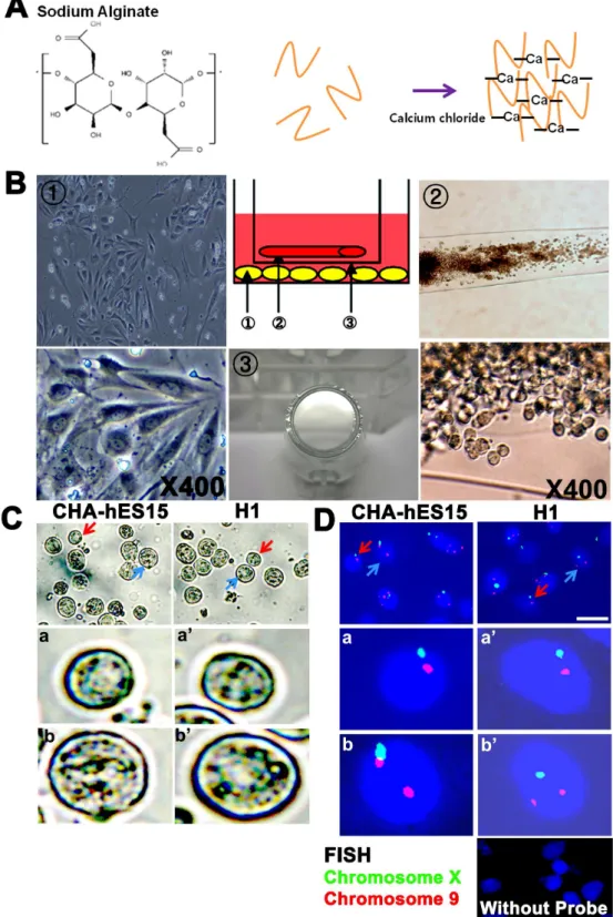
The structure of alginate-encapsulation containing GSC-like cells was not changed during in vitrodifferentiation (Fig. 4A and 4B). However, the populations of GSC-like cells in the inner wells and testicular cells in the outer wells increased. After 2 weeks of 3D-culture with or without co-culture with testicular cells, the encapsulated GSC-like cells were dissociated, and the morphology, FISH, gene expression and ploidy of these cells were analyzed. In the group co-cultured with testicular cells, although the numbers was very low, several types of GC-like cells ranging in size from 15 to 25mm were appeared afterin vitroculture. Their morphology was generally spherical with apparently intact plasma membranes and clear nuclear margins (Fig. 4C) and was looked similar to post-meiotic GC [22]. In FISH analysis, most of cultured cells showed non-meiotic (2n) cells and sperm-like cells were not identified. However, there were a few haploid (n) or tetraploid (4n) cells obtained after culturing (Fig. 4D and Fig. S7). In RT-PCR, TP-1 (a marker for spermatocyte and spermatid) expression and IAP (acrosome granule; a marker for spermatocyte and spermatid) expression were detected in differentiated germ cells derived from both types of ESCs (Fig. 5A and 5C). And integrin a6 (undifferentiated spermatogonia), c-Kit and TH2B (differentiating spermatogonia and spermatocytes) still remained in the 3D-co-culture group. The quantification analysis showed that the ratios of haploid germ cells were higher in the 3D-co-culture group than in the 3D-culture group (2.960.3% and 3.360.1%vs.0.960.1% and 1.160.3% in the CHA-hES15 and H1, respectively,p,0.05) (Fig. 5B).
In vivodifferentiation (propagation) of PGC-like cells in the testes
To determine whether the ESC-derived GSC-like cells main-tained their characteristics in vivo, the GSC-like cells were transplanted into recipient testes preceded by MACS sorting without in vitro differentiation. The transplantation of GSC-like cells resulted in the expression of GFP throughout the seminiferous tubule sections (Fig. S6). In 1–3 weeks after transplantation, the GSC-like cells derived from ESCs had a detectable GFP-positive population in some seminiferous tubules. At 3 weeks, an increase in GFP-positive cells was detected compared with at 1-week.
Analysis of gene expression
We performed microarray analysis to identify the similarity of gene expression between ESC-derived GSC-like cells and testis-derived spermatogonia. Two sets of separately purified undiffer-entiated hESCs (CHA-hES4 and H1), cultured GSC-like cells (derived from CHA-hES4 and H1) and spermatogonia (derived from the testes of two individual OA patients) were used for mRNA preparation and microarray hybridization. Of the 42,404 genes on the microarray, 9,534 transcripts were expressed at a level above the threshold defining differential expression (Fig. S4). The clustering tree showed that the differences (.2-fold) in the expression patterns between the ESC-derived GSC-like cells and the testis-derived spermatogonia were less pronounced than those between the ESC-derived GSC-like cells and the undifferentiated hESCs (p,0.05) (Fig. S5).
To evaluate the germ cell lineage further, 140 marker genes were selected based on previously published data [29]. A total of 22 genes were then excluded because of similar expression in the undifferentiated hESCs (Fig. 6A). Of the 118 remaining genes, 112 genes were expressed by both the ESC-derived PGC-like cells and the testis-derived spermatogonia. The other six genes were expressed by only the ESC-derived PGC-like cells or the testis-derived SSCs. Forty-eight of the remaining 118 genes, including TAF4B, GPR-125, GFRA1, POU3F1, DND1, and SEPTIN6, showed similar expression levels in both ESC-derived GSC-like cells and the testis-derived spermatogonia, and 52 of the 118 genes, including INTG5, INTGB1, EGR2, THY-1, CD9, SOX 13, and IFITM3, were up-regulated in testis-derived spermatogo-nia compared with ESC-derived GSC-like cells. Only 18 of the 118 genes, including NANOG, POU5F1, LIN28, UTF1 and SATB1, were up-regulated in ESC-derived GSC-like cells compared with testis-derived spermatogonia (Figure 6A). We used quantitative RT-PCR to validate the differential expression of selected spermatogonia-specific genes in ESC-derived GSC-like cells compared with testis-derived spermatogonia. The RT-PCR analysis showed that the mRNA levels of selected genes, including STARD13, NID2, PTPRM, DUSP6, NCH1 and NR0B1, in the two groups were similar to the levels detected in the microarray results (Fig. 6B).
Discussion
The pluripotency of ESCs provides an excellent model for studying differentiation and dedifferentiation in various lineages, including the production of germ cells. Previous studies have shown that ESC-derived PGC-like cells and germ cells have been spontaneously or directly differentiated by applying RA and/or BMP4 in culture, but a substantial number of these differentiated cells expressed specific markers for PGCs or germ cells onlyin vitro
systems. The results have confirmed that the addition of RA and BMP4 to cultures effectively induces the expression of GSC-specific markers in differentiated hESCs [30–32]. In addition, more enriched ESC-derived GSC-like cells were obtained using a
germ cell culture system, and the efficiency of haploid male germ cell production was increased using a 3D-co-culture system as compared to a 3D culture system alone (Fig. 5) and a 2D culture system(,1%, data not shown). Propagated ESC-derived GSC-like Figure 3. Proliferation of GSC-like cells through culture with GDNF, bFGF and LIF(A) Total numbers and proliferation activity of embryoid body (EB) cells (from CHA-hES15 and H1 cell lines) in cultured groups with EB medium+BMP4/RA, EB medium only or GC proliferation medium for 1 and 4 weeks. (B) Flow cytometric analysis of GFR-a1 expression in ESC-derived GSC-like cells (from CHA-hES15 and H1 cell lines) cultured in EB medium+BMP4/RA, EB medium only or GC proliferation medium for 1 and 4 weeks. (C) Immunocytochemical staining of GFR-a1 expression in ESC-derived GSC-like cells (from CHA-hES15 and H1 cell lines) cultured in EB medium+BMP4/RA, EB medium only or GC proliferation medium for 1 and 4 weeks.Note:avsbvsc,p,0.05, Scale bars are 50mm.
Figure 4.In vitroculture of GSC-like cells using a 3D-co-culture system and the potential for differentiation into germ cell lineagesI
(A) Schematic of calcium-crosslinked alginate; Soluble alginate in saline was mixed with CaCl2solution. The diffusion of calcium ions into the surrounding solution induces the crosslinking of the soluble alginate and the formation of hydrogels. (B) 3D-co-culture system; The encapsulated GSC-like cells were passed through a strainer (insert), cultured in 6-well culture dishes and differentiated into testicular cells; (1) Human testicular cells (2) Encapsulated GSC-like cells (3) Strainer (insert) (C) Putative differentiated germ cell with a diameter of 15–25mm. Red arrows, a and a9indicated putative differentiated germ cell. And blue arrows, b and b9indicated non-meiotic cell in 3D-co-culture group (from CHA-hES15 and H1 cell lines). (D) FISH analysis ofin vitrodifferentiated GSC-like cells from hESCs; Red arrows, a and a9indicated normal nucleus with meiotic cells (haploid; n) of chromosomes X (spectrum green) and 9 (spectrum red). And blue arrows, b and b9indicated non-meiotic cells (diploid; 2n) of chromosomes X (spectrum green) and 9 (spectrum red) in 3D-co-culture group (from CHA-hES15 and H1 cell lines). Scale bars are 20mm.
cells maintained the potential for proliferation capacity, as confirmed through cell transplantation into the testes of host mice (Fig. S6). Moreover, in vitro differentiated GSC-like cells have
shown similar gene expression profiles in SSCs obtained from human testes.
To improve low differentiation efficiency, we identified the critical factors involved in the differentiation of GSCs into germ Figure 5.In vitroculture of GSC-like cells using a 3D-co-culture system and the potential for differentiation into germ cell lineages.
(A) Expression of stage-specific genes (integrina6, c-Kit, TH2B and TP-1) duringin vitrodifferentiation of human ESC-derived GSC-like cells using RT-PCR. Integrina6 (undifferentiated spermatogonia), c-Kit and TH2B (differentiating spermatogonia and spermatocytes), TP-1 (meiotic spermatocytes and spermatids) were used as stage-specific germ cell marker genes. 18S ribosomal RNA was used as a control gene. Line 1–3: ESC-derived GSC-like cells (0, 1, and 2-week culture in the 3D-co-culture system), N: no reverse transcriptase. (B) DNA content ofin vitrodifferentiated ESC-derived GSC-like cells. a; DNA content of cells from obstructive azoospermia (OA, normal spermatogenesis sample) b; Undifferentiated human embryonic stem cells (CHA-hES15). N1 represents haploid germ cells and N2 represents diploid cells. (C) Localization of intra acrosomal protein (IAP) in human testes (upper panel) andin vitrodifferentiated GSC-like cells from hESCs (middle and lower figure). IAP was stained in spermatid acrosomes. The yellow arrows indicate fluorescence signals.Note:**; significantly different (p,0.05). Scale bars: 50mm.
cells [33–34] and the proliferation of in vitro induced GSC-like cells. Indeed, the addition of BMP and RA has shown that some growth factors might affect the induction of differentiation into GSC-like cells and the enrichment of GSC-like cellsin vitro. Several groups have reported that mouse GSC clumps continuously proliferate under culture conditions with GDNF and bFGF, and these proliferating clump-forming cells maintain the properties of GSCs [20,35]. In human systems, GSC-like cells proliferate when certain growth factors are added to the culture medium [23]. In the present study, we applied GC-proliferation medium supple-mented with EGF, LIF, GDNF and bFGF to propagate and
enrich ESC-derived GSC-like cells, and the population of
GFR-a1-positive cells derived from cultured EBs was highly enriched compared with the control group (Fig. 3).
In our previous studies, we introduced an in vitro 3D-culture system using sodium alginate for differentiation into haploid male germ cells [21–22]. For the effective differentiation of GSC-like cells derived from human ESCs, we employed a modified 3D-co-culture system using testicular somatic cells (Fig. 4). Afterin vitro
differentiation and quantification using markers for haploid male germ cells, we observed that a 3D-co-culture system was more effective than a simple 3D-culture system for the differentiation of Figure 6. Comparative analyses between ESC-derived GSC-like cells and testis-derived spermatogonia using known spematogonial marker genes(A) The expression profiles of the 118 genes in the 2 types of cells are presented as a Venn diagram and table. a; A total of 52 genes were more strongly expressed in testis-derived SSCs than in ESC-derived GSC-like cells. b; A total of 48 genes were similarly expressed in the 2 groups. c; A total of 18 genes were more strongly expressed in ESC-derived GSC-like cells than in testis-derived spermatogonia. (B) The validation of randomly selected spematogonial marker genes using real-time RT-PCR.Note:In vitrocultured ESC-derived GSC-like cells (at 2ndpassage);In vitrocultured testis-derived spermatogonia (at 2ndpassage).
GSC-like cells from human ESCs (Fig. 5). In particular, approximately 3% of the GSC-like cells could be differentiated into haploid male germ cells, exhibiting specific markers after only two weeks of short-term culture, although morphologically normal or functional spermatozoa were not observed in the cell cultures. GFR-a1 is the receptor for GDNF, a growth factor that regulates the ratio of self-renewal and differentiation of GSCs. GDNF-knockout mice exhibited a depletion of GSCs, whereas mice over-expressing GDNF show an accumulation of GSCs. von Schonfeldt and colleagues reported that GFR-a1 is localized to spermatogonia in the adult mouse testis [29], suggesting that male GSCs could be highly purified using this marker. Although CD9 and THY-1 are generally accepted to be male GSC markers, these surface markers were also detected on undifferentiated stem cells. In the present study, GFR-a1 was used as a marker for sorting male GSCs, and the sorted GFR-a1-positive cells were cultured (Fig. 3). In contrast to previous studies showing a 16 to 32-fold expansion after culturing for 1 month [20], the expansion rate of our GSC-like cells was low (3–4-fold). The results suggest that these cells might be different from other GSCs obtained from the testes, although GSC-like cells derived from hESCs exhibited similarities in morphology and marker expression of human male GSCs. Therefore, we compared the gene expression profiles of ESC-derived GSC-like cells and male GSCs (spermatogonia) obtained from human testes [23] using microarray analysis. The gene sets over-expressed in GSC-like cells derived from hESCs showed a significant overlap with the reference gene lists used to characterize male GSC-specific genes in previous rodent studies [36]. The previously identified male GSC-specific genes, such as
ITGA6, FGFR3, EPB41L4A, DPPA5, DPPA3 (STELLA), CHEK2,
CHD7, CDH1,BCL6B, ABCG2,TERT, TACSTD1, SOX3, SOX2,
SOHLH2, SEMA4D, RELN, PUNC, PIWIL2, NOTCH1, LIN28B, and LHX1, showed similar expression levels in all human and rodent samples. However, some SSC-specific genes, such as
UPK1B, SOHLH1, SNAP91, TEX14, TEX101, NANOS2, MBP,
DAZL,MAGEA4andSTAR8, were not expressed in either human sample (data not shown). The absence of those genes could reflect the fact that the human samples (GSC-like cells from hESCs and testicular spermatogonia) did not exhibit the same gene expression patterns as the rodent samples. Using microarray analysis, we identified six male GSC-specific genes showing differential expression in both human samples: the expression of TSPAN8
and FOXA2in the testis and the expression ofSLC6AL,FOXA2,
ANKRD6 andABCG2in GSC-like cells from hESCs. The results revealed that cultured GSC-like cells derived from hESCs have high similarity with human spermatogonia (Fig. 6A).
In the validated gene analysis (Fig. 6B),protein tyrosine phosphatases (PTPs), namely,DUSP6,PTPN14,PTPRG,PTPRM, andPTPRD, regulate adhesion and differentiation. Dual specific phosphatase 6 (DUSP6orMKP-3) encodes a negative feedback regulator of ERK signaling and regulates FGFR signaling during mouse develop-ment.PTPRMis associated with adherent junctions and dephos-phorylates key signaling substrates, such as beta-catenin and cadherin adhesion molecules. PTPRG is transiently expressed during ESC-derived embryoid body differentiation and is required for male GSC lineage commitment [37–39]. TENC1 encodes a member of the tensin family that is a focal adhesion molecule that binds to actin filaments (ENC1) and participates in signaling pathways [40]. NID2 encodes a member of the nidogen family, and this protein is a cell-adhesion protein that binds collagen I and IV and laminin and might be involved in maintaining the structure of the basement membrane. These genes have been associated with the formation of male GSC clumps and showed similar expression levels in ESC-derived GSC-like cells. STARD13
encodes a protein that contains an N-terminal sterile alpha motif (SAM) for protein-protein interactions followed by an ATP/GTP-binding motif, a GTPase-activating protein (GAP) domain, and a C-terminal STAR-related lipid transfer (START) domain, sug-gesting that this protein might be involved in the regulation of cell proliferation. In addition,STARD13interacts with EPB41L and TENC1 [40–41]. These genes have been associated with the proliferation of male GSC clumps and exhibited lower expression in GSC-like cells from hESCs than in male GSCs from testes, potentially reflecting the low expansion rate of GSC-like cells.
NR0B1 encodes an orphan member of the nuclear hormone receptor family that is expressed in tissues involved in the production of steroid hormones and the reproductive function of the testes. Indeed, NR0B1 null homozygous male mice have adrenal insufficiency and testicular disorganization, as well as failed spermatogenesis [42–43]. All of the above genes are associated with spermatogenesis, reflecting the fact that ESC-derived GSC-like cells are more similar to germ cells than to undifferentiated hESCs.
Conclusions
We developed a sequential culture system to induce and propagate human GSC-like cells that strongly express the male GSC-specific maker GFR-a1 and differentiate into male germ cells. Although the GSC-like cells derived from hESCs were not identical to spermatogonia, this study provided insight into the induction and propagation of spermatogonia. In addition, a large number of GSC-like cells can be applied to 3D-co-culture for differentiation into male germ cells. Therefore, we suggest that a sequential differentiation system is a useful tool for the study of male germ cell development and for future clinical trials using patient specific stem cells after more improvement of male GSC induction and the completion of meiosis.
Supporting Information
Figure S1 Schematic representation of the three-step method. (JPG)
Figure S2 Characterization of undifferentiated human embry-onic stem cells (CHA-hES15) Immunocytochemical staining for OCT4, NANOG, SSEA-3, SSEA-4 and TRA1-60, as human ESC-specific markers, and staining for VASA and GFR-a1, as spermatogonia specific markers. The bright field images show the typical morphology of hESCs. Scale bar is 100mm.
(JPG)
Figure S3 Characterization of human spermatogonial cells. (A) Cellular localization of VASA and GFR-a1 as spermatogonia specific markers in fetal gonads and adult testes. VASA was abundantly expressed in fetal gonad cells. GFR-a1 was observed in the undifferentiated spermatogonial cells at the basement membrane within the seminiferous tubules. The figure on the right shows the magnification of VASA and GFR-a1-positive signals. Scale bars: 50mm. (B) Immunocytochemical characterization of cultured spermatogonial cells using human adult testes. Integrin
b1, a6 and GFR-a1 were used as spermatogonia markers. The bright field images show the typical morphology of cultured spermatogonial cells. OA testis: seminiferous tubules of OA (normal spermatogenesis) patients. Scale bars: 50mm. (C) RT-PCR-based characterization of hESCs, GSC-like cells and spermatogonial cells. Note: Mock 1stab: not treated with primary antibodies, MEF: mouse embryonic fibroblast.
Figure S4 Expression profiles of undifferentiated hESCs, hESC-derived GSC-like cells and testis-derived spermatogonial cells. The expression profiles of the 9534/42404 genes that were differentially expressed in the three types of cells were hierarchically clustered and are presented as a heat-map. The expression level of each transcript is indicated in the color code bar; red indicates high expression, and green indicates low expression. Note: hESCs, undifferentiated human embryonic stem cells; GSC-like cells (from hESCs),in vitro cultured human ESC-derived GSC-like cells at 2nd passage; spermatogonia (Testis), in vitro cultured testis-derived spermatogonial cells at 2ndpassage.
(JPG)
Figure S5 Differentially expressed gene profiles of undifferenti-ated hESCs, hESC-derived GSC-like cells and testis-derived spermatogonial cells. (A) The expression profiles of the 9534 (count of differentially expressed genes (DEG) and their hierar-chical clustering) genes that were expressed in the 3 types of cells. (B) Functional classification using gene ontology information. Note: hESCs, undifferentiated human embryonic stem cells; GSC-like cells (from hESCs),in vitrocultured human ESC-derived
GSC-like cells at 2nd passage; spermatogonia (Testis), in vitro
cultured testis-derived spermatogonial cells at 2ndpassage. (JPG)
Figure S6 In vivo propagation of GSC-like cells in recipient testis (A) Testes after transplantation with GSC-like cells using injection pipettes. GSC-like cells were suspended in DPBS containing trypan blue. Seminiferous tubules containing the blue cell suspension were observed. (B) GFP signaling in GSC-like cells from recipient testes. Scale bars: 50mm.
(JPG)
Figure S7 Different type of FISH results. Detection of X chromosome and 9 chromosome in the differentiated GSC-like cells; a and a9: diploid (2n), b and b9: tetraploid (4n), c and c9: haploid (n,X) d and d9: haploid type (n,Y); Upper panel indicated CHA-hES15 cell lines. Lower panel indicated H1 cell lines. (JPG)
Author Contributions
Conceived and designed the experiments: DRL. Performed the experi-ments: JJL MSS JEL. Analyzed the data: JJL. Contributed reagents/ materials/analysis tools: DRL. Wrote the paper: DRL JJL.
References
1. Thomson JA, Itskovitz-Eldor J, Shapiro SS, Waknitz MA, Swiergiel JJ, et al. (1998) Embryonic stem cell lines derived from human blastocysts. Science 282: 1145–1147.
2. Martin GR (1981) Isolation of a pluripotent cell line from early mouse embryos cultured in medium conditioned by teratocarcinoma stem cells. Proc Natl Acad Sci U S A 78: 7634–7638.
3. Hubner K, Fuhrmann G, Christenson LK, Kehler J, Reinbold R, et al. (2003) Derivation of oocytes from mouse embryonic stem cells. Science 300: 1251– 1256.
4. Geijsen N, Horoschak M, Kim K, Gribnau J, Eggan K, et al. (2004) Derivation of embryonic germ cells and male gametes from embryonic stem cells. Nature 427: 148–154.
5. Kerkis A, Fonseca SA, Serafim RC, Lavagnolli TM, Abdelmassih S, et al. (2007) In vitro differentiation of male mouse embryonic stem cells into both presumptive sperm cells and oocytes. Cloning Stem Cells 9: 535–548. 6. Starz-Gaiano M, Lehmann R (2001) Moving towards the next generation. Mech
Dev 105: 5–18.
7. Ying Y, Liu XM, Marble A, Lawson KA, Zhao GQ (2000) Requirement of Bmp8b for the generation of primordial germ cells in the mouse. Mol Endocrinol 14: 1053–1063.
8. Toyooka Y, Tsunekawa N, Akasu R, Noce T (2003) Embryonic stem cells can form germ cells in vitro. Proc Natl Acad Sci U S A 100: 11457–11462. 9. Kee K, Gonsalves JM, Clark AT, Pera RA (2006) Bone morphogenetic proteins
induce germ cell differentiation from human embryonic stem cells. Stem Cells Dev 15: 831–837.
10. Fujiwara T, Dunn NR, Hogan BL (2001) Bone morphogenetic protein 4 in the extraembryonic mesoderm is required for allantois development and the localization and survival of primordial germ cells in the mouse. Proc Natl Acad Sci U S A 98: 13739–13744.
11. Zhou Q, Li Y, Nie R, Friel P, Mitchell D, et al. (2008) Expression of stimulated by retinoic acid gene 8 (Stra8) and maturation of murine gonocytes and spermatogonia induced by retinoic acid in vitro. Biol Reprod 78: 537–545. 12. Koshimizu U, Watanabe M, Nakatsuji N (1995) Retinoic acid is a potent growth
activator of mouse primordial germ cells in vitro. Dev Biol 168: 683–685. 13. Clark AT, Bodnar MS, Fox M, Rodriquez RT, Abeyta MJ, et al. (2004)
Spontaneous differentiation of germ cells from human embryonic stem cells in vitro. Hum Mol Genet 13: 727–739.
14. Thomas S (2000) Alginate dressings in surgery and wound management–Part 1. J Wound Care 9: 56–60.
15. Boontheekul T, Kong HJ, Mooney DJ (2005) Controlling alginate gel degradation utilizing partial oxidation and bimodal molecular weight distribu-tion. Biomaterials 26: 2455–2465.
16. Shang Q, Wang Z, Liu W, Shi Y, Cui L, et al. (2001) Tissue-engineered bone repair of sheep cranial defects with autologous bone marrow stromal cells. J Craniofac Surg 12: 586–593; discussion 594–585.
17. Lee JE, Kang MS, Park MH, Shim SH, Yoon TK, et al. (2010) Evaluation of 28 human embryonic stem cell lines for use as unrelated donors in stem cell therapy: implications of HLA and ABO genotypes. Cell Transplant 19: 1383– 1395.
18. Gadue P, Huber TL, Paddison PJ, Keller GM (2006) Wnt and TGF-beta signaling are required for the induction of an in vitro model of primitive streak
formation using embryonic stem cells. Proc Natl Acad Sci U S A 103: 16806– 16811.
19. Nishikawa SI, Nishikawa S, Hirashima M, Matsuyoshi N, Kodama H (1998) Progressive lineage analysis by cell sorting and culture identifies FLK1+ VE-cadherin+cells at a diverging point of endothelial and hemopoietic lineages. Development 125: 1747–1757.
20. Kanatsu-Shinohara M, Ogonuki N, Inoue K, Miki H, Ogura A, et al. (2003) Long-term proliferation in culture and germline transmission of mouse male germline stem cells. Biol Reprod 69: 612–616.
21. Lee DR, Kaproth MT, Parks JE (2001) In vitro production of haploid germ cells from fresh or frozen-thawed testicular cells of neonatal bulls. Biol Reprod 65: 873–878.
22. Lee DR, Kim KS, Yang YH, Oh HS, Lee SH, et al. (2006) Isolation of male germ stem cell-like cells from testicular tissue of non-obstructive azoospermic patients and differentiation into haploid male germ cells in vitro. Hum Reprod 21: 471–476.
23. Lim JJ, Sung SY, Kim HJ, Song SH, Hong JY, et al. (2010) Long-term proliferation and characterization of human spermatogonial stem cells obtained from obstructive and non-obstructive azoospermia under exogenous feeder-free culture conditions. Cell Prolif 43: 405–417.
24. Huang Z, Fasco MJ, Kaminsky LS (1996) Optimization of Dnase I removal of contaminating DNA from RNA for use in quantitative RNA-PCR. Biotechni-ques 20: 1012–1014, 1016, 1018–1020.
25. Chladek D, Peknicova J, Capkova J, Geussova G, Tepla O, et al. (2000) [Use of human sperm protein monoclonal antibodies in the diagnosis of sperm pathology and selection of a suitable assisted reproduction method for fertilization]. Ceska Gynekol 65: 28–32.
26. Ogawa T, Arechaga JM, Avarbock MR, Brinster RL (1997) Transplantation of testis germinal cells into mouse seminiferous tubules. Int J Dev Biol 41: 111–122. 27. Kim S, Kim GJ, Miyoshi H, Moon SH, Ahn SE, et al. (2007) Efficiency of the elongation factor-1alpha promoter in mammalian embryonic stem cells using lentiviral gene delivery systems. Stem Cells Dev 16: 537–545.
28. Castrillon DH, Quade BJ, Wang TY, Quigley C, Crum CP (2000) The human VASA gene is specifically expressed in the germ cell lineage. Proc Natl Acad Sci U S A 97: 9585–9590.
29. von Schonfeldt V, Wistuba J, Schlatt S (2004) Notch-1, c-kit and GFRalpha-1 are developmentally regulated markers for premeiotic germ cells. Cytogenet Genome Res 105: 235–239.
30. Aflatoonian B, Moore H (2005) Human primordial germ cells and embryonic germ cells, and their use in cell therapy. Curr Opin Biotechnol 16: 530–535. 31. Bowles J, Knight D, Smith C, Wilhelm D, Richman J, et al. (2006) Retinoid
signaling determines germ cell fate in mice. Science 312: 596–600.
32. Ying Y, Qi X, Zhao GQ (2001) Induction of primordial germ cells from murine epiblasts by synergistic action of BMP4 and BMP8B signaling pathways. Proc Natl Acad Sci U S A 98: 7858–7862.
33. De Felici M, Dolci S, Pesce M (1993) Proliferation of mouse primordial germ cells in vitro: a key role for cAMP. Dev Biol 157: 277–280.
34. Resnick JL, Bixler LS, Cheng L, Donovan PJ (1992) Long-term proliferation of mouse primordial germ cells in culture. Nature 359: 550–551.
36. Hamra FK, Schultz N, Chapman KM, Grellhesl DM, Cronkhite JT, et al. (2004) Defining the spermatogonial stem cell. Dev Biol 269: 393–410. 37. Li C, Scott DA, Hatch E, Tian X, Mansour SL (2007) Dusp6 (Mkp3) is a
negative feedback regulator of FGF-stimulated ERK signaling during mouse development. Development 134: 167–176.
38. Wadham C, Gamble JR, Vadas MA, Khew-Goodall Y (2003) The protein tyrosine phosphatase Pez is a major phosphatase of adherens junctions and dephosphorylates beta-catenin. Mol Biol Cell 14: 2520–2529.
39. Brady-Kalnay SM, Mourton T, Nixon JP, Pietz GE, Kinch M, et al. (1998) Dynamic interaction of PTPmu with multiple cadherins in vivo. J Cell Biol 141: 287–296.
40. Kawai K, Kitamura SY, Maehira K, Seike J, Yagisawa H (2010) START-GAP1/DLC1 is localized in focal adhesions through interaction with the PTB domain of tensin2. Adv Enzyme Regul 50: 202–215.
41. Nagaraja GM, Kandpal RP (2004) Chromosome 13q12 encoded Rho GTPase activating protein suppresses growth of breast carcinoma cells, and yeast two-hybrid screen shows its interaction with several proteins. Biochem Biophys Res Commun 313: 654–665.
42. Zanaria E, Muscatelli F, Bardoni B, Strom TM, Guioli S, et al. (1994) An unusual member of the nuclear hormone receptor superfamily responsible for X-linked adrenal hypoplasia congenita. Nature 372: 635–641.