Mariana Isabel Lopes Joaquim
Licenciada em Biologia
Exploring cell reprogramming
techniques for Angelman Syndrome
disease modelling
Dissertação para obtenção do Grau de Mestre em
Genética Molecular e Biomedicina
Orientador: Doutor Simão Teixeira da Rocha, FCT
Investigator, Instituto de Medicina Molecular
Exploring cell reprogramming techniques for Angelman Syndrome disease modelling
Copyright Mariana Isabel Lopes Joaquim, FCT/UNL, UNL
i
Agradecimentos
Em primeiro lugar quero agradecer ao meu orientador, Dr. Simão Teixeira da Rocha, por todo o conhecimento que me transmitiu, por toda a paciência, pelo apoio incansável e por acreditar em mim e nas minhas capacidades desde início. Este ano sob a sua orientação foi, sem dúvida, um privilégio, quer a nível profissional, quer a nível pessoal.
De seguida, agradeço à Professora Maria Carmo-Fonseca, chefe do laboratório RNA &
GeneRegulation, por me ter aceite no seu grupo, permitindo-me desenvolver este trabalho.
Gostaria de agradecer a todos os colaboradores deste projecto: à Dra. Sofia Duarte (IMM/Centro Hospitalar de Lisboa Central) por possibilitar o acesso a pacientes com Síndrome de Angelman; ao Jerome Mertens (Salk Institute, San Diego, USA) pelo envio dos plasmídeos essenciais ao desenvolvimento de parte deste trabalho, bem como pelo contacto estabelecido connosco de forma a esclarecer quaisquer dúvidas acerca do protocolo fruto do seu trabalho; à Teresa Silva (IMM/Lisboa) e à Dra. Cláudia Gaspar (IMM/Lisboa) pelos neurónios derivados de iPSCs que constituíram um controlo importante em parte deste trabalho; ao Dr. Edgar Gomes pelo plasmídeo GFP, também ele utilizado para um controlo essencial.
A todos os meus colegas de laboratório e de gabinete, um grande obrigado. Agradeço por toda a ajuda em momentos de stress laboratorial, pelos momentos de distracção e gargalhadas e por toda a companhia ao longo deste ano. Queria agradecer especialmente ao Duarte Brandão, não só por me acompanhar em grande parte do meu trabalho mas pela inestimável ajuda dada e dúvidas tiradas. Outro agradecimento especial à Ana Raposo, por ser a minha inigualável companheira de laboratório, por todas as vezes que me ajudou em momentos de apuros e, especialmente, por me fazer rir todos os dias. Um agradecimento também à Vanessa
Pires por me ter acolhido tão bem e por me ter aturado “verdinha” e ao João von Gilsa pelas
gargalhadas e inigualáveis cantorias.
A todos os meus restantes amigos, um enorme agradecimento, não só pelos momentos de distracção, mas também pelo apoio moral (mesmo quando não entendem nada do que estou a dizer) e por tolerarem as minhas ausências, especialmente durante a realização deste trabalho.
Ao Duarte Próspero, pela enorme paciência, pelo carinho incomparável, pelo apoio e pelo contínuo interesse no meu trabalho e no meu futuro.
Um agradecimento enorme a toda a minha família, que sempre acreditou e continua a acreditar em mim e, em especial, um obrigado aos meus tios Jorge e São e ao meu primo Rui, sem os quais este meu percurso no ensino superior não teria sido possível.
Por fim, agradeço aos maiores impulsionadores de tudo o que se relaciona com a minha vida, os meus pais e a minha irmã, que são, sem dúvida, quem de mais perto segue os meus
iii
Resumo
A Síndrome de Angelman (SA) é uma doença incurável do neuro-desenvolvimento causada pela ausência de expressão do gene UBE3A materno em neurónios. O UBE3A paterno é silenciado pelo transcrito antisense do UBE3A (UBE3A-ATS). No modelo de ratinho, a inibição da transcrição do UBE3A-ATS reactiva o UBE3A paterno e melhora funções cognitivas. Para avaliar se a mesma abordagem pode ser aplicada em pacientes com SA é necessário desenvolver um modelo celular desta doença.
Neste estudo visámos desenvolver esse modelo celular humano a partir de fibroblastos derivados de pacientes e avaliar o seu estado de imprinting.
Inicialmente foi tentado um protocolo de conversão neuronal directa baseado na expressão de dois factores de transcrição neuronais – ASCL1, NGN2 – e inibidores da via SMAD de forma a converter fibroblastos em neurónios. Apesar da elevada eficiência de infecção e detecção de expressão do ASCL1 transgénico, os iNs gerados não demonstraram sinais de identidade neuronal, baseado em resultados de RT-qPCR e IF. Este insucesso pode dever-se à falta de concentração dos lentivírus por ultracentrifugação, à falta de selecção com antibióticos e/ou ao destacamento das células durante a conversão. Seguidamente tentámos gerar NPCs a partir de iPSCs usando um protocolo comercial. No entanto, as “NPCs” geradas
não expressavam os marcadores genéticos correctos. Este insucesso pode dever-se à taxa de divisão inapropriada destas células durante a indução ou à falta de pluripotência destas iPSCs.
Apesar do insucesso na geração de neurónios, conseguimos optimizar a técnica
nascent-transcript RNA FISH, combinando a visualização do UBE3A com o SNORD116, unicamente
expresso pelo alelo paterno. Esta é uma ferramenta crucial para confirmar o estado de
imprinting do locus de Angelman nas células geradas.
No futuro, o estabelecimento de um modelo celular da SA servirá como uma plataforma de triagem de drogas para testar a reactivação do UBE3A paterno como alvo terapêutico para a SA.
v
Abstract
Angelman Syndrome (AS) is an imprinted neurodevelopmental disease with no cure caused by the lack of UBE3A expression, which, in neurons, is exclusively maternally expressed. The paternal UBE3A allele is silenced by the UBE3A antisense transcript ( UBE3A-ATS), which is only expressed from the paternal chromosome. In AS mouse model, inhibition of
the UBE3A-ATS transcription can reactivate paternal UBE3A. To translate such an approach to
humans, the development of a cellular model for this disease is necessary.
Here we sought to develop cellular model systems of AS from patient-derived fibroblasts and evaluate their imprinting status using RNA FISH-based single-cell approaches.
First, a neural direct conversion protocol based on expression of two neuronal transcription factors - ASCL1, NGN2– and SMAD pathway inhibitors was tried in order to convert fibroblasts into neurons. Despite high infection efficiency and detection of transgenic ASCL1 expression, the generated “iNs” did not show signs of neuronal identity based on RT-qPCR and IF analysis. This failure might have been caused by lack of lentiviruses concentration by ultracentrifugation, antibiotic selection skipping and/or dislodging of the cells under conversion. Second, we tried to generate NPCs from iPSCs using a commercially available differentiation protocol. Based on RT-qPCR and IF analysis, the generated “NPCs” failed to express the correct genetic markers.
This failure might be explained by inappropriate accelerated division rate of the cells during induction or lack of pluripotency of the newly-generated iPSCs used.
Despite unsuccessful generation of neuronal cells, we were able to optimize nascent-transcript RNA FISH, combining UBE3A and paternally expressed SNORD116, which is a crucial tool to confirm the imprinting status of the Angelman locus in newly-generated cells.
With future efforts, the establishment of AS cellular model systems will serve as drug screening platform to test paternal UBE3A reactivation as a therapeutic target for AS.
vii
Table of Contents
Agradecimentos... i
Resumo ... iii
Abstract ... v
List of Figures ... ix
List of Tables ... xi
Abbreviations ... xiii
1. Introduction ... 1
1.1. Epigenetics ... 1
1.1.1. Genomic Imprinting ... 1
1.2. Angelman Syndrome ... 4
1.2.1. Symptoms ... 4
1.2.2. UBE3A and the 15q11-q13 imprinted cluster ... 4
1.2.3. Causes ... 6
1.2.4. Diagnosis ... 6
1.2.5. Treatment ... 7
1.2.5.1. Therapeutic approaches under investigation ... 7
1.3. Cellular models of human neuronal diseases ... 8
1.3.1. Pluripotent stem cells ... 9
1.3.2. Induced pluripotent stem cells ... 9
1.3.2.1. Reprogramming techniques ... 10
1.3.3. iPSCs in disease modelling ... 10
1.3.3.1. iPSCs in neuronal disease modelling ... 11
1.3.4. Direct conversion into induced neurons ... 12
1.3.4.1. Direct conversion techniques ... 13
1.3.4.2. iNs in neuronal disease modelling ... 14
1.4. Aims of the study ... 14
2. Material and Methods ... 15
2.1. Cell culture ... 15
2.1.1. Punch-skin biopsy fibroblasts ... 15
2.1.2. NPCs differentiation from iPSCs ... 16
2.1.2.1. iPSCs expansion ... 16
2.1.2.2. Neural Progenitor cells generation and expansion ... 16
2.1.3. Neuronal direct conversion ... 17
2.1.3.1. HEK 293T expansion and transfection ... 17
viii
2.1.3.3. Neural direct conversion ... 18
2.2. Molecular Biology Techniques ... 18
2.2.1. Competent cells transformation ... 18
2.2.2. Plasmid DNA extraction... 19
2.2.3. Plasmid restriction digestion ... 19
2.2.4. RNA isolation from adherent cells and cDNA synthesis ... 19
2.2.5. Reverse transcriptase polymerase chain reaction (RT-PCR) ... 20
2.2.6. Reverse-transcriptase quantitative polymerase chain reaction (RT-qPCR) ... 20
2.3. Cellular characterization ... 21
2.3.1. RNA Fluorescent in situ hybridization (RNA-FISH) ... 21
2.3.2. Immunofluorescence (IF)... 22
3. Results and Discussion ... 23
3.1. Neural Direct Conversion ... 23
3.2. iPSCs differentiation ... 32
3.3. Evaluation of imprinting status of genes in the Angelman locus by nascent-transcript RNA FISH ... 36
4. Concluding Remarks and Future Perspectives ... 41
ix
List of Figures
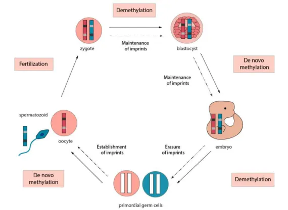
Fig. 1.1 – Representative scheme of the cycle of mammalian methylation and imprinting. ... 2
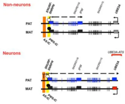
Fig. 1.2 – Map of the human 15q11-q13 imprinted region in non-neurons and neurons ... 5
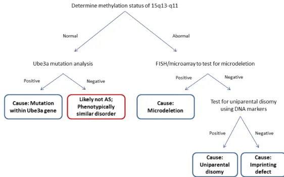
Fig. 1.3 – Representative scheme of Angelman Syndrome molecular diagnosis ... 6
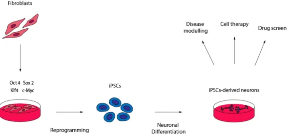
Fig. 1.4 – Representative scheme of iPSCs reprogramming and neuronal differentiation ... 10
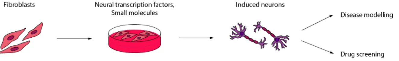
Fig. 1.5 – Representative scheme of neuronal direct conversion ... 13
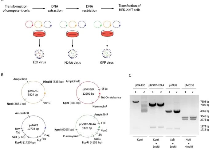
Fig. 3.1 - Characterization of the plasmid vectors used for neural conversion ... 24
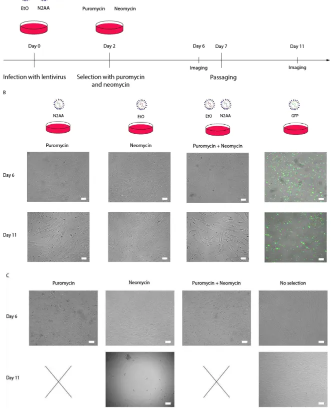
Fig. 3.2 - Infection and selection of AS 3y fibroblasts with lentivirus for neural conversion... 25
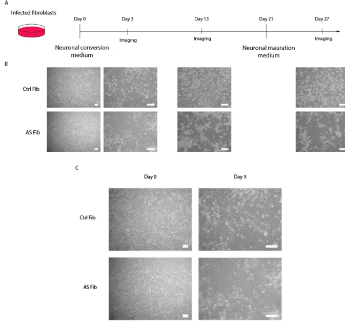
Fig. 3.3 - Neural direct conversion of control and AS 30y fibroblasts ... 27
Fig. 3.4 - RT-PCR for transgenic ASCL1 in control 30y iNs, AS 30y iNs, control 30y fibroblasts and AS 30y fibroblasts ... 28
Fig. 3.5 - RT-qPCR analysis of the relative expression of the fibroblast-specific genes DKK3 and THY1 and the late neuronal-specific gene MAP2 in control 30y fibroblasts, AS 30y fibroblasts, control 30y iNs and AS 30y iNs ... 28
Fig. 3.6 - 2nd round of neural direct conversion of control and AS 30y fibroblasts ... 31
Fig. 3.7 - iPSCs reprogramming and neural differentiation ... 33
Fig. 3.8 - Representative pictures of IF assay in iPSCs and/or NPCs ... 34
Fig. 3.9 - RT-qPCR analysis of the relative expression of the stem cells markers OCT4, NANOG, SOX2, the early neuronal-specific gene TUJ1 and the late neuronal-specific gene MAP2 in control iPSCs and control NPCs ... 35
xi
List of Tables
Table 1.1- Summary of the 12 imprinted disorders with the affected chromosome region and main clinical features ... 3 Table 2.1 - Primers used for RT-PCR. ... 20 Table 2.2 - Primers used for RT-qPCR. ... 21 Table 3.1 - Percentage of cells with two, one or no signal for UBE3A and SNORD116 probes in StellarisTM RNA FISH in control fibroblasts, AS fibroblasts, control iPSCs, AS iPSCs and control
xiii
Abbreviations
AS - Angelman Syndrome
BDNF – Brain-derived neurotrophic factor DAPI - 4',6-diamidino-2-phenylindole
DMEM –Dulbecco’s modified Eagle medium DMR – Differentially methylated region DMSO - Dimethyl sulfoxide
DNA – Deoxyribonucleic acid EtOH – Ethanol
FBS - Fetal bovine serum
GDNF – Glial cell line-derived neurotrophic factor GFP – Green fluorescent protein
ICR – Imprinting control region
iPSCs – Induced pluripotent stem cells iNs - Induced neurons
lncRNA – Long non-coding RNA
mASO – modified anti-sense oligonucleotide NC - Neural conversion
NEAA - Non-essencial aminoacids
NM - Neural maturation
NPCs – Neural progenitor cells PBS - Phosphate buffered saline
Pen/Strep - Penincilin/Streptomicin
PFA – Paraformaldehyde PWS – Prader-Willy Syndrome snoRNA – Small nucleolar RNA SSC - Saline-sodium citrate
1
1. Introduction
1.1. Epigenetics
Epigenetics is classically defined as the field of research that studies mitotically and/or meiotically heritable changes in gene activity that does not involve alterations in DNA sequence (Sadakierska-Chudy et al, 2014). More recently, the concept of epigenetics has broaden and could be defined as the study of “both heritable changes in gene activity and expression and also stable, long-term alterations in the transcriptional potential of a cell that are not necessarily heritable” (Overview of the Roadmap Epigenomics Project)
Mechanistically, epigenetic regulation operates at several different levels: DNA, RNA, histones and nucleosomes. More specifically, epigenetic marks are sustained through chemical modifications at the level of DNA (e.g. methylation of cytosines at CpG sites) and post-transcriptional modifications of histones (methylation, acetylation, etc), RNA-associated silencing and remodelling of the nucleosomes (Egger et al, 2004; Rajender et al, 2011; Sadakierska-Chudy et al, 2014). Among the several epigenetic modifications, DNA methylation is one of the best studied cases. DNA methylation is a stable, persistent and heritable mark and influences gene expression not only by impeding binding of transcription factors but also by attracting specific methyl-binding proteins or by affecting the interaction between histone and DNA (Sadakierska-Chudy et al, 2014). It is regulated by both DNA methyltransferases and demethylases. DNA methyltransferases are responsible for methylation by
adding methyl groups to 5’ position of cytosine residues of CpG dinucleotides (reviewed in Rajender et al, 2011), whereas demethylases like ten-eleven translocation (TET) enzymes are responsible for converting 5-methylcytosines to 5-hydroxymethylcytosines, therefore, demethylating DNA (reviewed in Kalish et al, 2014).
Many biological systems such as genomic imprinting, X-chromosome inactivation, heterochromatinization and transcriptional regulation are dependent on epigenetic machinery (Sadakierska-Chudy et al, 2014). Disruption of this machinery can lead to incorrect expression or silencing of genes, resulting in the so-called epigenetic diseases (Egger et al, 2004). For example, mutations in the DNMT3b gene causes ICF (immunodeficiency, centromeric region instability and facial anomalies) syndrome while an expansion and inappropriate methylation of a CGG repeat in the
FMR1 5’ region leads to X-Fragile syndrome (Egger et al, 2004). It is currently known that these changes are potentially reversible and, therefore, epigenetic modifications are being explored as therapy targets for several diseases (Sadakierska-Chudy et al, 2014).
1.1.1. Genomic Imprinting
2
wave of epigenetic reprogramming into a pluripotent state after fertilization, characterized by the removal of several epigenetic marks such as DNA methylation and chromatin modification, followed byde novo genomic methylation after embryo implantation (Bartolomei and Ferguson-Smith, 2011;
Morgan et al, 2005). Although resistant to the epigenetic reprogramming after fertilization, in the germline of the new organism, imprints are erased at an early stage and re-established at a later stage of germ cell development according to the sex of the contributing parent for the next generation (Reik and Walter, 2001; Soellner et al, 2017).
Fig. 1.1 – Representative scheme of the cycle of mammalian methylation and imprinting.
Most imprinted genes are present in clusters that are about 1Mb in length and contain both maternally and paternally expressed genes (Kalish et al, 2014). The imprinting of these clusters is under the control of short DNA elements named Imprinting Control Regions (ICR). ICR are typically differentially methylated regions (DMR) in which DNA is inherited from one parental germline but not from the other (Bartolomei and Ferguson-Smith, 2011; Kalish et al, 2014). Interestingly, there are typically long noncoding RNAs (lncRNA) in these clusters, some of which are believed to regulate the imprinting of nearby genes (Kalish et al, 2014). An example is the Angelman Syndrome 15q11-q13 imprinted cluster that is presented of Fig. 1.2.
3
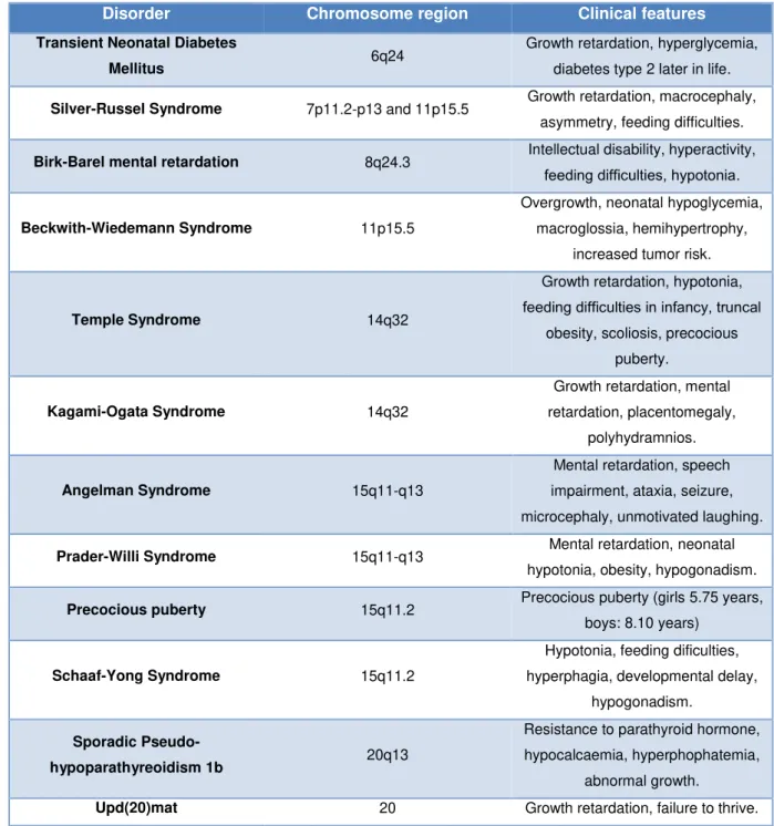
prenatal growth control, brain function and resource acquisition (Charalambous et al, 2007; Kalish etal, 2014). Many of these genes appear to be dosage sensitive and, therefore, functional consequences arise from changes in their expression levels (Bartolomei and Ferguson-Smith, 2011). Indeed, deletions or mutations in these genes lead to imprinted disorders. There are a group of currently 12 congenital imprinted diseases with similar underlying epi- and genetic etiologies and overlapping clinical features affecting mainly growth, development and metabolism (Soellner et al, 2017) (Table 1.1). For example, failure to express the paternal allele or maternal allele of genes within
the SNRPN imprinted domain results in Prader-Willi Syndrome (PWS) and Angelman Syndrome (AS),
respectively (Kalish et al, 2014).
Table 1.1- Summary of the 12 imprinted disorders with the affected chromosome region and main clinical features. Adapted from Bartolomei and Ferguson-Smith (2011) and Soellner et al (2017).
Disorder Chromosome region Clinical features
Transient Neonatal Diabetes
Mellitus 6q24
Growth retardation, hyperglycemia, diabetes type 2 later in life.
Silver-Russel Syndrome 7p11.2-p13 and 11p15.5 Growth retardation, macrocephaly, asymmetry, feeding difficulties.
Birk-Barel mental retardation 8q24.3 Intellectual disability, hyperactivity,
feeding difficulties, hypotonia.
Beckwith-Wiedemann Syndrome 11p15.5
Overgrowth, neonatal hypoglycemia, macroglossia, hemihypertrophy,
increased tumor risk.
Temple Syndrome 14q32
Growth retardation, hypotonia, feeding difficulties in infancy, truncal
obesity, scoliosis, precocious puberty.
Kagami-Ogata Syndrome 14q32
Growth retardation, mental retardation, placentomegaly,
polyhydramnios.
Angelman Syndrome 15q11-q13
Mental retardation, speech impairment, ataxia, seizure, microcephaly, unmotivated laughing.
Prader-Willi Syndrome 15q11-q13 Mental retardation, neonatal
hypotonia, obesity, hypogonadism.
Precocious puberty 15q11.2 Precocious puberty (girls 5.75 years,
boys: 8.10 years)
Schaaf-Yong Syndrome 15q11.2
Hypotonia, feeding dificulties, hyperphagia, developmental delay,
hypogonadism.
Sporadic
Pseudo-hypoparathyreoidism 1b 20q13
Resistance to parathyroid hormone, hypocalcaemia, hyperphophatemia,
abnormal growth.
4
1.2. Angelman Syndrome
Angelman Syndrome is a neurodevelopmental disorder characterized by four cardinal features: severe developmental delay, profound speech impairment, movement and balance disorder and easily excitable personality with an inappropriately happy affect (Lossie et al, 2001). AS is caused by disruption of the maternally expressed imprinted UBE3A gene in the 15q11-q13 imprinted locus in neurons (Margolis et al, 2015).
1.2.1. Symptoms
Angelman Syndrome was first described in 1965 by the English paediatrician Harry Angelman. He described three patients who presented a stiff, jerky gait, absence of speech, excessive laughter
and seizures, referring to them as “puppet children” (Angelman, 1965; Margolis et al, 2015). This disease presents a prevalence ranging from 1 in 12000 to 1 in 20000 (Buiting et al, 2016) and it is characterized by developmental delay, intellectual disability, absent speech, seizures, ataxic gait, easily excitable happy demeanor iniciated by social interaction and characteristic facies (reviewed in Kalsner and Chamberlain, 2016). Usually, infants with AS do not show any signs of the disease at birth, however delayed acquirement of motor skills, language and social skills are evident within the first year of life (Bird, 2014). The clinical problems associated with AS that develop in childhood persist into adulthood, hence adults with this condition are not capable of independent living, although many can perform tasks with supervision (Kalsner and Chamberlain, 2016; Buiting et al, 2016). The average life expectancy of AS patients is reasonably long excepting some early deaths due to severe seizures or accidental events (Buiting et al, 2016).
1.2.2.
UBE3A
and the 15q11-q13 imprinted cluster
E3A ubiquitin ligase gene (UBE3A) encodes E3A protein, a member of the large family of E3 ubiquitin ligase proteins (LaSalle et al, 2015), ubiquitously expressed in human tissues (Condon et al, 2013). UBE3A is involved in the process of marking proteins for degradation, by transferring the ubiquitin from E2 ubiquitin conjugation enzymes to the substrate protein (Chamberlain, 2013). In neurons, UBE3A protein localizes in pre- and post-synaptic neuronal compartments and in both cytoplasmic and nuclear locations (Dindot et al, 2008). UBE3A is a gene of interest due to its implication in both Angelman Syndrome and Chromosome 15q11.2–q13.3 Duplication Syndrome and due to its regulation through imprinting and non-coding RNAs.
Genomic imprinting in 15q11-q13 locus is controlled by a bipartite ICR composed by two elements: (1) the Prader-Willi syndrome imprinting center (PWS-IC) that includes the major promoter and exon 1 of the SNURF-SNRPN gene. Within the PWS-IC lies a differentially-methylated region that is methylated on the maternally-inherited allele and unmethylated on the paternally-inherited allele; (2) the Angelman syndrome imprinting center (AS-IC), that is thought to establish the maternal imprint of the PWS-IC in the maternal germline by driving expression from the upstream exons of
SNURF-SNRPN, (Chamberlain, 2013) (Fig. 1.2). The SNURF-SNRPN gene is expressed from the paternal
5
1999). SNURF is a nuclear localized protein of unknown function that is encoded by the three fist exons of SNURF-SNRPN (Runte et al, 2001). SNRPN is a small nuclear ribonucleoprotein that functions in pre-mRNA processing and thought to be involved in alternative splicing (Chamberlain, 2013). Downstream of the SNURF-SNRPN locus, and spanning a region of around 600kb of DNA, there is more than 148 exons which encode for non-coding transcripts (Runte et al, 2001), as, for example, the IPW long non-coding RNA (lncRNA) and the small nucleolar RNAs (snoRNAs) such asSNORD116 and SNORD115 clusters (Sato, 2017).
In somatic cells the paternally expressed SNURF-SNRPN drives the expression of polycistronic transcript that is terminated at the IPW region (Chamberlain, 2013) leading to the expression of both
SNURF-SNRPN and SNORD116 snoRNAs cluster. However, in neurons, the transcription of this
polycistronic transcript continues further, leading also to the expression of the SNORD115 snoRNAs cluster and of a non-coding anti-sense transcript which partially overlaps with the UBE3A gene, known
as UBE3A antisense transcript (UBE3A-ATS) (Meng et al, 2013). It is believed that UBE3A-ATS,
which is expressed from the paternal allele (Rougeulle et al, 1998), is required for the silencing of the paternal UBE3A (Meng et al, 2013), resulting in exclusively maternal expression of UBE3A in these cells. Indeed, Meng et al showed that premature termination of murine Ube3a-Ats leads to unsilencing of paternal Ube3a in multiple brain regions (Meng et al, 2013). To explain this repression, Buiting et al proposed a transcriptional collision model where two opposing polymerases for Ube3a and Ube3a-Ats on the paternal chromosome collide into each other around intron 4 of Ube3a, which would provoke stalling and dissociation of both polymerases, thereby terminating the transcription of Ube3a and its antisense (Buiting et al, 2016). However, formal proof of this mechanism remains to be tested.
6
1.2.3. Causes
Four molecular events can be at the origin of the maternal UBE3A lack of function in Angelman Syndrome: large deletions (around 5-7 Mb), also known as microdeletions, within the maternal chromosomal region 15q13-q11 (70-80%); mutation in the maternally inherited copy of UBE3A (10-20%); imprinting defect causing lack of expression of the maternal copy of UBE3A (3-5%); paternal uniparental disomy (UPD) (3-5%) (Lossie et al, 2001; Margolis et al, 2015). The diverse etiologies correlate with gradual differences in the severity of the disorder: large deletions result in loss of several other genes in the same region, therefore, patients with deletion within the maternal chromosomal region 15q13-q11 typically present a more severe phenotype than, for example, patients carrying point mutations affecting the UBE3A gene alone (Mertz et al, 2014; Stanurova et al, 2016).
1.2.4.
Diagnosis
Angelman Syndrome can only be confirmed by molecular diagnosis, which starts with the determination of the DNA methylation status of the SNURF-SNRPN promoter region (Williams et al, 2006), since the majority of Angelman patients will be positive for this test. Indeed, absence of maternal methylation pattern secures the diagnosis of AS (Margolis et al, 2015). The diagnosis proceeds to unravelling the cause of the disease as depicted in Fig. 1.3. Fluorescent in situ hybridization searches for a deletion within 15q11-q13 region and microarray allows the determination of the deletion size (Margolis et al, 2015). If a deletion is not found, diagnosis proceeds to DNA marker analysis of parent’s chromosome 15q11-q13 to confirm or exclude UPD. In the presence of two paternal copies, UPD is confirmed (Kalsner and Chamberlain, 2016) If both deletion and UPD are excluded, AS is probably due to an imprinting defect, caused by epigenetic phenomena or imprinting center point mutations or deletions (Margolis et al, 2015). For those patients with AS-related symptoms but a negative methylation test, UBE3A sequencing is performed and should detect a mutation within this gene. If not, AS is unlikely to be the diagnosis.
7
1.2.5. Treatment
Currently there is no cure for Angelman Syndrome and the treatment for this disease is exclusively symptomatic. The management of AS requires a many-sided approach and is based on
therapies that can improve the quality of patient’s life (Sachdeva et al, 2016) targeting epilepsy, sleep disturbance, muscle tone and gait, diet, speech and others (reviewed Kalsner and Chamberlain, 2016). Usually, seizures are treated with anticonvulsants, sleep problems are approached with a combination of pharmacology and therapies to mitigate gross and fine motor delay are used. At the level of communication, alternative strategies are tried such as the use of devices, picture exchange cards and modified sign language (Margolis et al, 2015).
1.2.5.1. Therapeutic approaches under investigation
AS is incurable but some therapeutic approaches are currently under investigation. Since AS is caused by UBE3A deficiency, recent research towards therapeutic approaches have focused on methods enabling the restoration of UBE3A expression in the mouse model, either by direct gene therapy or by un-silencing the paternal allele (Bi et al, 2016).
Injection of recombinant adeno-associated virus carrying the mouse Ube3a into the hippocampus of AS mice was attempted by Daily et al in 2011. This approach restored local Ube3a expression and improved hippocampus-dependent learning and memory. However, Ube3a expression in the cerebellum was not increased and there was no effect on motor dysfunction (Daily et al, 2011). One concern of this approach is the control of UBE3A expression, since high levels of UBE3A constitute a risk factor for autism spectrum disorder (Bi et al, 2016). Since this experiment in 2011 there has been no follow-up or any advancement using this approach.
In 2012, Huang and his co-workers showed that topoisomerase inhibitors can unsilence the dormant allele of murine Ube3a in neurons (Huang et al, 2012). They developed a high-content screen using primary mouse cortical neurons from Ube3a-Yellow Fluorescent Protein knockin mice (Huang et
al, 2012). They identified twelve topoisomerase I inhibitors and four topoisomerase II inhibitors that unsilenced the paternal Ube3a allele. Topotecan, a topoisomerase I inhibitor, was found to be the most effective, even at nanomolar concentration (Huang et al, 2012). It was latter shown that topotecan acts by stabilizing the formation of RNA:DNA hybrids at repeat elements within paternal
Snord116, which leads to an increase in chromatin decondensation and inhibition of Ube3a-Ats
expression (Powell et al, 2013). The inhibition of transcriptional progression of Ube3a-Ats leads to un-silencing of the paternal copy of Ube3a in AS model mice (Huang et al, 2012). Since topotecan is an FDA-approved anti-cancer drug, these results encouraged the study of this drug as a therapeutic approach for AS (Bi et al, 2016). However, in 2016, a study exploring the specificity of topotecan showed that the expression of many more genes was altered in the topotecan-treated wild-type neurons than in those neurons with topoisomerase I deletion (Mabb et al, 2016). These results raised the concern of topotecan unintended off-target effects (Tan and Bird, 2016).
8
RNaseH cleaves the RNA strand of the ASO–RNA heteroduplex which results in subsequent target RNA degradation by exonucleases (Wu et al, 2004). Meng et al, in 2015, administered phosphorothioate-modified chimaeric 29-O-methoxyethyl DNA ASOs, complementary to a 113 kilobase pair region of mouse Ube3a-Ats downstream of the Snord115 cluster of snoRNAs, in AS mice via intracerebroventricular injection. ASO treatment achieved not only specific reduction ofUbe3a-Ats but also sustained unsilencing of paternal Ube3a in neurons (Meng et al, 2015). Moreover,
Snrpn, Snord116 and Snord115 expression was not affected by the ASO treatment, neither by
increasing dose or time of the treatment (Meng et al, 2015). Restoration of Ube3a protein was only partial but it was sufficient to ameliorate some cognitive deficits such as memory impairment, although motor deficits did not seem to be rescued at any level (Bi et al, 2016). Meng et al postulated that complete phenotypic reversal might require treatment before a critical developmental window, a longer recovery time or a higher UBE3A induction level. Actually, a study investigating the effects of reinstating Ube3a expression during distinct neurodevelopmental windows of mice showed that AS-relevant phenotypes are only fully rescued during a very early time window, in the embryonic stage (Silva-Santos et al, 2015). The reinstatement of Ube3a in juvenile mice rescued the motor coordination deficits, which was not seen at later stages (Silva-Santos et al, 2015).
In any case, the use of modified ASOs (mASOs) against UBE3A-ATS is a promising therapeutic approach for AS. However, whether downregulation of the UBE3A-ATS is achievable using mASOs in humans and in which developmental time window ameliorates AS symptoms remains to be investigated.
1.3. Cellular models of human neuronal diseases
The study of human neurological disorders and the basic mechanisms behind those diseases have been limited for a long time by the lack of human brain cells for experimental purposes (Mertens
et al, 2016). Many studies on certain neuronal dysfunctions have been restricted to analysis of
post-mortem tissues of patients. In addition of being poorly preserved, these tissues usually represent the
end-stage of the disease (Nikoletopoulou and Tavernarakis, 2012). Although animal models, mainly mouse models, have contributed greatly to the better understanding of disease mechanisms, they do not fully recapitulate the human phenotype of the disease (Onuki and Takahashi, 2015). Also, most human neurological diseases arise from multiple factors, which are very often not represented by the model organisms (Mertens et al, 2016).
Recently, technologies for deriving human neurons in vitro have upgraded our ability to study cellular and molecular aspects of human neurons (Vadodaria et al, 2016). These promising technologies allow the generation of patient-specific cell lines which may serve as tools for understanding disease pathogenesis, for drug screens and, potentially, for cell replacement therapies (Pfisterer et al, 2011a).
9
1.3.1. Pluripotent stem cells
Human pluripotent stem cells (hPSCs) are normal primary cell lines with intrinsic capability for indefinite self-renewal and with the competence to, potentially, adopt any cellular fate through differentiation (Avior et al, 2016). hPSCs comprise human embryonic stem cells (hESCs) and induced pluripotent stem cells (iPSCs) (Mertens et al, 2016).
hESCs are originated from the late human blastocyst and have the unique potential to endlessly divide while maintaining an undifferentiated state and the capacity to differentiate into all germ layers as well as extra-embryonic tissues or placental cells (Menon et al, 2016). Because of these features, hESCs have emerged as an attractive model system to understand embryonic development and a promising source for cell-based therapies, drug studies and disease modelling (Murry and Keller, 2008). However, advances in embryonic stem cell technologies are limited by the controversial source of hESCs (Menon et al, 2016). Hence, the isolation of hESCs from human embryos raised serious ethical concerns, prompting efforts to find alternative sources of pluripotent cells (Sommer and
Mostoslavsky, 2012). Furthermore, hESCs are not patient-specific and therefore not amenable for cell-replacing therapies due to possible immune rejection.
1.3.2. Induced pluripotent stem cells
10
Fig. 1.4 – Representative scheme of iPSCs reprogramming and neuronal differentiation.1.3.2.1.
Reprogramming techniques
The original method for generating iPSCs used retroviral transduction to obtain expression of the four factors (Immamura and Inoue, 2012). Retroviral transduction has already been successfully used for reprogramming several cell types such as mouse and human fibroblasts, neural stem cells, keratinocytes, adipose cells, liver cells and blood cells (reviewed in Menon et al, 2016). In order to enhance the reprogramming efficiency, Sommer and colleagues used instead a lentiviral vector which led to a ten-fold increase of the reprogramming efficiency (Sommer et al, 2009). Nevertheless the use of integrating retroviruses or lentiviruses to deliver the reprogramming factors constitutes a drawback of iPSCs reprogramming since resulting iPSCs clones can display proviral integrations that increase the risk of insertional mutagenesis (Sommer and Mostoslavsky, 2012). Additionally, C-MYC is a known proto-oncogene that, with prolonged infection with retroviruses, may be aberrantly expressed and may induce oncogenic transformation (Immamura and Inoue, 2012; Sommer and Mostoslavsky, 2012). In any case, the use of lentivirus for iPSCs reprogramming remains the most used method in iPSCs research.
There are alternative reprogramming techniques available that circumvent the potential risks of viral approaches such as replication-defective adenoviral vectors, self-replicating episomal vectors and non-viral minicircle DNA vectors (reviewed in Menon et al, 2016). Despite being non-integrating approaches, these techniques yields very low reprogramming efficiencies.
1.3.3. iPSCs in disease modelling
11
using iPSCs occurred in 2008, when Park et al generated iPSCs from patients with a variety of genetic diseases with either Mendelian or complex inheritance (Park et al, 2008).One drawback of iPSCs generation is that it is a very laborious and time-consuming process (Ohnuki and Takahashi, 2015.) In fact, differentiation of fibroblasts into neural cells via iPSCs reprogramming usually takes 4-6 months before functional neurons are generated (Mertens et al, 2016). Another core aspect of the reprogramming protocols is the importance of monitoring of the iPSCs epigenetic state. In fact, Nazor et al identified iPSC-specific epigenetic and transcriptional aberrations in genes linked to X chromosome inactivation and genomic imprinting, which were not corrected during differentiation (Nazor et al, 2012). Hiura et al examined the status of imprinted genes in five iPSCs lines and found abnormalities such as loss of imprinting, although at low levels (Hiura et
al, 2013). Nevertheless, these results demonstrate that the analysis of the epigenetic status during reprogramming and differentiation is a critical safety step for iPSCs-based epigenetic disease models (Nazor et al, 2012 and Hiura et al, 2013).
1.3.3.1.
iPSCs in neuronal disease modelling
The modelling of neuronal diseases can be done through the differentiation of iPSCs into specific
neuronal cell types, with the first step being their differentiation into neuronal progenitor cells (NPCs) (Immamura and Inoue, 2012). NPCs, unlike iPSCs, are proliferative cells with limited capacity for self-renewal, giving origin to neuronal and glial progeny (Seaberg and van der Kooy, 2003). Neuronal differentiation of iPSCs has been efficiently achieved by using the knowledge gained from studying neurulation and the patterning of the early nervous system, namely, using neuronal inductive cues (Nikoletopoulou and Tavernarakis, 2012). The goal was to artificially recapitulate the signalling environment that the region-specific progenitors normally experience, which induces the expression of a combinatorial set of transcription factors characteristic of the desired neuronal cell type (Tamburini and Li, 2017). More specifically, the inhibition of activin, Nodal, TGF-β and bone morphogenetic protein signalling through SMAD signalling inhibitors such as Noggin, dorsomorphin and SB431542 has allowed the efficient neural induction of iPSCs (Immamura and Inoue, 2012).
The first reports of neural disease modelling occurred in 2008. Dimos et al generated iPSCs from an 80 years-old woman with amyotrophic lateral sclerosis and differentiated them into motor neurons (Dimos et al, 2008). In another study, Lee et al modelled Familial dysautonomia, an autosomal recessive congenital neuropathy, from reprogramming of fibroblasts from juvenile patients (Lee et al, 2009). More recently, studies have been developed with the aim of enhancing the efficiency of previous neural differentiation protocols and of directing the differentiation into defined types of neurons equivalent to in vivo cell populations (Nikoletopoulou and Tavernarakis, 2012).
12
1.3.4. Direct conversion into induced neurons
Direct conversion is a process that converts somatic cells into cells of different lineages, bypassing an intermediate pluripotent stage (Gopalakrishnan et al, 2017). This approach utilizes the overexpression of cell type-specific transcription factors to activate lineage changes and direct cellular identity towards the desired cell type (Mertens et al, 2016). Neurons can also be generated by direct conversion, being fibroblasts the most common source cells for neural direct conversion. The earliest report of direct conversion took place in 2010 when Vierbuchen et al identified, from a pool of nineteen, three neural-lineage specific transcription factors –ASCL1, BRN2 and MYT1L (BAM factors) - able to convert embryonic and postnatal mouse fibroblasts into functional neurons in vitro (Vierbuchen et al, 2010). The resulting induced neurons (iNs) expressed neural-specific proteins, generated action potentials and formed functional synapses (Vierbuchen et al, 2010). This study provided the first proof that accessible cells like dermal fibroblasts can be converted to functional neurons (Kim et al, 2012). Only one year later, several laboratories reported the generation of iNs from human fibroblasts (reviwed in Mertens et al, 2016). The iNs generated by neural direct conversion can potentially be used for multiple applications such as disease modelling and drug screening (Fig. 1.5).
Lineage reprogramming technique represents a time-saving process, when compared to other reprogramming approaches, since iNs are obtained within two to three weeks upon transcription factor overexpression, which constitutes the major advantage of this approach (Mertens et al, 2016). The resulting iNs have the ability to give rise to multiple neuronal subtypes, allowing to generate neuronal cells that are affected in many different neuronal diseases (Kim et al, 2011). One common aspect observed among studies generating different subtypes of neurons is that the conversion occurs within a short period upon factor introduction and neuronal identity is rapidly acquired, however subsequent functional maturation takes several weeks (Kim et al, 2012). Lineage reprogramming of somatic cells can be successfully performed on parental cells with different ages, although iNs derived from embryonic or neonatal human cells seem to functionally and physiologically mature much faster than adult cell derived iNs (Kim et al, 2011). Direct conversion benefits from common advantages with iPSCs reprogramming such as development of disease-specific lines that recapitulate the pathologic human condition in vitro and absence of immunological response due to host derived donor cells (Kim
et al, 2011). A very important aspect of converted neurons, regarding regenerative medicine, is that
they are directly reprogrammed into the target cells, which means that in vivo teratoma formation should not be a problem, contrary to iPSCs-derived cells (Kwon et al, 2016).
13
precursor stages and gives origin to neurons that have never been in an NPC-like stage. This can constitute a drawback in the case of modelling diseases whose phenotype is thought to develop from precursor cells (Mertens et al, 2016). Therefore, a useful approach that may resolve the two drawbacks referred above is direct conversion to induced neural stem/progenitor cells (iNSCs/iNPCs) since these recapitulate some stages of neurodevelopment as well as they are expandable cell lines (Kim et al, 2012). This has already been successfully attempted by several groups, who were able to directly convert adult human fibroblasts into expandable iNPCs (Mitchell et al, 2014; Meyer et al, 2015; Capetian et al, 2016). However, using this approach, the final goal, which is to obtain neurons, gets delayed since it adds one more cell-stage prior to neurons, instead of directly convert the fibroblasts into mature neuronal cells.Fig. 1.5 – Representative scheme of neuronal direct conversion.
1.3.4.1.
Direct conversion techniques
Most neural direct conversion protocols start with fibroblasts as the donor cell, given the fact that these cells are easily obtained and can stay proliferative in vitro for a reasonable number of passages (Pang et al, 2011; Pfisterer et al, 2011a; Ladewig et al, 2012). Many different conversion protocols were successful at generating iNs, raising the number of available protocols for this technique. For example, in 2011, Pfisterer et al successfully converted human postnatal fibroblasts using the three BAM factors previously used by Vierbuchen et al in the mouse fibroblasts (Pfisterer et al, 2011a). On the other hand, Pang et al combined this strategy with NEUROD1 transcription factor and observed an improving in the efficiency of generating human TUJ-1 positive neuronal cells two to three fold when compared with the BAM factors technique (Pang et al, 2011). Besides ectopic expression of transcription factors to mediate lineage conversion, other approaches have been explored such as miRNAs or induction of cellular reprogramming using small molecules (Gopalakrishnan et al, 2017). miRNAs have been shown to play an important role in direct reprogramming since they function as repressors of target mRNAs and post-transcriptional regulation of gene expression (An et al, 2016). Ambasudhan et al showed, in 2011, that the combination of miRNA-124 with BRN2 and MYTL1 directly converts postnatal and adult human fibroblasts into functional neurons (Ambasudhan et al, 2011). At the same time, Yoo et al were able to convert human fibroblasts into neurons using miRNA-9* and miRNA-124 (Yoo et al, 2011). Moreover, the addition of the transcription factors NEUROD2,
ASCL1 and MYTL1 improved the conversion efficiency as well as the maturation of the obtained iNs
14
spatial and temporal control of its action, through control of the concentration administered (Li et al, 2013). Actually, Ladewig et al showed that the efficiency of ASCL1/NGN2-induced neuronal conversion was higher upon combination with three molecules that inhibit SMAD, GSK-3β and BMP receptor pathways (Ladewig et al, 2012). Liu et al were also able to successfully convert human fibroblasts into mature neurons using the transcription factors NGN2 and SOX11 along with the small molecules forskolin and dorsomorphin (Liu et al, 2013).1.3.4.2.
iNs in neuronal disease modelling
Neuronal cells derived from direct conversion provide a novel platform for diverse applications, including disease modelling (Pfisterer et al, 2011a). Characteristics such as speed of conversion, possibility to generate patient-specific cell lines, recapitulation of age-related and disease-related aspects of the patient-derived original cells make direct conversion a very suitable approach for disease modelling. In fact, to the date, several subtypes of neuronal cells have already been converted from human fibroblasts (reviewed in Mertens et al, 2016). One neuronal type of clinical importance is motor neurons, which are affected in patients with disorders such as spinal muscular atrophy and amyotrophic lateral sclerosis (Gopalakrishnan et al, 2017). In 2011, combining BAM factors with subtype-specific transcriptional cues, Son et al generated spinal motor neurons from human fibroblasts, which expressed functional voltage-gated channels and were able to fire action potentials. Another clinically important neuronal subtype is dopaminergic neurons which are affected
in patients with Parkinson’s disease (Gopalakrishnan et al, 2017). Regarding this neuronal subtype it was not only possible to derive human dopaminergic neurons using direct conversion, which may
allow to model Parkinson’s disease (Pfisterer et al, 2011b), but also to derive mouse dopaminergic neurons that were transplanted and able to provide symptomatic relief in a Parkinson’s disease mouse
model (Kim et al, 2011). These studies hold promises for human modelling disease as well as for cell replacement therapy.
At present, neuronal disease modelling is still mostly based on iPSCs system. In any case, it is expected that, within the next few years, the number of disease models using directly converted neurons will increase at a big rate.
1.4. Aims of the study
The aim of the project is to develop a robust human disease modelling system to study Angelman Syndrome. Such a system will serve as a drug testing platform to evaluate, for example, mASO-mediated downregulation of UBE3A-ATS to reactivate paternal UBE3A gene. For that we have two major objectives:
1. Development of a human model system of Angelman Syndrome, either through neural direct conversion or iPSCs neural differentiation;
15
2. Material and Methods
2.1. Cell culture
2.1.1.
Punch-skin biopsy fibroblasts
3 year-old Angelman fibroblasts (AS 3y) were obtained from a punch-skin biopsy to a 3 year-old AS patient. After biopsy, skin sample was washed with phosphate-buffered saline 1x (PBS; Sigma Aldrich-Aldrich, Catalog# P3813) with gentle agitation. In a P100 mm petri dish (TPP, Catalog# TPP93100) the subcutaneous tissue was removed by scraping the dermal side using two forceps. The sample was then sectioned into approximately 0.5cm width stripes using a surgical scalpel and it was moved into 6-well plates (TPP, Catalog# TPP92006). 30 year-old Angelman fibroblasts (AS 30y) were previously obtained and expanded the same way by Duarte Brandão (IMM/ MC Fonseca’s Lab). Age-matched control fibroblasts (control 30y) were previously derived and provided by Dra. Sofia Duarte (IMM/Centro Hospitalar de Lisboa Central).
Both control 30y and AS 30y fibroblasts were thawed in a 37ºC bath and transferred to a Falcon® tube containing 5mL of fibroblast medium constituted by Dulbecco’s Modified Eagle Medium (DMEM;
Life Technologies, Catalog# 41966-029) supplemented with 10% Fetal Bovine Serum (FBS; Life Technologies, Catalog# 10270-106), 1mM L-glutamine (Life Technologies, Catalog# 25030-024) and 1% Penincillin/Streptomycin (Pen/Strep; Life Technologies, Catalog# 15070-063). The cells were centrifuged at 1000rpm for 5 minutes and the supernatant was discarded. The pellet was ressuspended in 5mL of fibroblast medium and seeded on a T25 flask (Starsted, Catalog# 833910 ). When the fibroblasts were approximately 80-90% confluent, the cells were passaged using TrypLE™
Express solution (Life Technologies, Catalog# 12605028). For that, the medium was removed and the cells were washed with 5 mL of PBS and then dislodged through incubation with TrypLE™ Express
solution at 37ºC for 3-5 minutes. Cells were then ressuspended with fibroblast medium and seeded in one T75 flask (VWR, Catalog# NUNC156499). Cells continued to be passaged using a 1:2 split ratio to expand the lines. Part of these cells were collected for RNA extraction (see 1.2.4), pelleted or frozen (explained below).
In order to make cell pellets for DNA or RNA extraction, fibroblasts were dislodged using a
TrypLE™ Express solution as explained above, transferred to a Falcon® tube and centrifuged at
1000rpm for 5 min. The supernatant was discarded and the pellet was ressuspended in 1mL of PBS and transferred to an Eppendorf tube. After centrifugation at 1000rpm for 5min, the supernatant was discarded. The pellet was snap frozen in liquid nitrogen for a few seconds and stored at -80ºC.
To cryopreserve the cells, after dislodging and centrifugation, the supernatant was discarded and the pellet was ressuspended in freezing medium: 10% dimethyl sulfoxide (DMSO; Sigma Aldrich-Aldrich, Catalog# D2438) in FBS. 1mL of ressuspended cells was transferred to each cryovial (Nunc, Catalog# 366656) and placed at -80ºC. For long storage the cells were placed in liquid nitrogen.
16
2.1.2. NPCs differentiation from iPSCs
2.1.2.1. iPSCs expansion
iPSC reprogramming of control and AS fibroblasts was performed by Isabel Onofre and Dr. Ana Rita Álvaro in the laboratory of Professor Luís Pereira de Almeida at CNC/UC, using a previously published protocol (Warlich et al, 2007). iPSCs expansion and adaptation to feeder-free conditions was performed by Duarte Brandão (IMM/ MC Fonseca’s Lab).
For iPSCs expansion in feeder-free conditions, cells were maintained in 6-well plates previously
coated with Matrigel (Corning, Catalog# 354230) in mTeSR™1 medium (STEMCELL Technologies,
Catalog# 5850) supplemented with 0.5% Pen/Strep. For Matrigel coating, Matrigel was diluted in cold DMEM/F12 (1:30), carefully ressuspended and 1mL of Matrigel-DMEM/F12 was placed in each well of a 6-well plate. The plate was incubated for 2 hours at room temperature or for 30 min at 37ºC before use. Prior to seeding of the cells, Matrigel was removed. Upon high confluency, iPSCs were passaged. For that, cells were washed with 1.5 mL of PBS and incubated 3 minutes with 1mL of 0.5mM EDTA (VWR, Catalog# 0105-1KG) in PBS. After incubation, EDTA was removed, 1.5 mL of
mTeSR™1 medium was added to each well and cells were scrapped from the well with a cell scraper.
The scrapped cells were collected and transferred to a Falcon® tube containing the volume of
mTeSR™1 medium necessary for the desired dilution (usually 1:3). Cells were then seeded in Matrigel-coated wells.
For iPSCs freezing, cells were dislodged using the approach described above and after scrapping cells were transferred to a Falcon® tube containing 1.5 mL of Washing medium [DMEM-F12 (Life Technologies, Catalog# 11039-021) supplemented with 10% of KnockOut Serum Replacement (Life Technologies, Catalog# 10828-028), 1% of non-essential aminoacids (Life Technologies, Catalog# 11140-035), 1mM of L-Glutamine, 0.1mM of β-Mercaptoethanol (Life Technologies, Catalog# 31350-010) and 1% of Pen/Strep]. Cells were centrifuged at 1000 rpm for 3 min, the
supernatant was removed and the pellet ressuspended in 250 μL of Freezing medium [10% dimethyl
sulfoxide (DMSO; Sigma-Aldrich, Catalog# D2438) in FBS]. This volume was transferred to a cryovial, which was stored at -80ºC. For long storage cells were preserved in liquid nitrogen.
2.1.2.2. Neural Progenitor cells generation and expansion
Neural Progenitor cells (NPCs) were derived from iPSCs following the protocol described in the STEMCELL Technologies Technical Manual – Generation and Culture of Neural Progenitor Cells using the STEMdiffTM Neural System, with adaptations. The Monolayer Culture Protocol was used.
Briefly, iPSCs cultured in mTeSR™1 medium in a P100 mm dish were washed once with PBS and dislodged with EDTA at 37ºC for 10 min. After incubation, 7 mL of DMEM-F12 were added and cells were dislodged by pipetting up and down. For cell counting, 50μL of the cell suspension was mixed with 50μL of Trypan Blue solution (Sigma-Aldrich, Catalog# T-6146) and 10μL were added to a
Neubauer chamber. 2x10⁶ cells were seeded in a well of a 6-well plate, previously coated with Matrigel, in 2mL of STEMdiffTM Neural Induction Medium (STEMCELL Technologies, Catalog# 05835)
17
was changed daily without ROCKi. Cell passaging was performed upon 80-90% confluency (usually every three days). Dislodging of cells was done through incubation with 1mL of ACCUTASETM (STEMCELL Technologies, Catalog# 07920) at 37ºC for 10 min. 5mL of DMEM-F12 were added to the cells and they were centrifuged at 300g for 5 min. After discarding the supernatant, pellet was ressuspended in STEMdiffTM Neural Induction Medium with ROCKi. 2x10⁵ cells were seeded into another Matrigel-coated well of a 6-well plate. This process was repeated until passage 2. From passage 3 on, the same protocol was followed, with a split ratio of 1:2.For NPCs expansion, cells were switched to a Complete STEMdiffTM Neural Progenitor Medium [Basal Medium Catalog# 05834), Supplement A (#05836), Supplement B (#05837)
]
and passaged upon confluency into a 1:2 split ratio as before. This way NPCs were expanded and part of the generated NPCs was freezed. Usually, cells from a 6-well plate were dislodged and centrifuged. The pellet was ressuspended in 1 mL of NPCs freezing medium (STEMdiffTM Neural Progenitor Medium supplemented with 10% DMSO) and transferred to one cryovial, which was stored at -80ºC and later in liquid nitrogen.For thawing the cells, one cryovial was thawed into one well of a 6-well plate. For that, the cryovial was thawed in a 37ºC water bath and cells were transferred to a Falcon® tube containing 10 mL of DMEM/F-12 medium. After centrifugation at 300g for 5 min, the pellet was ressuspended in 2mL of Complete STEMdiffTM Neural Progenitor Medium and cells were plated in a previously Matrigel-coated well of a 6-well plate.
2.1.3. Neuronal direct conversion
2.1.3.1. HEK 293T expansion and transfection
HEK 293T cells were thawed and passaged using a 1:2 split ratio following the same procedure as above in HEK medium: DMEM supplemented with 10% FBS, 1mM L-glutamine, 1% Pen/Strep and 1% Non-essencial Aminoacids (NEAA; Thermo Fisher, Catalog# LTID 41966-029). For transfection with lentiviral vectors, cells were dislodged and counted. 2.5x10⁶ HEK 293T cells were seeded per P100 mm dishes previously coated with gelatine (Sigma Aldrich-Aldrich, Catalog# G1890). The day after, transfection was performed: 10μg of each transfer vector - EtO and N2AA - and 5μg of both each packaging vectors - psPAX2 and pMD2G - were mixed and added to 600μL of DMEM and 50μL
of X-treme Gene 9 DNA Transfection Reagent (Roche, Catalog# 6365787001). This mixture was vortexed and added to each P100 mm petri dish containing the HEK 293T cells and the transfection took place at 37ºC overnight. After overnight incubation the medium was removed and replaced with fresh medium. 48h later the medium of the transfected HEK 293T cells containing the lentiviruses was
collected and filtered using a 0.45μM filters (VWR, Catalog# 514-0075). HEK medium was replaced for a second collection of viral medium for 24 hours.
2.1.3.2. Fibroblasts transduction
18
medium and replacing it with the viral medium collected from the transfected HEK 293T cells. To enhance the infection efficiency, 1μL of polybrene (Santa Cruz Biotechnology, Catalog# SC134220) was added to each 1mL of medium. 24 hours later a second infection was performed with new viralmedium. Transduced fibroblasts were expanded in the presence of 200μg/ml G418 (neomycin; Merck. Catalog# 345810) and 1μg/ml puromycin (Sigma-Aldrich, Catalog# P8833) in the first experiment, but given the high efficiency of infection, selection with puromycin and neomycin was not repeated in further experiments.
2.1.3.3. Neural direct conversion
The neural direct conversion was performed according to the protocol by Ladewig et al (2012). In summary, the transduced fibroblasts were passaged until high confluency was reached and 24 hours later the medium was changed to Neuron Conversion (NC) medium: DMEM:F12 (Life Technologies, Catalog# LTI11039-021) and Neurobasal medium (Thermo Fisher, Catalog# LTI21103-049) supplemented with N2 supplement (Stem Cell Techonolgies, Catalog# 07152), B27 supplement (Thermo Fisher, Catalog# LTI 17504-044), doxycycline (Sigma Aldrich, Catalog# D9891-1G), laminin (reference), dibutyryl cyclic-AMP (Sigma Aldrich, Catalog# D0627-100MG), human recombinant Noggin (Peprotech, Catalog# 120-10C-100µG), LDN-193189 (Sigma Aldrich, Catalog#SML0559-5MG) A83-1 (Sigma Aldrich, Catalog#SML0788-Catalog#SML0559-5MG), CHIR99 021 (Sigma-Aldrich, Catalog# SML 1046-5MG), Forskolin (Sigma Aldrich, Catalog#93049-10MG) and SB-431542 (Sigma Aldrich, Catalog# S4317-5MG). This medium was maintained for three weeks and it was changed every third day. After three weeks, the medium was replaced with Neural Maturation (NM) medium: DMEM:F12/Neurobasal (1:1) supplemented with N2, B27, GDNF (Peprotech, Catalog#450-10-10µG), BDNF (Peprotech, Catalog# 450-02-10µG), dibutyryl cyclic-AMP, doxycycline and laminin for two weeks. Images of the cells undergoing neural conversion were taken at day 3, day 13 and day 27 using a Zeiss Primo Vert microscope.
2.2. Molecular Biology Techniques
2.2.1. Competent cells transformation
Competent cells previously prepared in the lab were transformed with either pLVX-EtO, pLVXTP-N2AA, psPAX2, pMD2.G [kindly provided by J. Mertens (Salk Institute, San Diego, USA)] or GFP plasmids [kindly provided by Edgar Gomes’s Lab (IMM, Lisboa, Portugal)]. For that, 2μL of the
respective plasmid DNA were added to 100μL of competent cells and the mixture was placed on ice for 30 min. For the heat shock, the mixture was placed at 42ºC for 45 seconds and immediately moved to ice for 2 minutes. 1mL of Luria-Bertani medium (LB) was added and the mixture was incubated at
37ºC for 1 hour, with agitation. After incubation, 100μL of each plasmid mixture was added and
scattered in 0,1% ampicillin LB-agar plates (Grisp, Catalog# GAB03.0005), which were left at 37ºC overnight.
19
The day after, 150 μL of glycerol (Sigma-Aldrich, Catalog# G6279-500ML) were added to 850 μL ofbacteria. The mixture was vortex and stored at -80ºC.
2.2.2. Plasmid DNA extraction
Starting from either 5mL or 400mL culture of bacteria collected from the glycerol stock, plasmid DNA was extracted using the NZY Miniprep Kit (NZYTech, Catalog# MB01002) or the Genopure Plasmid Maxi Kit (Roche, Catalog# 3143422001), respectively, following the manufacturer’s protocols.
For plasmid restriction digestion to confirm the presence of the correct sequence in the packaging and transfer vectors, DNA was prepared using the NZY Miniprep Kit. On the other hand, for transfection of HEK 293T cells for lentivirus production, plasmid DNA was prepared using the Genopure Plasmid Maxi Kit since this protocol generates much higher yields and better quality of plasmid DNA than NZY Miniprep Kit.
2.2.3. Plasmid restriction digestion
To confirm that the packaging or transfer plasmids glycerol stocks had the correct sequence, each plasmid was digested with restriction enzymes. pLVX-EtO was digested with KpnI (Thermo Fisher, Catalog# FD0524). pLVXTP-N2AA was digested with EcoRI (Thermo Fisher, Catalog# FD0275) and KpnI. psPAX2 was digested with EcoRI and SalI (Thermo Fisher, Catalog# FD0644). pMD2.G was digested with HindIII (Thermo Fisher, Catalog# FD0505) and NotI (Thermo Fisher, Catalog# FD0595). All restriction digestions were performed for 1 hour at 37ºC.
The digestion products were separated on a 0.8% agarose (NZYTech, Catalog# MB05202) gel in 1x Tris-acetate-EDTA (TAE). Digital images were obtained using the Chemidoc XRS+ system (BioRad) and analysed using the Image Lab 5.2 software (BioRad).
2.2.4. RNA isolation from adherent cells and cDNA synthesis
For RNA extraction, cell’s medium was removed and 1mL of NZYol reagent (NZYTech, Catalog# MB18501) was added to the cells per 10cm2 of culture dish surface and incubated at room temperature for 5 minutes. The mixture was pipetted up and down, transferred to an Eppendorf tube and stored at -80ºC.
For the RNA isolation, 200μL of chloroform were added to the Eppendorf tube. The tube was vigorously shaken for 15 seconds and incubated for 3 min at room temperature, followed by
centrifugation at 1200g for 15 min at 4ºC. The aqueous phase was collected into a new tube and 1μL of Glycogen Blue and 500μL of 100% isopropanol were added. The mixture was incubated 10 min at room temperature and centrifuged at 1200g for 10 min at 4ºC. The supernatant was discarded, the pellet was washed with 1mL of 75% EtOH and centrifuged at 7500g for 5 min at 4ºC. The pellet was air-dried for 15 min and ressuspended in 30μL of RNase-free water.
DNase I treatment (Roche, Catalog# 4716728001) was performed on 5μg of RNA according to
20
sample was centrifuged at 13000 rpm for 30 min at 4ºC. The pellet was washed with cold 70% EtOH and centrifuged at 13000 rpm for 5 min at 4ºC. After air-dried, the pellet was ressuspended in RNase-free water. RNA concentration was quantified using Nanodrop 2000 (Thermo Scientific).cDNA synthesis from 500ng of RNA was performed using Transcriptor High Fidelity cDNA synthesis Kit (Roche, Catalog# 5081963001) according to the manufacturer’s protocol.
2.2.5.
Reverse transcriptase polymerase chain reaction (RT-PCR)
1/15 diluted cDNA product was used as template for RT-PCR in a 25μL reaction volume with
BIOTAQTM DNA polymerase (Bioline, Catalog# BIO-21060) according to the manufacturer’s instructions. The primer pairs used are described in Table 2.1. The cycling conditions were: 95ºC for 5 min, then 35 cycles of 95ºC for 30 sec, 60ºC for 30 sec, 72ºC for 20 sec and, finally, 72ºC for 10 min.
RT-PCR products were separated on a 1.5% agarose gel in TAE. The molecular weight marker 1Kb Plus DNA ladder (Invitrogen, Catalog# 10787018) was used. Digital images were obtained and analysed as in section 2.2.3.
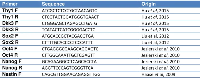
Table 2.1 – Primers used for RT-PCR.
Primer
Sequence
Origin
Ascl1 trans F
AGCAGGAGCTTCTCGACTTCACCA
Ladewig
et al
, 2012
Ascl1 trans R
AAGCGCATGCTCCAGACTGCC
Ladewig
et al
, 2012
2.2.6. Reverse-transcriptase quantitative polymerase chain reaction (RT-qPCR)
1/15 diluted cDNA product was used as template for RT-qPCR in a 25 μL reaction volume with -iTaqTM Universal SYBR® Green Supermix (BioRad, Catalog# 1725125) according to the
manufacturer’s instructions. The primer pairs used are described in Table 2.2. The cycling conditions were: 50ºC for 2 min, 95ºC for 10 min, 95ºC for 15 sec, 60ºC for 1 min, 95ºC for 15 sec,60ºC for 1 min and 95ºC for 15 sec. RT-qPCR was conducted in Real-Time thermal cycler ViiA7 96-well format or 384-well format (Applied Biosystems). Data was analysed in QuantStudioTM Real-Time PCR Software (Applied Biosystems).
Table 2.2 – Primers used for RT-qPCR.