Melanoma A375 Cell Migration
Chunmei Zhang2, Chao Yang1, Ruifei Wang1, Yang Jiao1, Khamal Kwesi Ampah1, Xiaoguang Wang3*, Xianlu Zeng1*
1Institute of Genetics and Cytology, Northeast Normal University, Changchun, Jilin Province, China,2Department of Cell Biology, Norman Bethune College of Medicine, Jilin University, Changchun, Jilin Province, China,3Department of Bioscience, Changchun Teachers College, Changchun, Jilin Province, China
Abstract
Integrins are heterodimeric transmembrane receptors that physically link the extracellular matrix (ECM) to the intracellular actin cytoskeleton, and are also signaling molecules that transduce signals bi-directionally across the plasma membrane. Integrin regulation is essential for tumor cell migration in response to growth factors. c-Abl kinase is a nonreceptor tyrosine kinase and is critical for signaling transduction from various receptors. Here we show that c-Abl kinase is involved in A375 cell migration mediated by avb3 integrin in response to PDGF stimulation. c-Abl kinase colocalizes with avb3 integrin dynamically and affects avb3 integrin affinity by regulating its cluster. The interaction between c-Abl kinase and avb3 integrin was dependent on the activity of c-Abl kinase induced by PDGF stimulation, but was not dependent on the binding ofavb3integrin with its ligands, suggesting that c-Abl kinase is not involved in the outside-in signaling ofavb3integrin. Talin head domain was required for the interaction between c-Abl kinase andavb3integrin, and the SH3 domain of c-Abl kinase was involved in its interaction with talin andavb3integrin. Taken together, we have uncovered a novel and critical role of c-Abl kinase inavb3integrin mediated melanoma cell migration.
Citation:Zhang C, Yang C, Wang R, Jiao Y, Ampah KK, et al. (2013) c-Abl Kinase Is a Regulator ofavb3Integrin Mediated Melanoma A375 Cell Migration. PLoS
ONE 8(6): e66108. doi:10.1371/journal.pone.0066108
Editor:Chih-Hsin Tang, China Medical University, Taiwan
ReceivedFebruary 9, 2013;AcceptedMay 2, 2013;PublishedJune 21, 2013
Copyright:ß2013 Zhang et al. This is an open-access article distributed under the terms of the Creative Commons Attribution License, which permits unrestricted use, distribution, and reproduction in any medium, provided the original author and source are credited.
Funding:This study was supported by grants from the National Nature Science Foundation of China (81071726, 81172014), the specialized Research Fund for the Doctoral Program of Higher Education (20100043110007), and the Fundamental Research Funds for the Central Universities (10SSXT129). The funders had a role in decision to publish.
Competing Interests:The authors have declared that no competing interests exist.
* E-mail: [email protected] (XZ); [email protected] (XW)
Introduction
The metastatic sequence of tumor cells is understood to involve detachment of cell within primary tumor, local migration and intravasating into the bloodstream, and extravasating into tissue, further local crawling, migration and invasion, generation of new colonies. Migration is a critical process for tumor cell to overcome this remarkable set of challenges [1–3]. Cell migration is a highly complex and regulated process, in which intracellular and extra cellular signals conjoin to produce a coordinated response. The direction of cell migration is controlled by growth factors and ECM gradients. Cells respond to local activation and amplification of signaling events on the side facing the attractant, which results in the orderly rearrangement of adhesive structures that connect the cell to the ECM [4,5]. There are several adhesion receptor families involved in the migration of cells, the best-studied adhesion receptors, and of particular interest in migration, are integrins. Integrins, the heterodimers consisting of a and b
subunits, contribute in multiple ways to the process of cell migration. First, integrin form connection between the intracellu-lar actin cytoskeleton and the ECM, which is critical for many cellular processes including efficient cell movement besides providing structural support for cells [6]. Second, integrins also mediate signal transduction. They mediate signal transduction through the cell membrane in both directions: binding of ligands to integrins transmits signals into the cell and results in cytoskeletal re-organization, gene expression and cellular differentiation
(outside-in signaling); on the other side, signals from within the cell (in response to local stimuli) can also propagate through integrins and regulate integrin ligand-binding affinity and cell adhesion (inside-out signaling) [7,8]. This bidirectional signaling is mainly mediated by the short cytoplasmic tails of the two integrin subunits [9]. Integrin avb3 is known to be responsible for cell
attachment and spreading, as well as cell locomotion. The expression of integrin avb3 has been detected in different types
of tumor cells, including breast, prostate, ovary, melanomas and gliomas, this expression has been reported to correlate with an aggressive phenotype and metastatic dissemination. Specifically, the increase of migration in tumor cell is due in part to integrin
avb3[10,11].
(nuclear localization signals) and DNA-binding sequences which are important for nuclear functions [14–16].
The mutant forms of c-Abl gene are well known to be involved in hematopoietic malignancies such as chronic myeloid leukemia (CML). To date, extensive evidence concerning the role of c-Abl kinase in the integrin signaling transduction has been accumulat-ed. Earlier reports indicated that integrin can regulate c-Abl kinase activity and cytoplasmic-nuclear transport in fibroblastic cells adhering to fibronectin [17], meanwhile, c-Abl kinase contributes to the activation of MAPK in cells plated to fibronectin [18]. Furthermore, c-Abl is a key intracellular molecule-mediating angiogenesis induced by bFGF which associates withb3integrin,
and c-Abl can mediate endothelial cells apoptosis when integrins
avb3and avb5were inhibited [19,20]. Adhesion-activated c-Abl
kinase phosphorylates cytoskeletal protein paxillin, which is a component of focal adhesion [21]. In addition, c-Abl protein levels, as assessed by immunohistochemistry, are increased in many solid tumors, but the increased expression is not consistently correlated with disease grade [22,23]. Some other findings demonstrated that the activated c-Abl kinase may play a role in some solid tumors, such as non-small cell lung cancer (NSCLC) and in aggressive human breast cancer [24,25]. Only more recently, it was reported that c-Abl kinase promotes melanoma cell invasion and drives metastatic progression via inducing transcrip-tional upregulation and activation of MMPs [26]. However, little is known about the role of c-Abl kinase in integrin signaling of solid tumor. These data prompted us to examine whether c-Abl kinase can interact with integrin to play a role in solid tumor progression.
In the present study, we have explored the role of c-Abl kinase in regulatingavb3integrin signaling in the migration of melanoma
cells. We demonstrated that c-Abl kinase was involved in avb3
integrin mediated A375 cell migration induced by PDGF. Endogenous c-Abl kinase andavb3 integrin co-localized
dynam-ically in response to extracellular PDGF stimulation, and PDGF stimulation also enhancedavb3integrin binding to its ligands.
c-Abl kinase interacted withavb3integrin by using its SH3 domain,
and this interaction was dependent on c-Abl kinase activity but not related to F-actin or ligand-integrin binding. The head domain of talin protein played a critical role in this interaction of c-Abl kinase withavb3integrin, and the activated c-Abl kinase directly linked to
talin head to regulate integrin activity. Therefore, our work has defined a novel role for c-Abl kinase as a regulator of inside-out integrin signaling in melanoma cell and supports the development of therapeutic interventions geared towards inhibiting c-Abl kinase in the regulation of metastasis in melanoma patients.
Materials and Methods
Cell culture and plasmids
Human melanoma A375 cells and human prostate carcinoma DU-145 cells were purchased from the Cell Bank of Type Culture Collection of Chinese Academy of Science (Shanghai, China). All of the cells were cultured in DMEM (Invitrogen) supplemented with 10% heat-inactivated fetal bovine serum (FBS), 100U penicillin and 100mg/ml streptomycin at 37uC in the presence of 5% CO2. A375 and DU-145 cells were detached with
trypsin-EDTA (0.05% Trypsin).
The GST-CrkII-C-terminal domain (CTD) plasmid was pro-vided by Dr. Giorgio Scita (European Institute of Oncology, Milan, Italy). The GST-talin2-head domain was kindly given by Dr. Pietro De Camilli (Yale University, School of Medicine, New Haven, CT). The pCMV-human-c-Abl was provided by Dr. Thomas Rudel (Max Planck Institute for Infection Biology, Berlin
Germany) and the sequences encoding the domains of c-Abl were amplified by PCR using the expression vector as templates.EcoRI and XhoI restriction sites were introduced by the PCR primers, and the fragments were subcloned into pGEX vector. pET28-c-Abl-SH3 was constructed as described above. The mutants of tyrosine sites in the SH3 domain of c-Abl kinase were introduced using the specific PCR primers.
Antibodies and reagents
LM609 (anti-avb3blocking mAb) and TDM29 (anti-b1blocking
Ab) were purchased from Millipore; TA205 (anti-talin mAb) was from Serotec; K12 (anti-c-Abl polyclonal Ab, rabbit IgG, sc-131) was purchased from Santa Cruz Biotechnology; Human PDGF-BB was purchased from R&D systems; ECL Plus western blotting detection reagents (RPN2132) and glutathione Sepharose 4B (17-0756-01) were purchased from Amersham Biosciences. G7781 (anti-glutathione S-transferase antibody, rabbit IgG), Cytochalasin D (CD) and PY20 (p-Tyr Antibody) as well as other chemicals were from Sigma-Aldrich. STI571 (Gleevec, Imatinib. Inhibitor of c-Abl kinase) was from Novartis (Basel, Switzerland); Rhodamine-conjugated phalloidin was purchased from Molecular Probes, and Calcein-Am was purchased from invitrogen.
In vitro protein binding assay
E. coli BL21 transformed with pET28-talin2-head was grown and induced with isopropylb-D-thiogalactoside. His-talin2-head was purified with Ni2+Sepharose (Qiagen) according to the manufacturer’s instructions. GST or GST fusion protein was eluted from glutathione-Sepharose 4B beads. Approximately 1mg of His-talin2-head immobilized to 10ml of Ni2+
Sepharose was incubated with 0.5mg of GST or GST fusion protein in 500ml of modified GBT buffer (10% glycerol, 50 mM HEPES-NaOH (pH 8.0), 170 mM KCl, 7.5 mM MgCl2, 0.1 mM EDTA, 1 mM
DTT, 1% Triton X-100) containing 1% BSA. After 2 h, the resins were extensively washed with GBT buffer, and the bound proteins were separated by 12% SDS-PAGE and immunoblotted with anti-GST antibody.
Far-western blot analysis
SDS-PAGE and western blotting were performed as described previously with little modifications. Briefly, immunoprecipitates from A375 cells were separated by 10% SDS-PAGE under reducing conditions and then transferred onto polyvinylidene difluoride (PVDF) membranes (Millipore, Bedford, MA, USA). After nonspecific binding was blocked with PBS containing 5% nonfat milk, the blots were incubated with blocking buffer containing 10mg/ml GST or the indicated GST-fusion proteins at 4uC overnight. Bound proteins were detected by ECL detection. [27].
Flow cytometric assay
A375 cells were washed twice and resuspended in PBS. To identify the molecules on cell surface, A375 cells were incubated with isotype IgG and monoclonal antibody (LM609 or TDM29) respectively. After incubation with isotype IgG, LM609 or TDM29, the cells were stained with FITC-conjugated goat anti-mouse IgG (2mg/ml) for 30 min at 22uC. The labeled cells were detected by a FACScan (Beckman-Counter, USA).
RNA interference
according to the manufacturers’ protocols. The extent of suppression and specificity for c-Abl were evaluated by western blotting with anti-c-Abl antibody, anti-actin antibody was used as control.
In vitro kinase assay
A375 cells were serum starved overnight, and then stimulated and lysed. The lysates were incubated with anti-c-Abl antibody (K12). After 2 h, 20ml of protein A-Sepharose beads (50% slurry) was added to the antibody/lysates mixture. After 1 h, the immunoprecipitates were washed at least three times with lysis buffer, and then washed three times with the kinase buffer (25 mM Tris, pH 7.5, 2 mM DTT, 5 mM b-glycerophosphate, 1 mM Na3VO4, 10 mM MgCl2). After 5 min preincubation at 30uC,
30ml of reactions were initiated by adding 3mg of GST-CrKII-CTD and 5mM ATP. After 30 min, reactions were terminated by adding 20ml of 36SDS sample buffer and resolved by
SDS-PAGE.
Protein expression and GST pull-down assay
Production of GST and GST-fusion proteins was induced in E.coli strain BL-21 transformed with corresponding plasmids by adding 0.3 mM isopropyl b-D thiogalactoside at 37uC for 3 h. Fusion proteins were purified by using glutathione-Sepharose 4B beads according to the manufacturer’s instructions. The isolated proteins were stored at 4uC no more than 1 week for experiments.
A375 cells were stimulated and lysed as mentioned above. Cell lysates were incubated with 10ml of glutathione-Sepharose beads coated with GST or GST fusion proteins. After 2 h, the beads were collected by centrifugation and washed four times with lysis buffer. The bound proteins were eluted by boiling the beads in SDS sample buffer and analyzed by SDS-PAGE.
Cell migration assay
Wound healing assay: twelve-well plates were coated with 5mg/ ml fibronectin (FN) at 37uC for 1 h, followed by blocking with 1% BSA for 1 h at 37uC. Transfected or control A375 cells were plated in serum-free DMEM until the cells formed a confluent monolayer. Then the cells were serum starved overnight and monolayers were wounded with pipette tip. After washing with PBS for two times, cells were incubated with DMEM containing 20 ng/ml recombinant human PDGF for indicated times and then imaged under phase-contrast microscope (Nikon).
Transwell plates assay: cell migration experiment was performed in transwell plates (8mm pore size). In brief, the overnight serum starved cells (2.06104cells) suspended in 150ml of DMEM containing 1% fetal bovine serum and antibody or inhibitor were added to the upper chamber, which was precoated with fibronectin on the lower side of the membrane. The lower well was filled with 500ml of DMEM containing 20 ng/ml recombinant human PDGF (R&D Systems). Cells were allowed to migrate for 8–12 h at 37uC. After migration, the cells on the upper side of the membrane were removed, and the migrated cells on the lower side of the membrane were fixed with methanol, stained with Crystal Violet and dried. The average number of migrated cells in five randomly chosen fields per well was taken to quantify the extent of migration. In addition, each set of experiments was performed in triplicate. Cells were imaged by phase-contrast microscopy.
Three dimensional migration in the agarose drop assay: twelve-well plates were coated with fibronectin (5mg/ml) at 37uC for 1 h, followed by adding serum-free DMEM medium containing 20 ng/ml recombinant human PDGF, STI571 with different
concentrations and 0.3% low melting point agarose (Invitrogen), and then placed at 4uC for 25 min to allow the agarose to solidify. Cells were trypsinized and 3ml centrifugal deposits were dropped into the middle of the solid medium. Cells drops were imaged by phase-contrast microscopy. The cell migration distance was detected by measuring the semidiameter.
Cell adhesion assay
The adhesion assay was performed as previously described. Briefly, 48-well plates were coated with fibronectin (5mg/ml) at 37uC for 1 h, and then blocked with 1% BSA for 1 h at 37uC. Overnight serum starved cells were resuspended in DMEM containing 20 ng/ml PDGF, and antibody or inhibitor remained in the medium throughout the assay period. Control cells were not stimulated with PDGF or wells were not coated with fibronectin. Cells were allowed to adhere at 37uC for different time. After incubation, unbound cells were removed by washing three times with PBS, and the adherent cells were fixed with 4% paraformal-dehyde at room temperature. The numbers of firmly adherent cells were imaged by phase-contrast microscopy and then quantitated.
Immunoprecipitation and immunoblotting
Overnight serum starved A375 cells were trypsinized, washed with PBS and suspended in DMEM at a density of 107cells/ml. For treatment, cells were divided into aliquots of 1 ml, and treated with PDGF or inhibitors as indicated. Cells were incubated for the required time ranging from 5 to 30 min, the stimulation of cells was terminated by placing the plates or tubes on ice. Then the treated cells were washed with ice-cold PBS and lysed in RIPA buffer (50 mM Tris-HCl, pH 7.6, 500 mM NaCl, 1% Triton X-100, 0.5 mM MgCl2, 1 mM PMSF, 10mg/ml leupeptin, 10mg/ml aprotinin). After incubation on ice for 15 min, the lysates were centrifuged at 14,000gfor 30 min. The supernatants were incubated with the indicated antibodies at 4uC for 2 h, and then 20ml of protein A/G-Sepharose beads (50% slurry) was added. After incubation for another 1 h at 4uC, the immune complexes were washed three times with lysis buffer and resolved by SDS-PAGE. After protein transfer, the nitrocellulose membranes were incubated with 5% nonfat milk in TBST (20 mM Tris-Hcl, pH 7.5, 50 mM NaCl, 0.05% Tween 20), and then with the indicated primary antibodies and the HRP-conjugated secondary antibodies at 37uC for 1 h, respectively. Chemiluminescent detection was performed by using ECL Plus western blotting reagents.
Immunofluorescence microscopy
A375 cells were cultured on fibronectin-coated coverslips. After serum starvation for 3 h, cells were stimulated with PDGF for required time. In inhibition experiments, cells were preincubated with STI571 for 10 min, and the inhibitor remained in the medium throughout the assay. The cells were subsequently washed twice with PBS and then fixed for 10 min at room temperature with 4% paraformaldehyde. After washing with PBS, cells were incubated for 1 h at room temperature with primary antibody followed by secondary antibody conjugated with either Alexa Fluor 488 or Alexa Fluor 647 (Invitrogen). All of these stained cells were observed under a confocal microscope.
Statistical analysis
determined by one-way analysis of variance.Pvalue less than 0.05 was considered statistically significant.
Results
PDGF induced melanoma cell migration isavb3integrin
dependent.
To characterize the function of c-Abl tyrosine kinase in tumor cell migration, two kinds of malignant tumor cells, human melanoma A375 cells and human prostate carcinoma DU-145 cells, were chosen in our experiments. We first examined the protein level of endogenous c-Abl kinase in these cells. The results showed that both A375 cell and DU-145 cell express c-Abl (data not shown). It is documented that cytoplasmic c-Abl kinase can be activated by various growth factors, such as PDGF, EGF, TGF-b
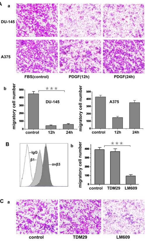
and AT-1. Because the PDGF could regulate lamellipodia formation and cell migration, and c-Abl kinase has a functional role in the morphological response to PDGF [13], we chose PDGF-BB as the migration inducer. To investigate the effect of PDGF on tumor cell migration, we treated the two kinds of tumor cells with 20 ng/ml PDGF for 12 h. Fig. 1A shows the migratory potential of these two cell lines in respond to PDGF. Compared with fetal bovine serum, PDGF specifically induced A375 cell migration but not DU-145 cell, so A375 cell was used in the following experiments. Since integrins have crucial role in cell migration, we subsequently tested the expression of integrinavb3
and b1 on A375 cells. As shown in Fig. 1B, bothavb3 and b1
integrins were expressed on A375 cell surface. To determine the roles of these two integrins in the migration induced by PDGF,
avb3 and b1 were respectively blocked with antibodies in the migration assay. As shown in Fig. 1C, LM609 (blocking anti-avb3
antibody) could dramatically inhibit the migration, whereas TDM29 (b1-blocking antibody) had no obvious inhibitory effect
on the migration, indicating that A375 cell migration induced by PDGF is mainly mediated by integrinavb3.
c-Abl kinase is required for melanoma cell migration induced by PDGF
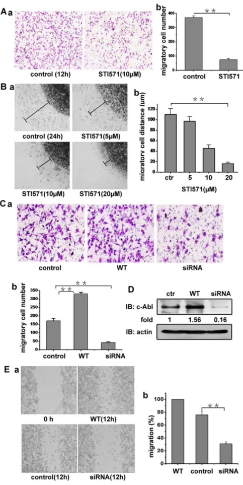
We next explored whether c-Abl kinase is involved in the A375 cell migration induced by PDGF based on the above results that PDGF can induce A375 cell migration and the accumulating evidences that PDGF can regulate c-Abl kinase activity. As shown in Fig. 2A, c-Abl kinase inhibitor STI571 (10mM) substantially inhibited the PDGF induced A375 cell migration. Furthermore, by testing the migration in 3D agarose drop model, STI571 was shown to reduce theavb3-mediated A375 cell migration in a dose
dependent manner (Fig. 2B). To confirm the role of c-Abl kinase in melanoma cell migration triggered by PDGF, WT-c-Abl kinase expression vector or specific siRNA targeting c-Abl kinase was transfected into A375 cells. In migration assay, the overexpression of wild type c-Abl kinase increased A375 cell migration and siRNA-mediated c-Abl kinase knockdown effectively inhibited the cell migration, compared with the non-transfected A375 cells (Fig. 2C). The c-Abl kinase protein level in the transfected A375 cells are shown in Fig. 2D. Similar results were obtained from the wound healing assay. As shown in Fig. 2E, the c-Abl-transfected cells migrated to fill about most of the wounded area within 12 h, while c-Abl knockdown cells showed little migration compared with the control cells, indicating that c-Abl kinase is involved in the melanoma cell migration induced by PDGF.
c-Abl kinase colocalizes withavb3integrin and
potentiatesavb3integrin mediated A375 cell adhesion The above results imply that c-Abl kinase may has some relations with integrin because integrin plays a crucial role in cell migration, pseudopod formation, spreading and adhesion [6]. We next investigated the relationship between c-Abl kinase andavb3
integrin. The subcellular localization of c-Abl kinase and avb3
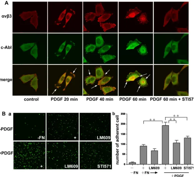
integrin was examined in A375 cells. As shown in Fig. 3A, c-Abl kinase and avb3 integrin were dynamically associated with each other in serum-starved A375 cells in response to PDGF stimulation. At the early stage of stimulation (20 min), c-Abl kinase andavb3integrin were mainly situated at the lamellipodia
of the spreading cell. Then (40 min), with the cell shape changing during spread, c-Abl kinase andavb3integrin both localized to the
focal contacts at the edges of the cells. When cells were stimulated for 1 h, c-Abl kinase and avb3 integrin colocalized to form
clustered spots, which then distributed in the plasma. The above phenomena were not observed when the cells were pretreated with STI571 before PDGF stimulation, suggesting a possible associa-tion of endogenous c-Abl kinase andavb3integrin based on c-Abl
kinase activity. Integrin clustering can enhance the binding of integrin to its ligands [9]. To confirm whetheravb3integrin cluster
after PDGF stimulation and the role of c-Abl kinase in regulating the affinity ofavb3integrin, we performed cell adhesion assay. As
shown in Fig. 3B,avb3integrin antibody LM609 could inhibit the
adhesion, indicating that the adhesion was mediated by avb3
integrin. PDGF stimulation led to an increase in the adhesion of A375 cells on the fibronectin, and STI571 inhibited the adhesion dramatically. These results indicate that the increase in the adhesion of A375 cells in response to PDGF stimulation is dependent upon avb3 integrin engagement and c-Abl kinase is
responsible for the engagement.
c-Abl kinase interacts withavb3integrin in response to
PDGF
Based on the above results, we were interested to discover if c-Abl kinase is involved in the signal pathway from PDGF receptor toavb3integrin. Serum-starved A375 cells were stimulated with
PDGF for indicated time and the treated cells were lysed, then the lysates were used for coimmunoprecipitation. As shown in Fig. 4A, after PDGF stimulation, c-Abl kinase was present in the
avb3 integrin immunoprecipitated complexes, and the highest
level of c-Abl kinase was detected after 10 min of stimulation. The association of avb3 integrin and c-Abl kinase was
dramatically blocked by STI571 incubation. Additionally, avb3
Figure 1. PDGF induces tumor cell migration.(A) The cell suspension containing 0.05% serum was seeded onto 0.8mm diameter upper transwell chamber, which was coated with fibronectin. The lower chamber contained 3% serum (FBS) or 50 ng/ml PDGF to induce cell migration. Cells were cultured for 24 h in each chamber. The migrating cells were fixed and stained with crystal violet and photographed. The cell migration was determined by counting cells in randomly selected five microscopic field per well. Bars represent mean6S.D. (b was statistic value of a). (B) Expression ofavb3andb1integrin on A375 cell surface was evaluated by LM609 (blocking anti-avb3integrin antibody) and TDM29 (specific anti-b1 integrin antibody). IgG was used as negative control. (C) The cell suspension containing 0.05% serum and antibody LM609 or TDM29 (50mg/ml) was put into 0.8mm diameter upper transwell chamber, which was coated with fibronectin. Free-serum medium with 50 ng/ml PDGF was put into the lower chamber to induce cell migration. Cells were cultured for 24 h in the chambers and the migrated cells were fixed and stained with crystal violet and photographed (b was statistic value of a). Cell migration was determined by counting cells in randomly selected five microscopic field per well. Bars represent mean6S.D of three independent experiments. ***, p,0.001 with respect to the control.
betweenavb3integrin and c-Abl kinase, we prepared the
GST-c-Abl SH3, GST-c-GST-c-Abl SH2, GST-c-GST-c-Abl SH3-2 and GST-c-GST-c-Abl C-term fusion proteins (Fig. 4E). As shown in Fig. 4F, avb3
integrin was found in GST-c-Abl SH3 and GST-c-Abl SH3-2,
but not GST, GST-c-Abl SH2 or GST-c-Abl C-term pull-down complexes, and GST-c-Abl-SH3 and GST-c-Abl-SH3-2 bound equally to avb3 integrin. The results above suggest that the
PDGF was put into the lower chamber to induce cell migration. b, migrations were determined by counting cells in randomly selected five microscope field per well. Bars represent mean6S.D. (B) the effects of different concentration of STI571 on cell migration. The migration experiment of A375 cells out of agarose drop explants treated with or without STI571 was conducted. After 24 h, the distant of migration was measured using inverted microscope fitted with a rule in eyepiece. a, Cell migration rate was evaluated by the distance of the leading edge of migrating cells from the edge of agarose droplet. b, The distance of cell migration was measured, the extent of cell migration within the drop = [(total area/drop area)6100]
–100. (C) A375 cells were transfected with WT c-Abl expression vector or specific siRNA targeting c-Abl. The migration experiment of A375 cell or tansfected cells was performed in transwell plates (b was the statistic value of a). (D) A375 cells or cells transfected with WT c-Abl or c-Abl specific siRNA were lysed (For the transfected cells, cells were lysed after 24 h transfection). Lysates were blotted with c-Abl and actin antibodies. (E) a. A375 cells or the cells transfected with WT c-Abl or c-Abl specific siRNA were plated into 24-well plated and allowed to form a confluent monolayer. After overnight serum starvation, the cell monolayer was ‘‘wounded’’ with a pipette tip. The cells were incubated with DMEM containing 20 ng/ml PDGF and then imaged. b. Cell migration was determined as shrinkage of an average gap area. Bars represent mean6 S.D of three independent experiments. **, p,0.01, ***, p,0.001 with respect to control.
doi:10.1371/journal.pone.0066108.g002
Figure 3. c-Abl kinase mediatesavb3integrin engagement after PDGF stimulation.(A) A375 cells were serum starved for 12 h during
spreading, then cells were either untreated or treated with 20 ng/ml PDGF for the indicated times. Cells were fixed, labeled with the antibodies of c-Abl kinase andavb3integrin and imaged under confocal microscope. In inhibiting experiments, cells were preincubated with STI571 for 10 min, and the inhibitor remained in the medium throughout the assay period. (B) starved A375 cells were digested with EDTA and suspended in serum-free medium for 2 h to restore the state of the cells and then stained with Calcein-Am. Then the cells were resuspended in DMEM containing 20 ng/ ml PDGF, antibody or inhibitor. Cells were allowed to adhere at 37uC for 1 h. Then the unbound cells were removed, and the adherent cells were fixed and imaged by phase-contrast microscopy (a) and then quantitated (b). Controls were the cells not stimulated with PDGF or the wells not coated with fibronectin. Images were obtained from at least three separate random fields of view. Bars represent mean6S.D of three independent experiments. **, p,0.01 with respect to control.
activated c-Abl kinase interacts with the cytoplasmic domain of
avb3integrin via its SH3 domain.
The association of c-Abl kinase withavb3integrin is
independent of cell adhesion and F-actin
Integrins function in bidirectional signaling, to further assess the molecular mechanism of the interaction between c-Abl kinase and avb3 integrin, we investigated whether c-Abl kinase was
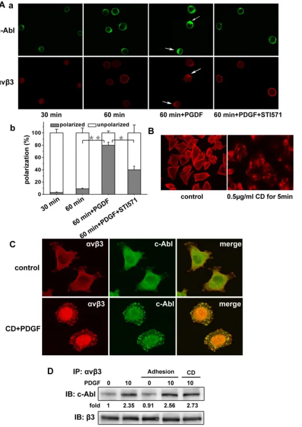
involved in the outside-in signaling. As shown in Fig. 5A, serum-starved A375 cells were detached and replated on fibronectin-coated coverslips, and then the cells were allowed to adhere for 30 min and 60 min respectively. Confocal immunofluorescence microscopy showed that c-Abl kinase and avb3 integrin were
approximately uniform in the cytosol after cell adhesion for 30 min or 60 min. However, in the presence of PDGF, c-Abl kinase was distributed and colocalized with avb3 integrin, and
STI571 could inhibit the polarization of c-Abl kinase and avb3
integrin. Here our results show that the binding ofavb3integrin
to ligands (cell adhesion) cannot affect the association of c-Abl kinase withavb3integrin. The redistributions of c-Abl kinase and avb3 integrin were produced by the PDGF stimuli, and the
redistribution of c-Abl kinase affect the redistribution of avb3
integrin.
c-Abl kinase plays important roles in F-actin dynamics, yet its role in growth-factor-induced cell motility is not well defined [13]. Considering c-Abl kinase can bind to F-actin, we next asked whether c-Abl kinase associates withavb3integrin via F-actin. We
first treated the spreading A375 cells with Cytochalasin D to inhibit the function of F-actin at different concentrations and time frames. We found that Cytochalasin D could significantly reduce the quantity of F-actin at concentration of 0.5mg/ml for 5 min and failed to make the cell detachable (Fig. 5B). To evaluate the impact of F-actin on the interaction between c-Abl kinase andavb3
integrin, A375 cells were treated with Cytochalasin D using the above concentration and time. Then the cells were stimulated with PDGF, and labeled with the antibodies of c-Abl kinase andavb3
integrin respectively. As shown in Fig. 5C, c-Abl kinase andavb3
integrin were highly colocalized and formed clustered spots in the cells. To further investigate the impact of cell adhesion and F-actin on the association of c-Abl kinase withavb3integrin, we examined
the interaction of c-Abl kinase andavb3integrin in A375 cells after
cell adhesion or pretreatment with Cytochalasin D by coimmu-nopreciptation assay. As shown in Fig. 5D, consistent with the Figure 4. c-Abl kinase activity is required for the interaction of c-Abl andavb3integrin.(A and B) Overnight serum starved A375 cells were
trypsinized, washed with PBS and untreated or treated with 20 ng/ml PDGF for the indicated times. Thereafter, the cells were lysed, and the lysates were immunoprecipitated with indicated antibodies and subjected to immunoblotting analysis with the appropriate antibodies. For inhibition assay, cells were pretreated with STI571 for 10 min and the inhibitor remained in the medium throughout the assay. (C) A375 cells were serum-starved and stimulated with PDGF for the indicated times. The lysates of cells were immunoblotted, and the activity of c-Abl tyrosine kinase was tested by immunoblotting with PY20. The loading control was immunoblotted with anti-actin antibody. (D) A375 cells were serum-starved and stimulated with PDGF for the indicated times. The activity of c-Abl kinase was determined by using an in vitro kinase assay with GST-CrkII-CTD as a substrate. Phosphorylation of GST-CrkII-CTD was tested by immunoblotting with PY20 (p-Tyr Antibody). The level of GST-CrkII-CTD was detected with anti-GST antibody to demonstrate equal loading. (E) A schematic diagram of GST c-Abl domains. (F) A375 cells were serum-starved and stimulated with PDGF for 10 minutes. The GST c-Abl domain pull-down assay was done, and the bound proteins were analyzed by SDS-PAGE and immunoblotted forb3 integrin. The immunoblotting bands were quantified by densitometry, and the data were normalized with respect to the controls.
Figure 5.b3integrin engagement and F-actin do not affect the interaction of c-Abl kinase andavb3integrin.(A) a. Serum-starved A375
above results, ligand binding toavb3integrin (cell adhesion) failed
to increase the association between c-Abl kinase andavb3integrin.
Also, inhibiting the de novo actin polymerization by Cytochalasin D failed to decrease the association between c-Abl kinase andavb3
integrin. These results strongly suggest that the association of c-Abl kinase withavb3integrin is cell adhesion-independent and F-actin
is not required for this association in A375 cells.
c-Abl kinase regulatesavb3integrin via talin
The results above showed that c-Abl kinase interacts with the cytoplasmic domain ofb3integrin after PDGF stimulation, and a
Far western blot analysis revealed that this interaction was not direct (data not shown). In addition, this interaction was not due to the cell adhesion and F-actin. It is unclear how c-Abl uses its SH3 domain to interact withavb3integrin. One possibility is that other
proteins are required for c-Abl kinase to interact with avb3
integrin. Talin is a major cytoskeletal protein which has an N-terminal head region of 50 kDa and an elongated helical rod of 220 kDa. Talin can bind to the cytoplasmic domains ofb-integrin with its head domain. Moreover, the binding of talin tob-integrin triggers a conformational change in the integrin extracellular domain which increases its affinity for ECM proteins [9]. We first detected the existent state of talin in serum-starved A375 cells in response to PDGF stimulation. Starved cells or stimulated cells were lysed and then separated by SDS-PAGE and immunoblotted with talin head domain antibody TA205. As shown in Fig. 6A, there was only the intact 270 kDa talin in the serum-starved cells. When cells were stimulated with PDGF, talin underwent proteolysis to generate a 50 kDa head, which increased with the stimulation prolongation. Pretreating cells with STI571 partially inhibited the generation of talin head. We also investigated the association of talin,b3integrin and c-Abl kinase in A375 cells by
coimmunoprecipitation assay. As shown in Fig. 6B, the intact 270 KDa talin was cleaved to generate the 50 kDa fragment in response to PDGF stimulation, and c-Abl kinase was present in the immunoprecipitated complexes of talin after PDGF stimulation. As a point of emphasis, the change in amount of c-Abl kinase was consistent with the generation of talin head. In addition, as expected, talin andb3integrin were constitutively present in the
same immunoprecipitated complexes. As shown in Fig. 6C, a pull-down assay by using GST- talin2-head also confirmed these interactions. c-Abl was found in the GST- talin2-head pull-down complexes and the amount increased with PDGF stimulation prolongation. The inhibition of c-Abl kinase activity reduced the interaction of talin head and c-Abl kinase. Meanwhile,b3integrin
was constitutively associated with talin head, and this association was not affected by PDGF stimulation. These results indicate that PDGF stimulation can induce the proteolysis of intact talin, and the phosphorylated c-Abl kinase interacts with talin head which constitutively associated withb3integrin.
To further reveal the mechanism of the interaction between c-Abl kinase and talin, we performed in vitro protein binding assay. Our above studies indicated that c-Abl kinase interacted with the cytoplasmic domain ofb3integrin via its SH3 domain.
There are tyrosine residue Y89 and Y134 in the SH3 domain of c-Abl kinase. To test whether these sites were responsible for the association of talin head and c-Abl kinase, GST-c-Abl-SH3 Y89F and Y134F were used for pull down assays. As shown in Fig. 6D, Y134F and Y89F both reduced the binding of talin to the SH3
domain of c-Abl kinase. To detect whether talin head domain directly binds to c-Abl kinase, a Far western blot analysis was performed. The results showed that GST-talin2-head directly interacts with c-Abl kinase (Fig. 6E). Thus, talin head was found to interact directly with c-Abl kinase and function in the activation of c-Abl kinase.
Discussion
The molecular mechanisms that induce cell migration are complex and involve the interplay of numerous receptors, including growth factor receptors and integrins, and their associated signaling intermediates. In this paper, we uncover a new function of c-Abl kinase in melanoma cell migration mediated byavb3integrin after activation of PDGF receptor. c-Abl kinase was mainly involved in the inside-out signaling ofavb3integrin
and potentiated the biological activity ofavb3 integrin. On the
contrary, c-Abl kinase exhibited low sensitivity to outside-in (ligand-binding) signaling of avb3 integrin. c-Abl kinase was
activated by PDGF and associated withb3 integrin cytoplasmic
domain via its SH3 domain, and this association was insusceptible to F-actin, but c-Abl kinase did not interact with b3 integrin
directly. Talin head domain constitutively interacted with b3
integrin cytoplasmic domain and linked c-Abl kinase and b3
integrin together.
Earlier reports demonstrated that c-Abl kinase has a critical role in the migration of human liver cells and NIH3T3 cells [20]. However, little is known if c-Abl kinase functions in cancer cell migration. We found that c-Abl kinase was involved in A375 cell migration induced by PDGF. This finding is supported adequately by a series of experiments. First, we found c-Abl kinase inhibitor STI571 suppresses A375 cell migration. The fact that c-Abl kinase is involved in tumor cell migration was also reflected in the transfected cells: targeting c-Abl with its specific siRNA inhibited A375 cell migration, andvice versa, the overexpression of wild-type c-Abl kinase increased A375 cell migration.
As we know, integrin plays a crucial role in tumor cell migration, especiallyb1and b3-integrins [8]. Using flow
cytom-etry, we showed thatavb3andb1integrin were expressed on A375 cell surface, but the antibody blocking experiments revealed that the PDGF induced A375 cell migration was mainly mediated by integrinavb3but notb1. This may be due to the facts that PDGF
cause aVb3 integrin activation but not b1 integrin and the
phosphorylated PDGF receptor is associated withaVb3integrin
but not withb1integrins [28–30]. Next we wanted to know if
c-Abl kinase had any relationship with avb3 integrin during cell
migration. Our microscopy data demonstrated that, in the serum-starved A375 cell, endogenous c-Abl kinase andavb3integrin were co-localized dynamically in response to PDGF stimulation, and c-Abl kinase and avb3 integrin formed clustered spots when cells
were stimulated with PDGF for 60 min. These data imply that c-Abl kinase possesses a potential effect on regulatingavb3integrin
and inhibition of c-Abl kinase affects the integrin binding activity (Fig. 3C). After PDGF stimulation, c-Abl kinase interacted withb3
integrin, and the interaction between c-Abl kinase andb3integrin
was attenuated after PDGF stimulation for 10 min. It is possible that c-Abl kinase phosphorylation was a transient process and the downstream activation of c-Abl kinase may serve as a negative feedback regulation to limit the duration of c-Abl activation, as previously suggested [32]. By using in vivo and in vitro kinase assays, we verified that PDGF stimulation increased c-Abl kinase activity within 10 min (Fig. 4), implying that kinase activity was required for c-Abl kinase to interact withb3integrin. The SH3
and SH2 domains of c-Abl kinase are thought to function by mediating the interaction between c-Abl kinase and its substrates [33]. The SH3 domain preferentially interacts with the proteins containing a proline-rich region to inhibit c-Abl kinase activity [34]. Here, we find that the b3integrin interacts with the SH3
domain of c-Abl kinase in A375 cells, but this interaction is not direct (Fig. 4E).
As a cytoplasmic tyrosine kinase, c-Abl is mainly linked to the transduction of extracellular signals through interaction with cell surface receptors. In our previous study, c-Abl kinase was shown to
be involved inb2integrin outside-in signaling in neutrophil [35].
Others reported that in fibroblasts, c-Abl kinase activation occurred after integrin-mediated cell attachment to fibronectin [21]. Here our results show that the binding ofavb3integrin to
ligands cannot affect c-Abl kinase redistribution and its association withavb3integrin (Fig. 5). Thus, c-Abl kinase was not involved in
the outside-in signaling ofavb3integrin. It became clear that most
leukocyte integrin (b2) and platelet integrin (aIIbb3), includingb1
-containing integrins, exist in a resting state until activated by stimulation. Whereas aVb3 integrin is constitutively active on
many cell types. Studies have been shown that the interactions between leg domains are strong for aIIbb3, but the association betweenaVandb3is weak, which may help to explain why the default state ofaIIbb3integrin on platelets is completely inactive
andaVb3integrin is constitutively active [9]. We suggest that the
constitutive activation of integrin failed to arouse c-Abl kinase activation. Collectively, c-Abl kinase functions in different integrin signaling in various manners.
There have been reports that c-Abl kinase can be activated by various stimuli and the activated c-Abl kinase has been shown to activate many cellular proteins to regulate cytoskeletal dynamics Figure 6. c-Abl kinase directly associates with talin head domain and regulatesb3 integrin ligation concomitantly after PDGF
stimulation.(A) Overnight serum-starved A375 cells were either untreated or treated with 20 ng/ml PDGF for the indicated times. Thereafter, the cells were lysed, and the lysates were immunoblotted with indicated antibodies. In inhibition experiments, cells were preincubated with STI571 for 10 min, and the inhibitor remained in the medium throughout the assay period. (B) Serum-starved A375 cells were stimulated as above. The lysates were immunoprecipitated with anti-talin antibody and immunoblotted with indicated antibodies. (C) Serum-starved A375 cells were treated as above. The lysates were incubated with GST or GST fusion proteins, and the bound proteins were separated by SDS-PAGE and immunoblotted with indicated antibodies. (D) GST or GST fusion proteins were incubated with Ni2+
[17,36]. The linkage between integrin and the actin cytoskeleton appears to be crucial for the integrin function [37]. We also asked if c-Abl kinase could regulate aVb3 integrin via actin. In CD treated cells, we noticed that c-Abl kinase still co-localizes with
aVb3integrin after PDGF stimulation, and CD did not attenuate
the interaction of c-Abl kinase and aVb3 integrin. So, we have
provided the evidence that c-Abl kinase regulates aVb3 integrin
clustering independent of actin (Fig. 5), and we believe there exist some other proteins close to aVb3 integrin that assisted c-Abl
kinase to regulate aVb3 integrin. To the best of our knowledge,
talin is one of few proteins directly linked to integrin cytoplasmic tail. In vertebrates, there are two talin genes (talin1 and talin2) that encode closely related proteins (74% identity). The intact talin protein (270 kDa) can be proteolysed by proteases such as calpain and trypsin to generate an N-terminal 50-kDa head domain and a 220-kDa C-terminal rod domain [38,39]. The head domain binds to the integrin cytoplasmic domain and is required for integrin activation. In our studies, we found that talin was cleaved into head domain and rod domain after PDGF stimulation (Fig. 6A). Although talin was cleaved after PDGF stimulation, the amount of
b3integrin associated with talin head domain (include intact talin)
were not affected by PDGF stimulation, whereas c-Abl kinase only interacted with cleaved talin head domain (Fig. 6B–C). So, we think that talin constitutively and directly associates with the cytoplasmic domain of b3 integrin in A375 cells, which may be
partially due to the constitutively active aVb3 integrin with low
affinity. When cells were stimulated with PDGF, the local concentration of active talin increased, the cellular environment regulated the local complex formation that made integrin clustering and then the integrin affinity became higher. Since the formation of a precise complex between theb-tail of integrin, talin and the membrane is the final key step in inside-out activation ofaVb3integrin [40], it is important to mention that
c-Abl kinase plays a necessary role in regulating talin activity. We found c-Abl kinase inhibitor STI571 suppressed talin activityin vivo
andin vitro(Fig. 6A–C). Thus, we consider that it is possible that c-Abl kinase is an upstream regulator of talin in aVb3 integrin
signaling in response to PDGF stimulation in A375 cell. In addition, our results show that c-Abl kinase directly associates with talin head domain (Fig. 6E), both the Y89F and Y134F mutants of c-Abl SH3 domain partially reduced the binding of talin head (Fig. 6D), indicating that these two sites are both important but neither one is unique.
The fact that growth factor receptors regulating integrin inside-out signals derived from G-protein, phosphatidylinositol-4,5-bisphosphate (PtdIns(4,5)P2) hydrolysis and PKC has been confirmed. However, the specific mechanisms involved are still under investigation [41]. Adding to the possible oncogenic roles of c-Abl kinase in both NSCLC and breast cancer. As we mentioned above, our studies have provided new insights into the nature of the crosstalk between growth factor receptors and integrin by measuring the novel role of c-Abl kinase in regulating melanoma cell migration: c-Abl kinase is critical for the formation of early clusters of integrin complex, and the complex might be the sites of PDGF receptor activation in the early stage of cell spreading in a new cycle of motile melanoma cells. However, the signaling pathways that c-Abl kinase regulate talin activity are only just beginning to emerge, and the structural basis for the activation of many ligand-binding sites in talin is not yet fully understood.
Acknowledgments
The authors thank Dr. Giorgio Scita (European Institute of Oncology, Milan, Italy), Dr. Pietro De Camilli (Yale University, School of Medicine, New Haven, CT), Dr. Thomas Rudel (Max Planck Institute for Infection Biology, Berlin Germany) for providing the plasmids that made this work possible.
Author Contributions
Conceived and designed the experiments: CMZ CY XGW XLZ. Performed the experiments: CMZ CY RFW. Analyzed the data: CMZ CY XLZ. Contributed reagents/materials/analysis tools: CMZ CY YJ XGW. Wrote the paper: CMZ XLZ KKA.
References
1. Kerbel RS (1995) Significance of tumor-host interactions in cancer growth and metastases. Cancer Metast Rev 14: 259–262.
2. Clark EA, Golub TR, Lander ES, Hynes RO (2000) Genomic analysis of metastasis reveals an essential role for RhoC. Nature 406: 532–535. 3. Geho DH, Bandle RW, Clair T, Liotta LA (2005) Physiological mechanisms of
tumor-cell invasion and migration. Physiology 20: 194–200.
4. Lauffenburger DA, Horwitz AF (1996) Cell migration: A physically integrated molecular process. Cell 84: 359–369.
5. Parent CA, Devreotes PN (1999) A cell’s sense of direction. Science 284: 765– 770.
6. Luo BH, Carman CV, Springer TA (2007) Structural basis of integrin regulation and signaling. Annu Rev Immunoled. 619–647.
7. Harburger DS, Calderwood DA (2009) Integrin signalling at a glance. J Cell Sci 122: 159–163.
8. Vicente-Manzanares M, Choi CK, Horwitz AR (2009) Integrins in cell migration – the actin connection. J Cell Sci 122: 199–206.
9. Askari JA, Buckley PA, Mould AP, Humphries MJ (2009) Linking integrin conformation to function. J Cell Sci 122: 165–170.
10. Verbisck NV, Costa ET, Costa FF, Cavalher FP, Costa MDM, et al. (2009) ADAM23 Negatively Modulatesavb3 Integrin Activation during Metastasis. Cancer Res 69: 5546–5552.
11. Uhm JH, Gladson CL, Rao JS (1999) The role of integrins in the malignant phenotype of gliomas. Frontiers in Bioscience 4: D188–199.
12. Vanetten RA, Jackson PK, Baltimore D, Sanders MC, Matsudaira PT, et al. (1994) The COOH-terminus of the c-Abl tyrosine kinase contains distinct F- and G-actin binding domains with bundling activity. J Cell Biol 124: 325–340. 13. Woodring PJ, Hunter T, Wang JYJ (2003) Regulation of F-actin-dependent
processes by the Abl family of tyrosine kinases. J Cell Biol 116: 2613–2626. 14. Sirvent A, Benistant C, Roche S (2008) Cytoplasmic signalling by the c-Abl
tyrosine kinase in normal and cancer cells. Bio Cell 100: 617–631.
15. Van Etten RA (1999) Cycling, stressed-out and nervous: cellular functions of c-Abl. Trends Cell Biol 9: 179–186.
16. Taagepera S, McDonald D, Loeb JE, Whitaker LL, McElroy AK, et al. (1998) Nuclear-cytoplasmic shuttling of C-ABL tyrosine kinase. Proc Natl Acad Sci 95: 7457–7462.
17. Lewis JM, Baskaran R, Taagepera S, Schwartz MA, Wang JYJ (1996) Integrin regulation of c-Abl tyrosine kinase activity and cytoplasmic-nuclear transport. Proc Natl Acad Sci 93: 15174–15179.
18. Renshaw MW, Lewis JM, Schwartz MA (2000) The c-Abl tyrosine kinase contributes to the transient activation of MAP kinase in cells plated on fibronectin. Oncogene 19: 3216–3219.
19. Yan W, Bentley B, Shao R (2008) Distinct angiogenic mediators are required for basic fibroblast growth factor- and vascular endothelial growth factor-induced angiogenesis: The role of cytoplasmic tyrosine kinase c-Abl in tumor angiogenesis. Mol Biol Cell 19: 2278–2288.
20. Lewis JM, Schwartz MA (1998) Integrins regulate the association and phosphorylation of paxillin by c-Abl. J Biol Chem 273: 14225–14230. 21. Xu J, Millard M, Ren X, Cox OT, Erdreich-Epstein A (2010) c-Abl mediates
endothelial apoptosis induced by inhibition of integrinsavb3 andavb5 and by disruption of actin. Blood 115: 2709–2718.
22. O’Donovan M, Russell JM, O’Leary JJ, Gillan JA, Lawler MP, et al. (1999) Abl expression, tumour grade, and apoptosis in chondrosarcoma. J Clin Pathol-Mol Pathol 52: 341–344.
23. Singer CF, Hudelist G, Lamm W, Mueller R, Czerwenka K, et al. (2004) Expression of tyrosine kinases in human malignancies as potential targets for kinase-specific inhibitors. Endocr Related Cancer 11: 861–869.
24. Lin J, Arlinghaus R (2008) Activated c-Abl tyrosine kinase in malignant solid tumors. Oncogene 27: 4385–4391.
25. Zhao H, Ou-Yang F, Chen IF, Hou M-F, Yuan S-SF, et al. (2010) Enhanced Resistance to Tamoxifen by the c-ABL Proto-oncogene in Breast Cancer. Neoplasia 12: 214–223.
invasion via distinct pathways, and drive metastatic progression. Oncogene 31: 1804–1816.
27. Wu Y, Li Q, Chen XZ (2007) Detecting protein-protein interactions by Far western blotting. Nat Protoc. 2(12): 3278–84.
28. Liden A˚ , Berg A, Nedrebo T, Reed RK, Rubin K (2006) Platelet-derived growth factor BB-mediated normalization of dermal interstitial fluid pressure after mast cell degranulation depends onb3 but notb1 integrins. Circ Res 98: 635–641. 29. Schneller M, Vuori K, Ruoslahti E (1997)avb3 integrin associates with activated insulin and PDGFbreceptors and potentiates the biological activity of PDGF. Embo Journal 16: 5600–5607.
30. Woodard AS, Garcia-Cardena G, Leong M, Madri JA, Sessa WC, et al. (1998) The synergistic activity ofavb3 integrin and PDGF receptor increases cell migration. J Cell Sci 111: 469–478.
31. Shattil SJ, Kim C, Ginsberg MH (2010) The final steps of integrin activation: the end game. Nat Rev Mol Cell Biol 11: 288–300.
32. Mitra S, Beach C, Feng G-S, Plattner R (2008) SHP-2 is a novel target of Abl kinases during cell proliferation. J Cell Sci 121: 3335–3346.
33. Mayer BJ, Baltimore D (1994) Mutagenic analysis of the roles of SH2 and SH3 domains in regulation of the Abl tyrosine kinase. Mol Cell Biol 14: 2883–2894.
34. Cicchetti P, Mayer BJ, Thiel G, Baltimore D (1992) Identification of a protein that binds to the SH3 region of Abl and is similar to Bcr and GAP-rho. Science 257: 803–806.
35. Cui L, Chen C, Xu T, Zhang J, Shang X, et al. (2009) c-Abl Kinase Is Required forb2 Integrin-Mediated Neutrophil Adhesion. J Immunol 182: 3233–3242. 36. Plattner R, Irvin BJ, Guo SL, Blackburn K, Kazlauskas A, et al. (2003) A new
link between the c-Abl tyrosine kinase and phosphoinositide signalling through PLC-gamma 1. Nat Cell Biol 5: 309–319.
37. Plattner R, Kadlec L, DeMali KA, Kazlauskas A, Pendergast AM (1999) c-Abl is activated by growth factors and Src family kinases and has a role in the cellular response to PDGF. Gene Develop 13: 2400–2411.
38. Critchley DR, Gingras AR (2008) Talin at a glance. J Cell Sci 121: 1345–1347. 39. Rees DJG, Ades SE, Singer SJ, Hynes RO (1990) Sequence and
domain-structure of talin. Nature 347: 685–689.
40. Anthis NJ, Campbell ID (2011) The tail of integrin activation. Trends Biochem Sci 36: 191–198.