REVISTA
BRASILEIRA
DE
ANESTESIOLOGIA
PublicaçãoOficialdaSociedadeBrasileiradeAnestesiologiawww.sba.com.br
SCIENTIFIC
ARTICLE
Cytotoxic
effects
of
local
anesthesia
through
lidocaine/ropivacaine
on
human
melanoma
cell
lines
Ding-Kun
Kang
∗,
Li-Yan
Zhao,
Hong-Li
Wang
LuoyangOrthopedic---TraumatologicalHospital,DepartmentofAnesthesiology,Luoyang,China
Received22February2015;accepted15April2015 Availableonline20April2016
KEYWORDS
Lidocaine; Ropivacaine; Cytotoxicity; Aminoamidelocal anesthetics; Melanomacelllines; Flowcytometry
Abstract
Background: Localanesthetics(LAs)aregenerallyconsideredassafe,butcytotoxicityhasbeen reportedfor severallocalanestheticsusedinhumans,whichisnotwellinvestigated.Inthe presentstudy,thecytotoxicityoflidocaine,ropivacaineandthecombinationoflidocaineand ropivacainewere evaluated on humanmelanoma celllines. Melphalan, anitrogen mustard alkylatingagent,wasusedasacontrolagentforcomparisonofcytotoxicactivity.
Methods:Melanomacelllines,A375andHs294T,wereexposedto1htodifferentconcentrations ofaboveagents.Cell-viabilityafterexposurewasdeterminedbyflowcytometry.
Results:InvestigatedLAsshowed detrimentalcytotoxicityonstudiedmelanomacelllinesin time-(p<0.001),concentration-(p<0.001),andagentdependant.In bothA375 andHs294T celllines, minimum cellviability rateswere found after 72h ofexposure to theseagents. Lidocaine2%causedareductionofvitalcellsto10%± 2% and14%± 2%inA375andHs294T, respectivelyafter72hofexposure.Ropivacaine0.75%after72hreducedviablecellsto15%±3% and25%± 3%inA375andHs294T,respectively.Minimumcellviabilityafter72hexposuretothe combinationwas10%±2%and18%±2%inA375andHs294T,respectively.Minimumcellviability after72hexposuretomelphalanwas8%± 1%and12%± 2%,inA375andHs294T,respectively.
Conclusion:LAshavecytotoxicactivityonhumanmelanomacelllinesinatime-, concentration-and agent-dependant manner.Apoptosis in the celllines was mediated through activityof caspases-3andcaspases-8.
©2016SociedadeBrasileiradeAnestesiologia.PublishedbyElsevierEditoraLtda.Thisisan openaccessarticleundertheCCBY-NC-NDlicense( http://creativecommons.org/licenses/by-nc-nd/4.0/).
∗Correspondingauthor.
E-mail:[email protected](D-K.Kang). http://dx.doi.org/10.1016/j.bjane.2015.04.002
PALAVRAS-CHAVE
Lidocaina; Ropivacaína; Citotoxicidade; Anestésicoslocaisdo grupoamino-amida; Linhagenscelulares demelanoma; Citometriadefluxo
Efeitoscitotóxicosdeanestesialocalcomlidocaína/ropivacaínaemlinhagens
celularesdemelanomahumano
Resumo
Justificativa: Osanestésicoslocais(ALs)sãogeralmenteconsideradoscomoseguros,mas cito-toxicidadefoirelatadaemváriosanestésicoslocaisusadosemsereshumanos,aqualnãoébem investigada.Nopresenteestudo,acitotoxicidadedelidocaínaeropivacaínaedacombinac¸ão delidocaínaeropivacaínafoiavaliadaemlinhagenscelularesdemelanomahumano.Melfalano, umagentealquilantedemostardanitrogenada,foiusadocomoumagentedecontroleparaa comparac¸ãodaatividadecitotóxica.
Métodos: Linhagens celulares de melanoma, A375 e Hs294T, foram expostas por 1 hora a concentrac¸ões diferentes dos agentes mencionados acima. A viabilidade celular após a exposic¸ãofoideterminadaporcitometriadefluxo.
Resultados: OsALsinvestigadosmostraramcitotoxicidadeprejudicialnaslinhagenscelulares de melanomaestudadasdependentedotempo(p<0,001),daconcentrac¸ão (p<0,001)edo agente.EmambasaslinhagensdecélulasA375eHs294T,níveismínimosdeviabilidadecelular foramencontradosapós72horasdeexposic¸ãoaessesagentes.Lidocaínaa2%provocouuma reduc¸ãodascélulasvitaispara10%± 2%e14%± 2%emA375eHs294T,respectivamente,após72 horasdeexposic¸ão.Ropivacaínaa0,75%após72horasreduziuascélulasviáveispara15%±3% e25%± 3%,emA375eHs294T,respectivamente.Aviabilidadecelularmínimaapósexposic¸ão de72horasparaacombinac¸ãofoide10%±2%e18%±2%emA375eHs294T,respectivamente. Aviabilidadecelularmínimaapósexposic¸ãode72horasaomelfalanofoide8%± 1%e12± 2, emA375eHs294T,respectivamente.
Conclusão:Os ALs possuem atividade citotóxica em linhagens de celulares de melanoma humanodemododependentedotempo,daconcentrac¸ãoedoagente.Aapoptosenaslinhagens celularesfoimediadapormeiodaatividadedascaspases-3ecaspases-8.
©2016SociedadeBrasileiradeAnestesiologia.PublicadoporElsevierEditoraLtda.Este ´eum artigo OpenAccess sobumalicenc¸aCCBY-NC-ND( http://creativecommons.org/licenses/by-nc-nd/4.0/).
Introduction
A little is known about chemotherapy and anesthetics in general. In perioperative and ambulatory settings local anestheticsareadministeredasintra-articularinjections.1 Aminoamidelocalanesthetics exhibittheiractivity mainly by blocking the impulse conduction of nerve axons in a reversiblemanner. Lidocaineandropivacaine both belong toaminoamideclassof localanesthetics withtheformer having antiarrhythic activity (class-1b). In general, local anesthetics (LAs) prevent or relieve pain by binding to specific receptor sites on the sodium (Na+) channels in nerves and blocking the movement of ions through these pores.Boththechemical andpharmacologicpropertiesof individual local anesthetic drugs determine their clinical properties.2 Local anesthetics including lidocaine applied topicallyhavebeenshowntoproducegoodpaincontrolin patients withoral or rectalcancer. Lidocaine and ropiva-caineareusedataconcentrationof1.5%or2.0%and0.5% or0.75%,respectively,forsurgicalanaesthesiawithreduced cardiotoxictyandCNStoxicity.1,3,4TheseLAsarewidelyused forpaincontrolin patientswithhead andneckcancer,to inhibitthemetastasisandrelapseoftumoursandtoreduce surgicalstressinducedinhibition ofnaturalkiller(NK)cell activity. However, both in vitro and in vivo studies have showncytotoxicitytowardsseveralculturedcells.5,6 Previ-ous publications have reported that single dose injection
compounds to evaluate cytotoxic potential. The primary purposeofthestudywastoinvestigatewhetherthese com-monly used LAs have cytotoxic effects on the melanoma cell lines. In this study,effect of pH of local anesthetics oncelldeathofmelanomacelllineswasalsostudied. Cas-paseactivitytestwasperformedtoconfirmwhetherthecell deathwascausedbyapoptosis.Melphalan,anitrogen mus-tardalkylatingagent,isachemotherapydrugthatactsby alkylatingDNAnucleotideguanine.Melphalanwasreported tobecytotoxiconseveralcelllinesincludingmelanomaand thereforeisusedasastandardagenttoevaluatecytotoxic potentialandusedascomparator.19,20
Materials
and
methods
Materials
Lidocaine, ropivacaine, and melphalan (137-58-6; 98717-15-8 and 148-82-3, respectively) were purchased from Sigma---Aldrich, Germany. All these drugs and reagents were preservative free and dissolved in buffered saline solution (pH 7.0). 3-[4,5-Dimethylthiazol-2yl]-2,5-diphenyltetrazolium bromide (MTT) was purchased fromSigmaChem.Ind.,St.Louis,MO,USA.
Cellculture
Humanmalignantmelanomacell line A375waspurchased from Institute of Cell Biology, Shanghai Institute for Bio-logical Science, Chinese Academy of Science, China. The celllinewasculturedinDulbecco’sModifiedEagle’sMedium (DMEM) culture medium (Life Technologies, Carlsbad,CA, USA).Thecelllinewassupplementedwith4mMglutamine, the dipeptidel-alanyl-l-glutamine that prevents
degrada-tionandammoniabuildupinbothadherentandsuspension cultures, and 5% fetal bovine serum (Life Technologies). Hs294T,humanmelanomacellline(InstituteofCellBiology, ShanghaiInstituteforBiologicalScience,ChineseAcademy of Science,China) wasculturedin DMEM culture medium (Lonza, Basel, Switzerland) supplemented with 2mM glu-tamine(LifeTechnologies)and5%fetalbovineserum(Life Technologies).Thenormalhumandermalfibroblast(NHDF; Lonza) cell line was cultured in MEM␣ culture medium (Lonza)supplementedwith2mMglutamine(Life Technolo-gies)and10%fetalbovineserum(LifeTechnologies). Cells weregrowntoconfluenceandtransferredto48wellPlates 48hbeforeexperimentaltreatment.
All the media contained 0.1mg/mL streptomycin, 100U/mL penicillin, and 0.25g/mL amphotericin B (Lonza).Thecellswereculturedat37◦Cinahumid atmo-spheresaturatedwith5%CO2at95%relativehumidity.
Analysisofcellapoptosisbyflowcytometry
Lidocaineand ropivacaineform the group of aminoamide local anesthetics but differ in their analgesic potency, onset of action and duration of anaesthesia. Melanoma cells were exposed for 1h to 1mL of local anesthetics solutionswithfollowingconcentrations:0.03125%,0.0625%, 0.125%,0.25%,0.5%,1.0%and2%oflidocaine,and0.03125%,
0.0625%, 0.125%, 0.25%, 0.5%, and 0.75% of ropivacaine, melphalan (0.2%) and 0.03125%, 0.0625%, 0.125%, 0.25%, 0.5%,and0.75%oflidocaineandropivacainecombination. Theconcentrationoftestcompoundsrangedfrom0.03125% to2.0%.Controlcellsweretreatedwithnormalsaline solu-tionfor 1h.The treatedsolutions containingnonadherant melanoma cells were removed and centrifuged. Formed cell pellets were washed in buffered saline and returned totherespectivewellswithculturemedium.After24and 72h, cell viability was determined using flow cytometry. TodeterminetheinfluenceofpHonhumanmelanomacell lines,viabilityrates wereanalysed 24and72haftera1-h exposure to salinesolution withpH of 7.4, 7.0, 6.0, 5.0, and 4.0. Following general procedure was used for anal-ysis of melanoma cell viability: A375 cells (1×10⁶mL−1) wereseededintocultureplatesandwereallowedtoadhere and harvested after treatment with lidocaineropivacaine and combination of lidocaine and ropivacaine for 24h. Controlsincluded unstainedcellsanduntreatedcells (i.e. not vehicle-treated). All processing was done on ice. At the individual timepointsculture medium wascollected, pooledwiththesuspendedcellsandcentrifugedfor5minat 1200rpmtomakesurethatallthecellularmaterialwas col-lected.Thecellpelletsweresuspendedin500L1×binding bufferatadensityofapproximately1×106mL−1.Samples wereincubatedwith1LAnnexinV-FITCstainingand5L PI for exactly5minat room temperaturein the darkand thensamplesweretransferredto5mLFACStubesandthen measurementwascarriedoutonaFACSCalibur cytometer (Becton Dickinson, USA).PI and V-FITC fluorescence were detectedintheFL-1(green)andFL-2(red)channels, respec-tively,aftercorrectiontothespectraloverlapbetweenthe twochannels.CellQuestsoftware(BectonDickinson, Coun-try)wasemployedtoanalysethedata.1
Assayforcaspase-3/8activity
Caspase-3/8activitywasanalysed at12,24and72hafter exposure tothelocal anesthetics usingcolorimetricassay kits(KeygenCo.,China).Cells(1×10⁶well−1)wereseeded into 6-well plates,and treated with different concentra-tionsoflidocaine,ropivacaineormixtureoflidocaineand ropivacaineand melphalanfor 12,24 and72h. Caspase-3 andcaspase-8hydrolyzepeptidesubstrateAc-DEVD-PNAand Ac-IETD-PNA, respectively,whichleadstothereleaseofa p-nitroanilide (pNA)moiety.pNA concentrationcalculated according totheabsorbencymeasured atawavelength of 405nmandcalibration curvebasedonstandard pNA solu-tions.AllvaluesobtainedwereexpressedaspNA/mgoftotal protein.Further,allvaluesobtainedwerenormalizedtothe cellviabilityofcontrol.13
Statisticalanalysis
120
A
B
100
80
60
40
20
0
Control 0.0325 0.0625 0.125 0.250
Concentration of ropivacaine, % Ropivacaine
Cells analyz
ed (%) - 24 hours
120
100
80
60
40
20
0
Cells analyz
ed (%) - 72 hours
0.500 0.750 Standard Control 0.0325 0.0625 0.125 0.250
Concentration of ropivacaine, % Ropivacaine
0.500
*
0.750
*
Standard
# #
#
*
Figure1 EffectofropivacaineonA375melanomacellviability.Adose---responsecurveofcytotoxicityofropivacainewasmeasured (A)24hand(B)72haftera1hexposureusingflowcytometry.(AandB)Ropivacaineconcentrationsof0.50%and0.75%caused significantdecreasesincellviabilitycomparedwithcontrol(salinesolution)andwerecomparablewithstandardagent(melphalan 0.5%)after24and72h.Thebarsrepresentthemeanvaluesofexperimentswithstandarderroraserrorbars(*p<0.05;#p<0.01).
two-sidedtests.The statistical analysiswasperformedby usingSPSS12.0software.
Results
Analysisofcellviabilityusingflowcytometry
Inthisstudy,lidocaine,ropivacaineandcombinationof lido-caineandropivacainedemonstratedaconcentration-, time-andagent-dependantcytotoxicitytohumanmelanomacells (A375,Hs294T)andtheresults obtainedwerecomparable
tomelphalan,anitrogenmustardalkylatingagent,usedas astandardagentforcomparison(Figs.1---6).
Exposure toincreasing concentrations of lidocaineand ropivacaineresultedinadecreasingnumberofobservable viable cells. Treatment with a combination of lidocaine and ropivacaine at a concentration of 0.25% and higher causedasignificantdeclineofviablecellsinbothA375and Hs294T cell lines. The fractionof apoptotic and necrotic cellsincreased.Minimumviabilityofcellswasobservedata concentrationof 0.75% and viabilityrates at this concen-tration were 15%±2% (p<0.005) after 24h and 10%±2% (p<0.005)after72h,respectively(Fig.3AandB).InA375
120
A
B
100
80
60
40
20
0
Control 0.0325 0.0625 0.125 0.250
Concentration of lidocaine, % Lidocaine
Cells analyz
ed (%) - 24 hours
0.500 0.750 #
1.00 *
2.00 #
Standard #
120
100
80
60
40
20
0
Control 0.0325 0.0625 0.125 0.250
Concentration of lidocaine, % Lidocaine
Cells analyz
ed (%) - 72 hours
0.500 0.750 #
1.00 *
2.00 *
Standard #
Figure2 EffectoflidocaineonA375melanomacellviability.Lidocaineconcentrations of0.75%andhighercausedsignificant decreases incellviability compared with control(salinesolution) andwere cytotoxicity was comparablewith standardagent (melphalan0.5%)after24and72h.Thebarsrepresentthemeanvaluesofexperimentswithstandarderroraserrorbars(*p<0.05;
120
A
B
100
80
60
40
20
0
Control 0.0325 0.0625 0.125 0.250
*
Concentration of combination (%) (Lidocaine+Ropivacaine)
Lidocaine+Ropivacaine
Cells analyz
ed (%) - 24 hours
0.500
#
0.750
*
Standard
#
120
100
80
60
40
20
0
Control 0.0325 0.0625 0.125 0.250
#
Concentration of combination (%) (Lidocaine+Ropivacaine)
Lidocaine+Ropivacaine
Cells analyz
ed (%) - 72 hours
0.500
*
0.750
*
Standard
#
Figure3 EffectofcombinationoflidocaineandropivacaineonA375melanomacellviability.Viabilityratesweresignificantly loweredbycombinationoflidocaineandropivacaineconcentrations greaterthan0.25%andabove24and72haftertreatment. Thebarsrepresentthemeanvaluesofexperimentswithstandarderroraserrorbars(*p<0.05;#p<0.01;control,salinesolution;
standard,melphalan0.5%).
cellline, ropivacaineconcentrations of0.50% andgreater causedsignificantdecreasesofviabilityandincreased apo-ptosisandnecrosiscomparedwitha salinecontrolat any ofthe studytimepoints(Fig.1A andB). The cytotoxicity of ropivacaine rose in a concentration-dependent man-ner.Ropivacaine0.75%caused areductionof vitalcells to 22%±3%(p<0.005)after24hand15%±3%(p<0.005)after 72h,respectively.
Treatment with different concentrations of lidocaine also caused decline in the viability of A375 cell lines in adose-dependent manner (Fig.2Aand B). Lidocaineat a
concentrationof0.75%andhighercausedreductionofthe vitalcells in A375cellline and minimumviabilityofcells were25%±2%(p<0.001)and10%±2%(p<0.005)after24 and 72h, respectively. Melphalan was used asa standard agentanditcausedareductionofthevitalcellsto10%±2% (p<0.001)and8%±1%(p<0.001)after24and72h, respec-tively.
Similarresults wereobtainedin Hs294Tmelanoma cell lines,furtherconfirmingthecytotoxiceffectoftheseagents onmelanomacelllines(Figs.4---6).Inthisstudy,A375cell linewasmoresensitivetolocalanestheticswhencompared
120
A
100
80
60
40
20
0
Control 0.0325 0.0625 0.125 0.250
Concentration of ropivacaine, % Ropivacaine
Cells analyz
ed (%) - 24 hours
0.500
#
0.750
#
Standard
#
120
B
100
80
60
40
20
0
Control 0.0325 0.0625 0.125 0.250
Concentration of ropivacaine, % Ropivacaine
Cells analyz
ed (%) - 72 hours
0.500
#
0.750
*
Standard
#
120
A
100
80
60
40
20
Control 0.0325 0.0625 0.125 0.250 0.500
Concentration of lidocaine, % Lidocaine
Cells analyz
ed (%) - 24 hours
0.750 #
1.00 #
2.00 *
Standard #
0
120
B
100
80
60
40
20
Control 0.0325 0.0625 0.125 0.250 0.500
Concentration of lidocaine, % Lidocaine
Cells analyz
ed (%) - 72 hours
0.750 #
1.00 #
2.00 #
Standard #
0
Figure5 EffectsoflidocaineonHs294Tmelanomacellviability.Treatmentwith0.75%andhigherconcentrationsoflidocaine significantlyreducedviabilityafter24and72h.Thebarsrepresentthemeanvaluesofexperimentswithstandarderroraserror bars(*p<0.05;#p<0.01;control,salinesolution;standard,melphalan0.5%).
with Hs294T cell line. In Hs294T cell line, exposure with combinationoflidocaineandropivacaineat concentration of0.25%orgreatercausedcytotoxicity,andminimum viabil-ityrateswere20%±2%(p<0.005)and18%±2%(p<0.005) after72h,respectively(Fig.6AandB).Treatmentwith ris-ingconcentrationsoflidocainealsodemonstrateddeclinein theviabilityofHs294Tcelllinesinadose-dependent man-ner.Lidocaineataconcentrationof0.75%andhighercaused reductionofthevitalinA375andminimumviabilityrates were23%±5%(p<0.005)and14%±2%(p<0.001)after24 and72h,respectively(Fig.5AandB).
Exposuretoincreasingconcentrationofropivacainealso causeddeclineintheviabilityofHs294Tcellline.Treatment withaconcentrationof0.5%orgreatercausedreductionof
thevitalcellsin thecellline andminimumviabilityrates were32%±3%(p<0.001)and25%±3%(p<0.005)after24 and72h,respectively(Fig.4AandB).Melphalan,standard cytotoxicagent,ataconcentrationof0.2%causeda reduc-tionofthevitalcells to15%±2%(p<0.001) and12%±2% (p<0.001)after24and72h,respectively.
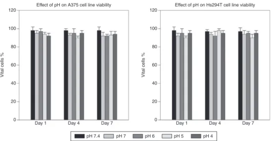
EffectofpHoncytotoxicity
Acidityofthelocalanestheticscouldbeoneofthecausesfor cytotoxiceffectandtoevaluatethesamethecelllineswere treatedwithsalinesolutionswithapHrangingbetween4.0 and7.4. Melanomacelllines’ (A375and Hs294T) viability
120
A
100
80
60
40
20
0
Control 0.0325 0.0625 0.125
Concentration of combination (%) (Lidocaine+Ropivacaine)
Lidocaine+Ropivacaine
Cells analyz
ed (%) - 24 hours
0.250
*
0.500
#
0.750
*
Standard
#
120
B
100
80
60
40
20
0
Control 0.0325 0.0625 0.125
Concentration of combination (%) (Lidocaine+Ropivacaine)
Lidocaine+Ropivacaine
Cells analyz
ed (%) - 72 hours
0.250
*
0.500
#
0.750
*
Standard
#
Figure6 EffectsofcombinationoflidocaineandropivacaineonHs294Tmelanomacellviability.Viabilityratesweresignificantly loweredbycombination oflidocaineandropivacaineconcentrationsgreaterthan0.25%andabove24and72haftertreatment. Thebarsrepresentthemeanvaluesofexperimentswithstandarderroraserrorbars(*p<0.05;#p<0.01;control,salinesolution;
120
100
80
60
40
20
0
Day 1
Vital cells %
Day 4
Effect of pH on A375 cell line viability
Day 7
120
100
80
60
40
20
0
Day 1
pH 7.4 pH 7 pH 6 pH 5 pH 4
Vital cells %
Day 4
Effect of pH on Hs294T cell line viability
Day 7
Figure7 EffectsofpHonmelanomacellviability.Celllines(A375andHs294T)werenotaffectedsignificantlyaftera1-hexposure tosalinesolutionswithpHof7.4,7.0,6.0,5.0,and4.0after1,4,and7daysusingflowcytometry.Theratiosofvitalcellsare shownaspercentageofthetotalcellnumberindifferenttreatmentgroups.Thebarsrepresentstandarderroraserrorbars.
wasnotaffectedsignificantlyaftera1-hexposuretosaline solutionswithpH of4.0, 5.0,6.0,7.0 and7.4 after1,4, and7daysusingflowcytometry.Theratiosofvitalcellsare shownaspercentageofthe totalcellnumberindifferent treatmentgroups(Fig.7AandB).
Caspase-3/8activityanalysis
Apoptosisismediatedbyacascadeofcaspasesor aspartate-specific cysteine proteases. Lidocaine, ropivacaine, and the combination of lidocaine and ropivacaine signifi-cantlyincreasedtheexpressionofactivatedcaspase-3and caspase-8 in a time-dependent manner in A375 cell line (Fig. 8A and B). Peak levels were achieved in all of the localanestheticsrapidlyafter12h.Attheirpeaks,allthese
agentsshowedsignificanthigherlevelofcaspasegeneration comparedwiththecontrolgroup(p≤0.004).
Discussion
Results fromthe conductedstudy supported the hypothe-sisthattheexaminedaminoamidelocalanestheticsposses’ cytotoxic effect on human melanoma cell lines. Local anesthetics have been used in the dermenhysis, spinal anaesthesia, and topical anesthesia to relieve pain in patients withcancer. Lidocaine andropivacaine are clini-callyusedataconcentrationof1.5%or2%and0.5%or0.75%, respectively,forsurgicalanesthesia.1,3Ropivacainehasalso beenreportedtobelesscardiotoxicandlesscentral ner-voustoxicitythanlidocaineandothercommonlyusedlocal anesthetics.Accordingtoapharmacodyanmicstudyoflocal
1.6
1.4
1.2
1.0
0.8
0.6
0.4
0.2
0.0
0 12 24
Time in hours Caspase-3 activity
Caspase-3 activity (
A
v
a
lue)
72
1.6
1.4
1.2
1.0
0.8
0.6
0.4
0.2
0.0
0 12 24
Time in hours
Ropivacaine Lidocaine Lidocaine+Ropivacaine
Caspase-8 activity
Caspase-3 activity (
A
v
a
lue)
72
anesthetics,ropivacainehasalongeractiontimeandhigher potencywhencomparedwithlidocaine.Furthermore, ropi-vacainehasahigherrecoveryratefromcardiacarrestthan lidocaine.21 However,lidocaineandropivacainehavebeen reportedto possesscytotoxicactivity.22 Studies have also beenreporteddose-andtime-dependentcytotoxiceffects oftheselocalanestheticsondifferentcancers.23---25But,not muchresearchhasbeenperformedtoevaluatetheeffectof lidocaineandropivacaine,commonlyusedlocalanesthetics, onhumanmelanomacelllinesandthisdriventheauthors tocarryoutthepresentstudy.Inaddition,thecombination studyoftwolocalanestheticswasalsocarriedoutto inves-tigatethesynergisticcytotoxicactiononhumanmelanoma celllines.Thecombinationofsameclassoflocal anesthet-ics,i.e.aminoamide,wasdevoidofanycompatibleissues andwasfoundtopossesssynergisticactivityandthe cyto-toxic effectfrom the combination were comparable with that of melphalan.However, in the present study ropiva-cainehasdemonstratedcytotoxicactivitywhencompared withnormalsalinesolutionat anyconcentration, butwas lesscytotoxicwhencomparedwithlidocaine,the combina-tionandmelphalan.
Several studies have shown cytotoxiceffects of differ-entconcentrationsoflocalanestheticsafterdifferenttimes of exposure and assessment.7---13 On comparison with an identicalexperimentalsettingoncancercelllines,human melanomacelllinesweremoresensitivetolocal anesthet-ics causing more cell death after treatment with equal concentrations oflocalanesthetics for thesameexposure andassessmenttimes.Afterexposuretolocalanesthetics, immediatecelldeathmighthavebeencausedbynecrosis, whereastheviabilityofcellsdecreasedinatime-dependent manneroverseveralhours.Publishedpapersreportedthat membrane permeability and cytotoxicity was maximum whenthelipophilicity,asdeterminedbytheoctanol---water partitioncoefficient (logp) approached-3.26,27 This finding was further supported by our work in which ropivacaine (logp=2.91)andlidocaine(logp=2.56)hadshowncytotoxic effectsonA375andHs294Tmelanomacelllines.Thisresult suggeststhatthecytoxicityoflocalanestheticsisverymuch relatedtotheirmembranepermeability.
Acidity of the local anesthetics could be one of the causesforcytotoxiceffectsandtoassessthesamethecell linesweretreated withsalinesolutions withapHranging between4.0and7.4.Viabilityassessmentshowedno differ-encesacrossthestudiedpHrangeandcytotoxicactivitydue toacidityoflocalanestheticscanbeexcludedinthisstudy (Fig.7AandB).
Caspaseactivitywasevaluatedtodifferentiatewhether apoptosisornecrosiswasresponsibleforcytotoxicactivity.13 Caspasesplayanimportantroleinapoptosis(programmed cell death), necrosis and inflammation. They are broadly classifiedby their rolesin apoptosis(caspase-3, -6,-7, -8 and-9inmammals)andininflammation(caspases-1,-4,-5, and-12inhumans).Further,caspasesinvolvedinapoptosis areclassifiedbased onmechanismof action aseither ini-tiatorcaspases(caspases-8and-9)orexecutionercaspases (caspases-3,-6,and-7).Intheapoptoticcell,caspases-3is activatedbyextrinsic(deathligand)andintrinsic (mitochon-drial)pathways.28,29Inthisstudy,activityofbothcaspases-3 andcaspases-8wasincreased,suggestingtheroleofthese caspasesintheapoptosisregulation(Fig.8AandB).
Inthisstudy,themethodwasdesignedtodecreaseand to approximately measure possible iatrogenic cell dam-age using an experimental setting in monolayer cultures. Localanesthetics exposure can impair cellularadherence toculture discs.So,non-adherent cells inthe studywere washed and returned back. Most commonly used instru-menttomeasurecellviabilityisflowcytometry,sothiswas employedforaccurateestimateoftheviability.4,25,30
Aminoamide local anesthetics used in this study are widelyusedtotreatirritation,soreness,itchinessandare injected as a dental anaesthetic, or used as local anes-theticsforminorsurgery.31,32Thestudiedlocalanesthetics, includingcombinationsof lidocaineandropivacaine,were morecytotoxicathigherconcentrationsthanatlower con-centrations.Theinvestigatedstudysuggeststhatlidocaine, ropivacaineandcombination of lidocaineand ropivacaine canbefurtherinvestigatedfor theiranticancerproperties for the treatment of melanoma patients, since presently availablechemotherapeuticagentspossessdevastatingside effects.The combination of lidocaineand ropivacaine,in particular,wasfoundtobeascytotoxicasthatofmelphalan, a nitrogen mustard alkylating agent (10% vs. 8%, respec-tively;p<0.01). The studiedlocalanesthetics canfurther beinvestigatedincombinationwithotheranticanceragents or with other local anesthetics for synergistic activity on melanomaand othercancers.On the other hand,authors basedontheresultsofthisstudyrecommendusing commer-ciallyavailablelowconcentrationsofthelesscytotoxiclocal anesthetics,suchasbupivacainefortreatmentofskinand relateddiseases or usingthe concentrationof theselocal anestheticsatwhichtheywerefoundtobelesscytotoxic.
Conclusion
Thecytotoxicactivityoftheinvestigatedaminoamidelocal anesthetics on melanoma cell lines (A375 and Hs294T) is dependentonconcentration,agentandexposuretime.Of the studied local anesthetics, ropivacaine was less cyto-toxicwhen comparedwithlidocaineand the combination oflidocaineandropivacaine.Apoptosisinthecelllineswas mediatedthroughactivityofcaspases-3andcaspases-8.Cell viabilitywasnotaffectedbytheacidityofthestudiedlocal anesthetics. This study had few limitations. In this study, onlytwocelllineswereinvestigatedandthisisaninvitro
study.Althoughthestudygivesanideaofcytotoxicactivity oftheseagentsininvitrosettings,toconfirmthisactivity clinicaltrialsinhumanpopulationarerequired.
Conflicts
of
interest
Theauthorsdeclarenoconflictsofinterest.
References
1.Breu A,EcklS,ZinkW,et al.Cytotoxicityoflocal anesthet-ics on humanmesenchymal stem cells invitro. Arthroscopy. 2013;29:1676---84.
3.SungCM,HahYS,KimJS,etal.Cytotoxiceffectsofropivacaine, bupivacaine,andlidocaineonrotatorcufftenofibroblasts.Am JSportsMed.2014;42:2888---96.
4.GrishkoV,XuM,WilsonG,etal.Apoptosisandmitochondrial dysfunctioninhumanchondrocytesfollowingexposureto lido-caine, bupivacaine, and ropivacaine. JBone Joint Surg Am. 2010;92:609---18.
5.Dregalla RC, Lyons NF, Reischling PD, et al. Amide-type local anestheticsand humanmesenchymal stemcells: clini-calimplicationsforstemcelltherapy.StemCellsTranslMed. 2014;3:365---74.
6.FedderC,Beck-SchimmerB,AguirreJ,etal.Invitroexposure ofhumanfibroblaststolocalanestheticsimpairscellgrowth. ClinExpImmunol.2010;162:280---8.
7.SakaguchiM,KurodaY,HiroseM.Theantiproliferativeeffect oflidocaineonhumantonguecancercellswithinhibitionof theactivityofepidermalgrowthfactorreceptor.AnesthAnalg. 2006;102:1103---7.
8.KimM,LeeYS,MathewsHL,etal.Inductionofapoptoticcell deathinaneuroblastomacelllinebydibucaine.ExpCellRes. 1997;231:235---41.
9.Unami A, Shinohara Y, Ichikawa T, et al. Biochemical and microarrayanalysesofbupivacaine-inducedapoptosis.JToxicol Sci.2003;28:77---94.
10.Perez-CastroR,PatelS,Garavito-AguilarZV,etal. Cytotoxic-ityoflocalanestheticsinhumanneuronalcells.AnesthAnalg. 2009;108:997---1007.
11.LeeHT,XuH,SiegelCD,etal.Localanestheticsinducehuman renalcellapoptosis.AmJNephrol.2003;23:129---39.
12.NakamuraK,KidoH,MorimotoY,etal.Prilocaineinduces apo-ptosisinosteoblasticcells.CanJAnaesth.1999;46:476---82. 13.ShenQ,TianF,JiangP,etal.EGCGenhancesTRAIL-mediated
apoptosisinhumanmelanomaA375cellline.JHuazhongUniv SciTechnolMedSci.2009;29:771---5.
14.RossBK,CodaB,HeathCH.Localanestheticdistributionina spinalmodel:apossiblemechanismofneurologicinjuryafter continuousspinalanesthesia.RegAnesth.1992;17:69---77. 15.AraiT,HokaS.Neurotoxicityofintrathecallocalanesthetics.J
Anesth.2007;21:540---1.
16.Kishimoto T, Bollen AW, Drasner K. Comparative spinal neurotoxicity of prilocaine and lidocaine. Anesthesiology. 2002;97:1250---3.
17.KamiyaY,OhtaK,KanekoY.Lidocaine-inducedapoptosisand necrosis inU937cells depending onitsdosage. BiomedRes. 2005;26:231---9.
18.BoselliE, Duflo F,Debon R, et al. The induction of apopto-sisbylocalanesthetics:acomparisonbetweenlidocaineand ropivacaine.AnesthAnalg.2003;96:755---6.
19.BauerTW,GutierrezM,DudrickDJ,etal.Ahumanmelanoma xenograftinanuderatrespondstoisolatedlimbperfusionwith TNFplusmelphalan.Surgery.2003;133:420---8.
20.HanssonJ,BerhaneK,CastroVM,etal.Sensitizationofhuman melanoma cellsto thecytotoxic effectof melphalanbythe glutathionetransferaseinhibitorethacrynicacid.CancerRes. 1991;51:94---8.
21.AliQE,ManjunathaL,AmirSH,etal.Efficacyofclonidineas anadjuvanttoropivacaineinsupraclavicularbrachial plexus block:aprospectivestudy.IndianJAnaesth.2014;58:709---13. 22.ChlebowskiRT,BlockJB,CundiffD,etal.Doxorubicin
cytotox-icityenhancedbylocalanestheticsinahumanmelanomacell line.CancerTreatRep.1982;66:121---5.
23.Lirk P, Berger R, Hollmann MW, et al. Lidocaine time- and dose-dependentlydemethylatesdeoxyribonucleicacidinbreast cancercelllinesinvitro.BrJAnaesth.2012;109:200---7. 24.MaletA,FaureMO,DeletageN,etal.Thecomparativecytotoxic
effectsofdifferentlocalanestheticsonahumanneuroblastoma cellline.AnesthAnalg.2015;120:589---96.
25.KarpieJC,ChuCR.Lidocaineexhibitsdose-andtime-dependent cytotoxiceffectsonbovinearticularchondrocytesinvitro.Am JSportsMed.2007;35:1621---7.
26.Fujisawa S, Atsumi T, Kadoma Y, et al. Antioxidant and prooxidantactionofeugenol-relatedcompoundsandtheir cyto-toxicity.Toxicology.2002;177:39---54.
27.Ishihara M, Yokote Y, Sakagami H. Quantitative structure---cytotoxicity relationship analysis of coumarin anditsderivativesbysemiempiricalmolecularorbitalmethod. AnticancerRes.2006;26:2883---6.
28.McIlwainDR,BergerT,MakTW.Caspasefunctionsincelldeath anddisease.ColdSpringHarbPerspectBiol.2013;5:a008656. 29.PorterAG,JanickeRU.Emergingrolesofcaspase-3inapoptosis.
CellDeathDiffer.1999;6:99---104.
30.ChuCR,CoyleCH,ChuCT,etal.Invivoeffectsofsingle intra-articularinjectionof0.5%bupivacaineonarticularcartilage.J BoneJointSurgAm.2010;92:599---608.
31.NeafseyPJ.Patchingpainwithlidocaine:newusesforthe lido-caine5%patch.HomeHealthcNurse.2004;22:562---4. 32.DiCroceDE,TrinksPW,deLaCC.Amide-typelocalanesthetics