Salmonella enterica
Serovar Typhi
Patricio Retamal1,2, Mario Castillo-Ruiz1, Guido C. Mora1*1Departamento de Ciencias Biolo´gicas, Universidad Andre´s Bello, Santiago, Chile,2Programa de Doctorado Gene´tica Molecular y Microbiologı´a, Pontificia Universidad Cato´lica de Chile, Santiago, Chile
Abstract
The MgtC is a virulence factor inSalmonellaTyphimurium that is required for growth at low-Mg2+concentrations and
intramacrophage survival. This gene is codified in a conserved region of theSalmonellapathogenicity island 3 (SPI-3), and is also present in the chromosome of otherSalmonellaserovars. In this study we characterized the MgtC factor inS.Typhi, a
human specific pathogen, by using mgtCand SPI-3 mutant strains. We found that MgtC is the most important factor
codified in the SPI-3 ofS.Typhi for growth in low-Mg2+media and survival within human cells. In addition, by using reporter
genes we determined that the low-Mg2+concentration, acidic media and PhoP regulator inducemgtCexpression in S.
Typhi. We suggest that MgtC is the most important virulence factor codified in the SPI-3 ofS.Typhi.
Citation:Retamal P, Castillo-Ruiz M, Mora GC (2009) Characterization of MgtC, a Virulence Factor ofSalmonella entericaSerovar Typhi. PLoS ONE 4(5): e5551. doi:10.1371/journal.pone.0005551
Editor:Dana Davis, University of Minnesota, United States of America
ReceivedJanuary 30, 2009;AcceptedApril 20, 2009;PublishedMay 14, 2009
Copyright:ß2009 Retamal et al. This is an open-access article distributed under the terms of the Creative Commons Attribution License, which permits unrestricted use, distribution, and reproduction in any medium, provided the original author and source are credited.
Funding:This study was supported by FONDECYT grant 1060999, UNAB grant 0404 and predoctoral fellowships from CONICYT and PUC. The funders had no role in study design, data collection and analysis, decision to publish, or preparation of the manuscript.
Competing Interests:The authors have declared that no competing interests exist. * E-mail: [email protected]
Introduction
The Salmonella enterica genome has at least five DNA regions associated with pathogenicity, referred to as the Salmonella
pathogenicity islands (SPI). One such island, SPI-3, is located in theselClocus ofS.Typhimurium and contains ten ORFs [1], among which some have been experimentally associated with virulence functions of this bacterium. This is the case for themgtCBoperon, for which there is evidence of involvement in intramacrophage survival and virulence in mice [2,3]. This operon is codified in allSalmonella
serovars in a very conserved SPI-3 region [4]. ThemgtCsequence seems to encode a virulence factor that has been repeatedly acquired by horizontal gene transfer throughout bacterial evolution, since it has also been associated with virulence in Mycobacterium tuberculosisand Brucella suis [5–7]. MgtC is a protein of unknown function of about 25 kDa in size. InS. entericaserovar Typhimurium (S.Typhimurium), the experimental evidence suggests that MgtC participates in adaptation to low-Mg2+
environments, supporting bacterial invasion and proliferation in macrophages [2]. Although is co-transcribed withmgtB, which encodes a Mg2+
transporter, MgtC is not required for MgtB function [8]. Indeed, a recently described polypeptide encoded by themgtCBoperon, named MgtR, promotes MgtC degradation by a bacterial protease, acting as a negative feedback that limits the amount of MgtC under certain conditions [9]. In addition, it has been shown that the two-component system PhoP-PhoQ induces the expression ofmgtC, in response to low Mg2+
levels and acidic pH [8,10].
Another SPI-3 gene involved in bacterial pathogenicity ismisL, which encodes an autotransporter protein involved in the adhesion ofS.Typhimurium to the extracellular matrix in mice and chicks, thereby acting as an intestinal colonization factor [11,12]. It has also been shown thatmarT, another sequence present in SPI-3,
encodes a transcriptional regulator that induces the expression of
misL [13]. There is no additional information on other SPI-3 ORFs, all of them remaining until now as sequences encoding conserved hypothetical proteins with unknown function [14].
S. Typhimurium is a wild host range serovar and has been extensively studied in a murine model of systemic infection to indirectly elucidate some microbiological and immunological traits of typhoid fever in humans [15], a life-threatening and systemic infection caused by theS. entericaserovar Typhi (S.Typhi). The latter, a human-restricted pathogen, is a facultative intracellular bacterium responsible for significant morbidity and mortality worldwide, and there are an estimated 21.5 million cases per year, most of which occur in developing countries [16].
The aim of this work was to characterize the role of the MgtC factor in the virulence ofS.Typhi by comparing the growth and survival of mgtC and SPI-3 mutant strains in different stressful conditions, and determining the signals and transcriptional regulators that command MgtC expression. We demonstrated that MgtC is the most important factor in S. Typhi SPI-3 for bacterial growth in a low-Mg2+
environment and for bacterial survival inside human cells. In addition, the PhoP regulator participates in inducing the expression ofmgtCinS.Typhi.
Materials and Methods
Bacterial strains and growth conditions
AllSalmonellaTyphi strains used in this study are derived from STH2370, a Chilean clinical isolate described previously [15]. Unless otherwise stated bacteria were grown at 37uC in Luria Bertani (LB) broth or in M9 minimal medium supplemented with either 10mM or 10 mM MgCl2, 0.2% glucose, tryptophan and
to pH 7.0 (NaHPO4/NaH2PO4 25 mM) or 5.0 (citric acid/
sodium citrate 0.1 M) and the following antibiotics were added: chloramphenicol (Cam; 20mg/mL), kanamycin (Kan; 50mg/mL), ampicillin (Amp; 100mg/mL) and gentamicin (Gem; 50mg/mL).
PCR amplifications and construction of mutant strains
PCR amplifications were performed in a standard volume of 25mL. Reaction mixes contained 16 PCR buffer, 1.5 mM MgCl2, each deoxynucleoside triphosphate (200mM), primers
(1mM), 100 ng of template DNA, and 2 U of Taq (Fermentas) DNA polymerase. Standard conditions for amplification were an initial step of 95uC for 5 min, 30 cycles of incubation at 96uC for 40 s, 60uC for 40 s, and 72uC for 2 min, followed by a final extension step at 72uC for 10 min. TemplateS.Typhi chromo-somal DNA was prepared by the phenol chloroform extraction method [17].
TheDmgtC::FRT (DmgtC) mutant strain was constructed using the lambda Red recombinase system [18]. Briefly, the CamR cassette (chloramphenicol resistance, codified in the pKD3 plasmid) was amplified using the primers MGW1 (59 -ATGGAG- GAACGTATGTTAATGTTTCCTTATATTTTAAATTTGTA-GGCTGGAGCTGCTTCG-39) and MGW2 (59 -TGACCCAC- GAGCTCGGCACGAATTTCTTTATAGCCCTGTTCATAT-GAATATCCTCCTTA-39). Once the DmgtC::cat mutant strain was obtained, the CamRdeterminant was removed and substitut-ed by the ‘‘FRT scar’’ [18], and the resulting colonies were testsubstitut-ed by PCR to confirm themgtCdeletion.
The DSPI-3::FRT mutant (DSPI-3) was constructed with the same procedure, amplifying the KanR cassette (kanamycin resistance, codified in the pKD4 plasmid) using the primers
SPW1 (59
-AACGCAGGCGCTACGTTTGTCGATGCCGTAA-CTTTCTGAATGTAGGCTGGAGCTGCTTCG-39) and SPW2
(59
-GCTAAATATAGCACGTACTTATTCTTCCAGAAAAAA-TGGACATATGAATATCCTCCTTA-39). Once theDSPI-3::aph
mutant strain was obtained, the KanRdeterminant was substituted by the ‘‘FRT scar’’ as described previously [18].
With the mutant strain EG14598 (S. Typhimurium 14028s DphoP::cat) [19], a P22 HT105/1 int201 phage lysate was made [20] and used for generalized transduction overS.Typhi strains.
Phenotypic analysis of theS.Typhi mutant strains
Growth in a Mg2+
-limiting environment was evaluated as described previously [21] with some modifications. Briefly, an overnight culture grown in M9 minimal media with 10 mM MgCl2was washed three times with Mg2+-free medium, diluted 1/
200 in culture media containing either 10mM or 10 mM MgCl2,
and incubated with shaking at 37uC for different lengths of time. Growth was measured with a spectrophotometer at an optical density of 600 nm (OD600).
To evaluate the effect of pH, overnight cultures were grown in LB broth at pH 7, then washed three times with LB at the desired pH (5 or 7), diluted 1/200 in the same medium and incubated with shaking at 37uC for different lengths of time. Growth was measured with a spectrophotometer at OD600.
The infection assays using monocytic (U937) and epithelial (HEp-2) human cells were carried out as described previously [15,22], with the following modifications. Cells were grown in Dulbecco’s modified Eagle’s medium supplemented with 10% (vol/vol) fetal bovine serum, seeded into 24-well tissue culture plates at a concentration of 105cells per well, and then incubated at 37uC in 5% CO2until confluent growth was achieved. Later the
cells were centrifuged and washed three times with PBS. Approximately 26106 to 56106 CFU of exponential-phase
(OD600, 0.15 to 0.20) anaerobically grown bacteria was pelleted,
washed twice with PBS, and resuspended in 1 mL of PBS. Aliquots (100mL) of bacteria were added to cells at a multiplicity of infection of 50:1(U937) and 100:1 (HEp-2). After 1 h of infection, cells were centrifuged and washed three times with PBS, and the medium was replaced with Dulbecco’s modified Eagle’s medium supplemented with 10% (vol/vol) fetal bovine serum containing gentamicin (200mg/mL). After additional incubation for 1 and 23 h (times 2 and 24 h respectively) U937 and HEp-2 cells were washed three times with PBS and lysed with 0.5% deoxycholate, and the titers of intracellular bacteria were determined by serial dilution of cell lysates on agar plates. The percentage of survival was calculated at 2 h considering the initial inoculate as 100%, and at 24 considering the CFU counted at 2 h as 100%.
b-Galactosidase assay
ThemgtCpromoter activity was evaluated by a transcriptional fusion to the Lac reporter, as described previously [23,24] using the pCE36 plasmid.b-Galactosidase activity was measured by a modification of the Miller’s method [25]. Fifty microliters of the bacterial culture were suspended in 950mL of Z buffer (60 mM Na2HPO4, 40 mM NaH2PO4, 10 mM KCl, 1 mM MgSO4,
50 mMb-mercaptoethanol, pH 7.0). Bacteria were permeabilized with 10mL chloroform and 10mL 0.1% SDS, incubated at 30uC for 10 min, and 200mL of o-nitrophenyl-b-D-galactopyranoside (4 mg/mL) was added. Reactions were stopped by addition of 500mL 1 M Na2CO3.b-Galactosidase activity was calculated in
Miller units, using the formula 1036(OD42021.756OD550)/
(mL6min6OD600).
RNA isolation and RT-PCR
Total RNA was extracted and purified using Trizol and was treated with RNase-free DNase I (amplification grade; Gibco-BRL). RT-PCR was performed with 500 ng of DNase-treated RNA using the Superscript reverse transcriptase (Invitrogen). Amplification was performed for 30 cycles (94uC for 40 s, 55uC for 40 s, and 72uC for 1.5 min, followed by a 10 min extension at 72uC). The primers used were RTMGC1 (59 -TCGGCG-TGTTATGCGGCTTA-39), RTMGC2 (59 -AGCCCTGTTCC-TGAGCGGGG-39) and RTMGB2 (59 -CACGGCGTAACGG-GAGCCAG-39) corresponding to an internal region of themgtC
(RTMGC1 and RTMGC2) and mgtC-mgtB
(RTMGC1-RTMGB2) sequences. In addition, the universal primers 8F and 1498R were used to amplify 16S rRNA [15]. Genomic DNA served as a positive control, and DNase-treated RNA that had not been reverse transcribed was used as a negative control. The PCR product was electrophoresed on 1% agarose gels and stained with ethidium bromide.
MgtC epitope tagging and immunoblot assay
A translational fusion of three copies of FLAG epitope (36FLAG) with the MgtC sequence was constructed using the method described by Uzzau et al. [26]. The 36FLAG epitope codified on
the pSUB11 plasmid was amplified using the primers FMG1 (59 - CGATAATATCACCGCAATTCACTGGAGCATTGATAGT-CAAGACTACAAAGACCATGACGG-39) and FMG2 (59 -ACT- GACCCCTGCCAGTGCCATCAGAACGTAAATAAACGGG-CATATGAATATCCTCCTTAG-39). Once inserted immediately preceding the translation stop signal, themgtC-36flag fusion was
confirmed by PCR and the functionality of the protein was verified by the growth of the strain in low-Mg2+
medium.
1 mL of 100 mM Tris–HCl (pH 8) and sonicated, the total protein was quantified by the Bradford method. The SDS-PAGE was made using 10 ng of total protein per sample.
Statistical analysis
Statistical analysis was performed using the one way ANOVA and Student’s t-test for independent samples. Values of P,0.05 were considered significant. These tests were performed using Microsoft ExcelHsoftware.
Results
1. MgtC is required forS.Typhi growth in a low-Mg+2 medium
To elucidate the role ofmgtCinS.Typhi, we investigated growth in low-Mg+2
minimal media as evaluated previously in S.
Typhimurium [2,8]. The Mg2+
concentrations used were 10mM or 10 mM, representing the intracellular and extracellular environment respectively [2,27]. At the 10mM concentration, theDmgtCmutant strain grew significantly less than the wild type strain (p,0.05), reestablishing its phenotype when complemented withmgtCcloned in pBBR-5 plasmid. In contrast, at 10 mM Mg2+
there was no difference among the tested strains (Fig. 1A and 1B). These results are in accordance with reports of the role of MgtC in the virulence ofS.Typhimurium [2,8] and other bacteria [5,6].
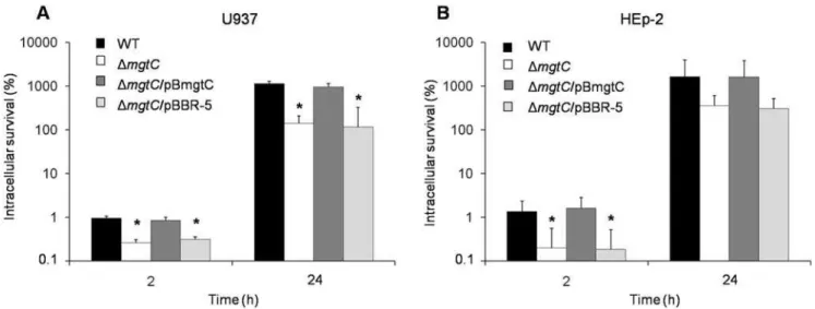
2. MgtC is required for growth ofS. Typhi within epithelial and monocytic human cells
To verify the role of MgtC in the intracellular survival ofS.
Typhi, we tested the DmgtCstrain in infection assays using both HEp-2 epithelial and U937 monocytic cell lines. Two post-infection times were evaluated, 2 h and 24 h, representing the early and late survival abilities respectively. As expected, MgtC is required for infection of monocytic cells, with significant differences (p,0.05) among wild type and DmgtC strains (Fig. 2A). Remarkably, inside HEp-2 epithelial cells there was a significant impairment (p,0.05) in the invasive phenotype of the
DmgtCmutant strain (Fig. 2B), suggesting that MgtC participates during the infection of this kind of human cell.
3. MgtC reestablishes the wild type phenotype of a SPI-3 mutant strain both in low Mg2+media and inside
monocytic cells
Previously it has been shown that MgtC can restore the wild type intramacrophage survival phenotype of a S. Typhimurium
mgtCB mutant strain [2]. In S. Typhi we wanted to determine whether MgtC is required for intracellular survival and growth at low-Mg2+ concentrations in the context of a SPI-3 deletion.
Therefore, we constructed the DSPI-3 mutant strain, the complemented DSPI-3/pBmgtC (mgtC+
) and DSPI-3/pBBR5 (mgtC2) strains, and repeated the assays of growth in low Mg2+
media (Fig. 3A) and survival inside human monocytic cells (Fig. 3B). The results suggest that MgtC can restore the phenotypes observed in a SPI-3 mutant strain by itself and can be considered the most important product codified on theS.Typhi SPI-3 island for bacterial response to those experimental conditions.
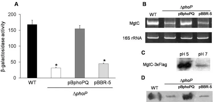
4. The PhoP regulator controlsmgtCexpression in a low Mg2+and acidic environment
In S. Typhimurium, the PhoP-PhoQ two-component system regulates the expression of many genes when bacteria are exposed to the intracellular environment, including SPI-2 and SPI-3 associated effectors [10,19]. The signals sensed by this regulatory system are the extracellular pH and Mg2+
concentrations. PhoQ is the sensor component that phosphorylates the PhoP regulator, which then modifies gene expression. By using a STH2370 DphoP::cat mutant strain (DphoP), we examined whether PhoP regulates MgtC in S. Typhi. The results obtained by b -galactosidase and RT-PCR assays show that mgtCtranscription is induced in a phoP-dependent manner by either low Mg2+
(10mM, data not shown) or pH 5 (Fig. 4A and 4B). Moreover, immunoblot assay shows an increase in the MgtC levels under the same conditions (Fig. 4C and 4D), a finding that differs from the
Figure 1. MgtC is necessary for growth at a low-Mg2+concentration.Strains WT (STH2370 wild type),DmgtC(mgtC2),DmgtC/pBmgtC (mgtC+
) andDmgtC/pBBR-5 (mgtC2) were grown in M9 minimal medium supplemented with 10 mM (A) or 10mM (B) MgCl
2. The OD600was
reported situation inS. Typhimurium where MgtC translation is not detected in many hours [8,9].
Discussion
In previous reports the Salmonella SPI-3 island has been associated with intramacrophage invasion by supporting survival when Mg2+
is scarce, a condition that seems a common strategy of the host to avoid the growth of intracellular bacteria [1,2,8]. Mg2+
is a divalent ion essential for living organisms that works as a regulator and co-factor in many proteins, stabilizing membranes, ribosomes and other cellular structures [28]. Salmonella contains several transport systems, both inducible and constitutive, that have functional complementarities with the aim of adjusting the Mg2+
concentration in different environmental conditions [29,30].
In addition, these systems are controlled by transcriptional and post-transcriptional regulatory networks to maintain strict control of the Mg2+
balance [31,32], stabilizing its concentrations as required for biological processes in Salmonella. In this context, MgtC seems to be the most important SPI-3 factor that supports the survival and growth ofSalmonellain low-Mg2+
concentrations, as observed in previous reports forS.Typhimurium and in this work withS.Typhi. This factor is codified in a SPI-3 conserved region [4] and probably exerts the same, although yet unknown, function in allSalmonellaserovars.
In this work, the decreased ability to survive within human monocytic cells observed with aS.TyphiDSPI-3 strain could be overcome with an mgtC-containing plasmid, which restored the wild type phenotype at 2 and 24 hours post-infection. This means that MgtC is a virulence factor playing a major role that is not
Figure 3. MgtC can restore the WT phenotype in a SPI-3 mutant strain.Strains WT (STH2370 wild type),DSPI-3 (SPI-32),DSPI-3/pBmgtC (mgtC+) andDSPI-3/pBBR-5 (SPI-32). (A) For growth in 10
mM MgCl2strains were incubated in M9 minimal medium and the OD600was measured at
the indicated times. (B) U937 cells were infected at a MOI of 50:1. Colonies were counted at time 2 h and 24 h and expressed as a percentage of intracellular survival. Values represent the mean of at least three independent experiments6SD (*p,0.05).
doi:10.1371/journal.pone.0005551.g003
Figure 2. MgtC has an important role in the growth of S. Typhi within human cells.Infection assays in U937 (A) and HEp-2 (B) human cells using WT (STH2370 wild type),DmgtC(mgtC2),DmgtC/pBmgtC (mgtC+
) andDmgtC/pBBR-5 (mgtC2) strains. Culture cells were infected at a MOI of 50:1 (U937) and 100:1 (HEp-2), respectively. Colonies were counted at time 2 h and 24 h and expressed as a percentage of intracellular survival. Values represent the mean of at least three independent experiments6SD (*p,005).
supplied by any other bacterial factor codified either inside SPI-3 or in the entire chromosome ofSalmonella.
A characteristic phenotype associated with S. Typhi MgtC was demonstrated by the early survival phenotype inside HEp-2 epithelial cells, in which theS.TyphimgtCmutant strain showed a significant lower survival (p,0.05) than the wild type strain (Fig. 2B). This difference has not been reported previously in any
Salmonella serovar and suggests that S.Typhi requires the MgtC function from the initial infective phase, when it colonizes the intestinal epithelium. Whether this requirement responds to particular conditions during bacterial entry or when bacteria are inside human epithelial cells are questions that remain to be elucidated.
However, these findings are indicating that both epithelial and monocytic human cells represent a variety of conditions that require the MgtC function for the bacterial survival. This hypothetical ‘‘multi-requirement’’ of MgtC is in accordance to several reports suggesting a connection of this virulence factor with the structural stability of Mg2+
channels in the bacterial cell membrane [7], linked to survival in Mn+2
depleted environments, or modifying the membrane potential of the host cell and affecting the host-pathogen interaction [33,34]. Since single amino acid substitutions can affect one role of MgtC without affecting others [35], it seems possible to assume a diversity of functions in which this internal membrane protein participates during infection. In addition, MgtC is important in other bacterial species that are able to invade, live and proliferate within host cells, as forMycobacterium tuberculosis,Brucella melitensisandYersinia pestis[5,7].
The expression assays showed that mgtC transcription and translation are induced at low Mg2+concentrations and acidic pH,
and that PhoP is the global regulator that participates in this process. These signals stimulate the expression of many genes associated with pathogenicity by inducing the PhoP-PhoQ system [10], and MgtC ofS.Typhi is one of these. In addition,mgtCand
mgtBare co-transcribed inS.Typhi (data not shown), suggesting the almost identical functionality of these sequences between S.
Typhimurium andS.Typhi, and probably in allSalmonellaserovars that contain themgtCBoperon in their chromosomes. Previously it has been shown in S. Typhimurium that MgtR induces the degradation of MgtC but not of MgtB, resulting in a downreg-ulation of MgtC when the operon is expressed [9]. Our results suggest that inS. Typhi this regulation could be different, since MgtC is detected after acidic or low magnesium stimuli.
In conclusion, in this work we determined that MgtC in S.
Typhi represents a mechanism of pathogenicity codified inside the SPI-3 that has a relevant role in the intracellular survival of bacteria, induced by the PhoP global regulator in response to a low-Mg2+ concentration and acidic pH. These findings suggest
that MgtC is a key factor in most, if not all, pathogenicSalmonella
serovars.
Acknowledgments
The authors thank Eduardo Groisman that kindly provided the mutant strain EG14598, and Juan Fuentes by the critical review of this paper.
Author Contributions
Conceived and designed the experiments: PR GCM. Performed the experiments: PR MCR GCM. Analyzed the data: PR GCM. Wrote the paper: PR GCM.
Figure 4. The PhoP regulator controls the expression ofmgtCinS. Typhi.The strains used were WT (STH2370 wild type),DphoP(phoP::cam),
DphoP/pBphoPQ (phoPQ+) andDphoP/pBBR-5 (phoPQ2). The cultures were grown in acidic LB medium (pH 5) for 5 h, and the samples taken had an OD600ranging from 0.3 to 0.6. The expression ofmgtCwas evaluated using three methods:A,b-Galactosidase assay with strains carrying the reporter
lacZgene downstream of themgtCpromoter. Values represent the mean of at least three independent experiments6SD (*p,0.05).B, RT-PCR assay.
C, Immunoblot method using a STH2370 MgtC-36Flag epitope-tagged strain grown on LB broth at both pH 5 and pH 7.D, Immunoblot method using strains carrying the MgtC-36Flag epitope tag and grown in acidic LB medium (pH 5) for 5 h.
References
1. Blanc-Potard AB, Solomon F, Kayser J, Groisman EA (1999) The SPI-3 pathogenicity island ofSalmonella enterica. J Bacteriol 181: 998–1004. 2. Blanc-Potard AB, Groisman EA (1997) The Salmonella selClocus contains a
pathogenicity island mediating intramacrophage survival. Embo J 16: 5376–5385.
3. Smith RL, Kaczmarek MT, Kucharski LM, Maguire ME (1998) Magnesium transport in Salmonella typhimurium: regulation of mgtA and mgtCB during invasion of epithelial and macrophage cells. Microbiology 144(Pt 7): 1835–1843. 4. Amavisit P, Lightfoot D, Browning GF, Markham PF (2003) Variation between pathogenic serovars withinSalmonella pathogenicity islands. J Bacteriol 185: 3624–3635.
5. Buchmeier N, Blanc-Potard A, Ehrt S, Piddington D, Riley L, et al. (2000) A parallel intraphagosomal survival strategy shared byMycobacterium tuberculosisand
Salmonella enterica. Mol Microbiol 35: 1375–1382.
6. Lavigne JP, O’Callaghan D, Blanc-Potard AB (2005) Requirement of MgtC for
Brucella suisintramacrophage growth: a potential mechanism shared bySalmonella entericaandMycobacterium tuberculosisfor adaptation to a low-Mg2+
environment. Infect Immun 73: 3160–3163.
7. Blanc-Potard AB, Lafay B (2003) MgtC as a horizontally-acquired virulence factor of intracellular bacterial pathogens: evidence from molecular phylogeny and comparative genomics. J Mol Evol 57: 479–486.
8. Moncrief MB, Maguire ME (1998) Magnesium and the role of MgtC in growth ofSalmonellatyphimurium. Infect Immun 66: 3802–3809.
9. Alix E, Blanc-Potard AB (2008) Peptide-assisted degradation of theSalmonella
MgtC virulence factor. Embo J 27: 546–557.
10. Groisman EA (2001) The pleiotropic two-component regulatory system PhoP-PhoQ. J Bacteriol 183: 1835–1842.
11. Dorsey CW, Laarakker MC, Humphries AD, Weening EH, Baumler AJ (2005)
Salmonella entericaserotype Typhimurium MisL is an intestinal colonization factor that binds fibronectin. Mol Microbiol 57: 196–211.
12. Morgan E, Campbell JD, Rowe SC, Bispham J, Stevens MP, et al. (2004) Identification of host-specific colonization factors ofSalmonella entericaserovar Typhimurium. Mol Microbiol 54: 994–1010.
13. Tukel C, Akcelik M, de Jong MF, Simsek O, Tsolis RM, et al. (2007) MarT activates expression of the MisL autotransporter protein ofSalmonella enterica
serotype Typhimurium. J Bacteriol.
14. Chaudhuri RR, Khan AM, Pallen MJ (2004) coliBASE: an online database for
Escherichia coli,ShigellaandSalmonellacomparative genomics. Nucleic Acids Res 32: D296–299.
15. Bucarey SA, Villagra NA, Martinic MP, Trombert AN, Santiviago CA, et al. (2005) TheSalmonella entericaserovar Typhitsxgene, encoding a nucleoside-specific porin, is essential for prototrophic growth in the absence of nucleosides. Infect Immun 73: 6210–6219.
16. Pang T, Levine MM, Ivanoff B, Wain J, Finlay BB (1998) Typhoid fever– important issues still remain. Trends Microbiol 6: 131–133.
17. Sambrook J, Fritsch F, Maniatis T (1989) Molecular Cloning: a Laboratory Manual. Cold Spring Harbor, N.Y.: Cold Spring Harbor Laboratory.
18. Datsenko KA, Wanner BL (2000) One-step inactivation of chromosomal genes inEscherichia coliK-12 using PCR products. Proc Natl Acad Sci U S A 97: 6640–6645.
19. Bijlsma JJ, Groisman EA (2005) The PhoP/PhoQ system controls the intramacrophage type three secretion system ofSalmonella enterica. Mol Microbiol 57: 85–96.
20. Schmieger H (1972) Phage P22-mutants with increased or decreased transduction abilities. Mol Gen Genet 119: 75–88.
21. Tu X, Latifi T, Bougdour A, Gottesman S, Groisman EA (2006) The PhoP/ PhoQ two-component system stabilizes the alternative sigma factor RpoS in
Salmonella enterica. Proc Natl Acad Sci U S A 103: 13503–13508.
22. Contreras I, Toro C, Troncoso G, Mora G (1997)SalmonellaTyphi mutants defective in anaerobic respiration are impaired in their ability to replicate within epithelial cells. Microbiology 143: 2665–2672.
23. Ellermeier CD, Janakiraman A, Slauch JM (2002) Construction of targeted single copy lac fusions using lambda Red and FLP-mediated site-specific recombination in bacteria. Gene 290: 153–161.
24. Bucarey SA, Villagra NA, Fuentes JA, Mora GC (2006) The cotranscribed
Salmonella entericasv. TyphitsxandimpX genes encode opposing nucleoside-specific import and export proteins. Genetics 173: 25–34.
25. Miller FD, Hershberger CL (1984) A quantitative beta-galactosidase alpha-complementation assay for fusion proteins containing human insulin B-chain peptides. Gene 29: 247–250.
26. Uzzau S, Figueroa-Bossi N, Rubino S, Bossi L (2001) Epitope tagging of chromosomal genes inSalmonella. Proc Natl Acad Sci U S A 98: 15264–15269. 27. Eriksson S, Lucchini S, Thompson A, Rhen M, Hinton JC (2003) Unravelling the biology of macrophage infection by gene expression profiling of intracellular
Salmonella enterica. Mol Microbiol 47: 103–118.
28. Maguire ME, Cowan JA (2002) Magnesium chemistry and biochemistry. Biometals 15: 203–210.
29. Chamnongpol S, Groisman EA (2002) Mg2+
homeostasis and avoidance of metal toxicity. Mol Microbiol 44: 561–571.
30. Smith RL, Maguire ME (1998) Microbial magnesium transport: unusual transporters searching for identity. Mol Microbiol 28: 217–226.
31. Spinelli SV, Pontel LB, Garcia Vescovi E, Soncini FC (2008) Regulation of magnesium homeostasis in Salmonella: Mg(2+)
targets themgtA transcript for degradation by RNase E. FEMS Microbiol Lett 280: 226–234.
32. Cromie MJ, Shi Y, Latifi T, Groisman EA (2006) An RNA sensor for intracellular Mg(2+)
. Cell 125: 71–84.
33. Alix E, Blanc-Potard AB (2007) MgtC: a key player in intramacrophage survival. Trends Microbiol 15: 252–256.
34. Gunzel D, Kucharski LM, Kehres DG, Romero MF, Maguire ME (2006) The MgtC virulence factor of Salmonella enterica serovar Typhimurium activates Na(+)
,K(+)
-ATPase. J Bacteriol 188: 5586–5594.