170 Rev Odonto Cienc 2012;27(2):170-173
Received: July 30, 2011 Accepted: April 11, 2012
Conflict of Interests: The authors state that there are no financial and personal conflicts of interest that could have inappropriately influenced their work.
Copyright: © 2011 Manzi et al.; licensee EDIPUCRS. This is an Open Access article distributed under the terms of the Creative Commons Attribution-Noncommercial-No Derivative Works 3.0 Unported License.
Case Report
Radiographic and imaging diagnosis of
rhinolith in dental clinics: A case report
Diagnóstico radiográfico e imaginológico do rinólito na
clínica odontológica: relato de caso
Flávio Ricardo Manzi a
Priscila Dias Peyneau a
Fernanda Pimenta Piassi a
Vinícius de Carvalho Machado b
Andréa Cristina Lopes c
a Dental School, Pontifical Catholic University of
Minas Gerais (PUC-MG), Belo Horizonte, MG, Brazil
b Dental School, São Leopoldo Mandic, Campinas,
SP, Brazil
c Private practice, Belo Horizonte, MG, Brazil
Correspondence: Flávio Ricardo Manzi
Av. Dom José Gaspar, 500. Prédio 45 Clínica de Radiologia Coração Eucarístico Belo Horizonte, MG – Brasil
30535-901
E-mail: [email protected] Abstract
Purpose: Rhinolith is a calcification that occurs in the nasal cavity and may be discovered during routine radiographic exam. This calcification is rare, generally single, exogenous or endogenous, unilateral, and asymptomatic. It can cause nasal obstruction, fetid odour and purulent secretion. This study reported a case of rhinolith and its radiographic features. Case description: A female, 14-year-old patient had a radiopaque mass in the right nasal cavity. The primary treatment plan included oral surgery to remove a supposed impacted supernumerary tooth, which diagnosis was based on conventional radiographic exams. After an unsuccessful surgery, additional exam with computed tomography showed images compatible with a rhinolith.
Conclusion: Computed tomographic scans are required for differential diagnosis of pathologies in the nasal cavity to provide proper treatment.
Key words: Rhinolith; computed tomography; oral radiology
Resumo
Objetivo: O rinólito é uma calcificação na cavidade nasal que pode ser descoberta durante o exame radiográfico odontológico de rotina. Esta massa calcificada é rara, pode exógena ou endógena, geralmente ocorre isoladamente, unilateral e assintomática. Pode causar obstrução nasal, odor fétido e secreção purulenta. Este estudo objetivou apresentar um caso de rinólito e suas características radiográficas.
Descrição do caso: Uma paciente do sexo feminino, 14 anos de idade, tinha uma massa radiopaca na cavidade nasal direita. O tratamento odontológico primário incluía uma cirurgia para a remoção de um suposto dente supranumerário impactado diagnosticado após exames de radiografia convencional. Devido ao insucesso da cirurgia, foi realizado um exame por tomografia computadorizada e foi diagnosticado o rinólito.
Conclusão: Exames de tomografia computadorizada adequados são necessários para o diagnóstico diferencial de patologias na cavidade nasal para proporcionar um tratamento adequado.
Rev Odonto Cienc 2012;27(2):170-173 171 Manzi et al.
Introduction
Rhinoliths are calciications that occur in the nasal cavity due to the deposition of mineral salts on a foreign body (1-2). This calciication can be exogenous or endogenous, is rare, generally single, unilateral and asymptomatic. Rhinoliths can cause nasal obstruction, fetid odour and purulent secretion. When asymptomatic, they are frequently discovered during routine radiographic examination (3-5).
Conventional radiographs are important for the differentiation of the rhinolith from other lesions with similar aspect, such as odontoma, impacted tooth, foreign bodies and other calciications. Nevertheless, computerized tomography provides better images to evaluate this condition and its relationship with adjacent structures.
The aim of this study was to report a clinical case of a rhinolith, describing its main characteristics for proper diagnosis by dental and medical imaging exams.
Case report
A female, 14-year-old patient was undergoing orthodontic treatment when the dentist referred her for removal of an impacted supernumerary tooth located in the right maxilla, close to tooth 13. This initial dental treatment plan was proposed as a result of diagnosis based on panoramic radiograph, lateral teleradiograph and occlusal radiograph of the maxilla. In these exams a well-deined, ovoid radiopaque image was observed close to the nasal fossa on the right side (Fig. 1-3). The patient presented no symptoms and was not aware of any mass inside her nasal cavity. During surgery, the attempt to ind the impacted tooth was unsuccessful. The patient was then submitted to an image exam with cone beam computed tomography. The panoramic and cross-sectional reconstructions enabled visualization of a hyper-dense mass, with unilateral hypo-dense areas in the central portion, measuring 4.56 high, 12.15 mm-wide (in the mesio-distal direction) and 7.93 mm-deep (in the labial-palatal direction) within the nasal fossa on the right side. The image was compatible with a calciication (rhinolith), which promoted a thickening of the nasal mucosa in this region and bone rarefaction in the nasal cavity loor. Tridimensional reconstruction was performed to improve analysis and location of the lesion (Fig. 4).
Fig. 1. Panoramic radiograph showing well-defined ovoid radiopaque image close to the nasal fossa floor on the right side (indicated by the arrow).
Fig. 2. Lateral teleradiograph, taken for orthodontic purposes, showing well-defined radiopaque image
(indicted by the arrow).
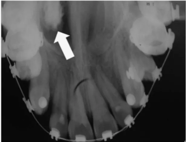
Fig. 3. Occlusal radiograph of the maxilla, showing a well-defined ovoid radiopaque image in the nasal cavity on the right side (indicated by the arrow).
Fig. 4. Tridimensional reconstruction of Computerized
Tomograph (3D): (A) posterior view and (B) inferior view
172 Rev Odonto Cienc 2012;27(2):170-173
Radiographic and imaging diagnosis of rhinolith
Because the lesion was asymptomatic and the patient did not have sinusitis or any other symptom that would justify the removal of the rhinolith, the treatment plan was to follow-up the case on a six-monthly basis to detect any possible alteration inside the nasal fossa and/or adjacent structures.
Discussion
The rhinolith is a mineralized mass located inside the nasal cavity, resulting from total or partial calciication
of an intranasal foreign body (1-2).It is a rare pathologic
condition, generally asymptomatic and unilateral, discovered accidentally during routine radiographic exam (3-5). The rhinolith grows slowly and can take years to be perceived. Its most common location is in the nasal cavity loor, between the anterior and posterior portion of the nostrils (2,7).
The aetiology of the rhinolith is still obscure. Its origin may be exogenous or endogenous, in which various substances may function as a potential nucleus for the deposition of mineral salts (3-5,8). Although its exact pathogenesis is not yet completely understood, the onset of reactional chronic inlammation occurs through the presence of a foreign body in the nasal cavity, which leads to the deposition of mineral salts of nasal, lachrymal or inlammatory origin around this foreign body (5,11). Blood clot remains, dried pus, epithelium peelings, leukocytes, bone fragments, products of cellular lysis, and necrosis of the mucosa are examples of endogenous precipitants. Fruits, seeds, granules, buttons, dirt, insects, sand, peas, parasites, wood, glass and residues of gauze plugs are examples of exogenous sources (4,5,7-11). The latter items must be introduced into the nasal cavity, and this can occur anteriorly through the nostrils (generally inserted by children) or reach the posterior opening through the rhinopharynx and choanals during episodes of vomiting,
sneezing and choking (3,9,11).In addition, other factors may
contribute to its formation, such as obstruction or stagnation of nasal secretions, chronic and acute inlammations, deposition of calcium or magnesium salts and the enzymatic activities of bacterial pathogens (3).
The growth and calciication of these objects are caused by contact with nasal luid and salt precipitations on their surfaces (9). Physical and chemical factors (changes in pH, hypersaturation of secretions, infections and chronic and acute inlammations) as well as mechanical factors (stasis of nasal secretions and alterations in air low) participate in the
rhinolith calciication process. Pinto et al. (4) reported that
the rhinolith more frequently affects young women, although other studies have demonstrated cases of the pathology in patients of both sexes and all ages (3,4,7).
In the present study, the patient’s lesion was asympto-matic, unilateral and discovered accidentally in the loor of the nasal fossa on the right side. The patient was not aware of the existence of the rhinolith and did not inform the origin of the material introduced into the nasal cavity. Also, this case in a young female subject is in agreement with the epidemiological features from the literature.
Although it is an accidental inding in the majority of cases, the rhinolith may be associated with various complaints, such as unilateral nasal obstruction, fetid odour, epistaxis (nosebleeding), asnomia (loss of sense of smell), nasal and facial swelling, pain, purulent rhinorrhea, epiphora (alteration in normal tear drainage) and headache (3-5,11-13). The presence of nasal sept deviation and perforation, destruction of the lateral wall of the nasal cavity, involvement of the maxillary sinus and production of oroantral or oronasal istula are rare complications that may occur with the growth of the rhinolith (5).
The importance of a correct understanding of the rhinolith is to allow early diagnosis by means of radiographic images in association with clinical aspects, such as signs and symptoms, previous history of foreign body in the nose and physical exam (9). The main differential diagnoses are made against osteogenic or odontogenic lesions, osteoma, odontoma, calciied nasal polyps, osteosarcomas, chondrosarcomas, epithelial cell carcinoma (ECC), impacted teeth and various categories of pathologic entities, such as syphilis and tuberculosis with calciication (3,5,11). In the present case at irst the lesion was thought to be an impacted supernumerary tooth, which led to an unnecessary surgery. The dentist may use conventional radiographs (Panoramic Radiography, Postero-anterior Radiography, Maxillary Occlusal Radiography) or Computerized Tomography for evaluation of rhinoliths. The precise location and dimensions of the lesion are limited when conventional radiographs are
used (9),and different radiographic techniques are necessary
for the inal diagnosis. Panoramic radiography, for example, produces distortion of the image and may provide false information about the exact location of the lesion (9). The conventional radiographic images of the clinical case here reported were not suficient for a deinitive diagnosis, due to the lack of reliable information provided by this type of image.
CT has been used to determine the size and location of rhinoliths, as well as to identify associated sinusitis (5,8) and complications (13). This advanced imaging method presents high sensitivity and speciicity for identifying calciications and foreign bodies, and can be applied most effectively for the
diagnosis of rhinoliths (5,9,11).In addition, the advantage of
this exam is the non superimposition of adjacent structures, and calciications in the head and neck can be differentiated from bone and normal cartilage.
The typical image of the rhinolith in the CT is a heterogeneous hyperdense mass with the central portion containing organic matter (hypodense), or the nucleus of a foreign body may be evident. In this case, a hyperdense mass and central hypodensity could be observed, showing a nucleus surrounded by calciied material (5,9,11).
Rev Odonto Cienc 2012;27(2):170-173 173 Manzi et al.
the maxillary sinus, and is rarely found in the nasal cavity. The ossifying ibroma is a well-delimited mixed lesion, with varying coniguration and size. It is important to remember that differential diagnosis is made using radiographs and imaging exams in association with the clinical indings of these lesions, considering that it is sometimes necessary to use different radiographic techniques to obtain the inal diagnosis (9).
Final considerations
Dentists should know how to recognize and diagnose a rhinolith, since a correct diagnosis of this pathologic entity helps in the differential diagnosis of severe pathologies of the nasal cavity, providing adequate treatment or only radiographic follow-up.
1. Hsiao JC, Tai CF, Lee KWl. Giant rhinolith: A case report. Kaohsiung J Med Sci 2005;21:582-5.
2. Patil K, Guledgud M, Malleshi S. Rhinolith. Indian J Dent Res 2009;20:114-6.
3. Orhan K, Kocyigit D, Kisnisci R. Rhinolithiasis: An uncommon entity of the nasal cavity. Oral Surg Oral Med Oral Pathol Oral Radiol Endod 2006;101:e28-32.
4. Pinto LSS, Campagnoli EB, Azevedo, RS, Lopes, MA. Rhinoliths causing palatal perforation: case report and literature review. Oral Surg Oral Med Oral Pathol Oral Radiol Endod 2007;104: e42-46.
5. Sumbullu M, Tozoglu U, Yoruk O, Yilmaz A, Ucuncu H. Rhinolithiasis: the importance of flat panel detector-based cone beam computed tomography in diagnosis and treatment. Oral Surg Oral Med Oral Pathol Oral Radiol Endod 2009;107:e65-67.
6. Ghanbari H, Mohammad F, Daneshi A. Report of a unusual cause of Rhinolithiasis: An ‘Opioma’. Ear Nose Throat J 2007;86:48-9.
7. Keck T, Liener K, Strgter J. Rhinolith of the nasal septum. Int J Pediatr Otorhinolaryngol 2000;53:225-8.
8. Hadi U, Ghossaini S, Zaytoun G. Rhinolithiasis: A forgotten entity. Otolaryngol Head Neck Surg 2002;126:48-51.
9. Barros CA, Martins RR, Silva JB. Rhinolith: A radiographic finding in a dental clinic. Oral Surg Oral Med Oral Pathol Oral Radiol Endod 2005;100:486-90.
10. Daneshbod Y, Khademi B, Janfeshan K. Intraoral presentation of rhinolith. Otolaryngol Head Neck Surg 2008;138:535-6.
11. Sudhakar S, Kumar B, Prabhat M. Rhinolith: A case report and review of literature. J Indian Acad Oral Med Radiol 2010;22:165-7.
12. Arora S, Garg L, Julaha M, Tuli B. Naso-oral fistula due to rhinolithiasis: a rare presentation. J Oral Sci 2009;51:481-3.
13. Aksungur EH, Binokay FB, Biçakçi K. A rhinolith which is mimicking a nasal benign tumor. Eur J Radiol 1999;31:53-5.
14. Royal SA, Gardne, RE. Rhinolithiasis: an unusual pediatric nasal mass. Pediatr Radiol 1998;28:54-5.