64
C
ASER
EPORTReceived for publication 05/02/2013 - Accepted for publication 20/06/2013 The authors declare no conflicts of interests.
Síndrome de Tolosa-Hunt
Tolosa-Hunt syndrome
Eduardo Scaldini Buscacio
1, Yoshifumi Yamane
2, Renata Nogueira
3Municipal Hospital of Piedade - Rio de Janeiro (RJ), Brazil.
Rev Bras Oftalmol. 2016; 75 (1): 64-6
1 Ophthalmology Resident, Hospital Municipal da Piedade - Rio de Janeiro (RJ), Brazil.
2 Doctor, Professor, Universidade Gama Filho in Rio de Janeiro (RJ), Brazil.
3 Neurology Resident, Hospital Universitário Clementino Fraga Filho - Rio de Janeiro (RJ), Brazil.
A
BSTRACTTolosa Hunt syndrome is a rare disease, whose etiology is unknown. It presents as a painful ophthalmoplegia of one or more oculomotor cranial nerves, which resolves spontaneously and responds well to treatment with corticosteroids. This study is a case report of a patient who had followed painful oftalmoplegias cases involving the oculomotor and abdcens nerves being treated with corticosteroids, obtaining a dramatic response. Another goal is to describe the pathophysiological, clinical, differential diagnosis, since it is a diagnosis of exclusion, and the therapeutic measures adopted according to the International Headache Society 2004 (ISH-2004) by presenting the case study conducted with the standards the study cited above.
Keywords: Tolosa-Hunt syndrome/diagnosis; Ophthalmoplegia/drug therapy; Prednisone/therapeutic use; Pain/classification; International Classification of Diseases; Case reports
R
ESUMOA Síndrome de Tolosa Hunt é uma doença rara, cuja etiopatogenia é desconhecida. Apresenta-se como uma oftalmoplegia dolorosa de um ou mais nervos cranianos oculomotores, que regride espontaneamente e responde bem ao tratamento com corticoides. O presente estudo trata-se de um relato de caso de um paciente que apresentou seguidos casos de oftalmoplegias dolorosas, envolven-do o nervo oculomotor e o abducente senenvolven-do trataenvolven-do com corticoesteroides obteve uma resposta dramática. Objetiva-se ainda descrever as características fisiopatológicas, clínicas, o diagnóstico diferencial, visto que é um diagnóstico de exclusão, e medidas terapêuticas instituídas de acordo com o International Headache Society 2004 (ISH-2004) através da apresentação do caso clínico conduzido com as normas do estudo supracitado.
Descritores: Síndrome de Tolosa-Hunt/diagnóstico; Oftalmoplegia/quimioterapia; Prednisona/uso terapêutico; Dor/classifica-ção; Classificação Internacional de Doenças; Relato de casos
65 Tolosa-Hunt syndrome
I
NTRODUCTIONT
he Tolosa-Hunt syndrome (THS) is a rare disorder characterized by hemicrania or periorbital pain associated to ophthalmoplegia of one or more cranial nerves and that has a dramatic response to corticosteroid therapy(1). It wasnitially described by Tolosa and Hunt in 1954 and 1961 as a stenosis of the cavernous portion of the internal carotid artery due to an idiopathic granulomatous inflammatory process(2,3).
The present study aims to describe the patho-physiological and clinical characteristics and the therapeutic measures instituted according to the International Headache Society 2004 (ISH-2004)(4) with the presentation of a clinical case conducted
with the standards of the aforementioned study.
A study is developed presenting a case report of a patient attended in the ambulatory with diagnostic criteria according to ISH-2004(5). After the exclusion of other differential diagnoses,
the patient was treated with prednisone 1 mg/kg/day and showed dramatic therapeutic response with two reincidences. The follow-up time for the patient was 12 months.
R
ESULTSCH, 56 years old, black, hypertensive, registers to the emergency service complaining of headache, vertical binocular diplopia without any associated mitigating or aggravating factors, and with hypotropia of the left eye (Figure 1). The condition evolves persistently for a month, when there was partial blepharoptosis in the left eye associated to left retroorbitary pain of moderate intensity that would not decrease with the use of common analgesics.
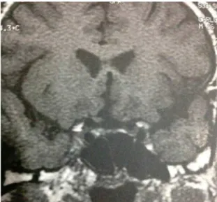
The following laboratory tests were requested with normal results: complete blood count, fasting blood glucose, glycated hemoglobin, coagulation, HSS, PCR, ANA, anti-HIV, VDRL, FTA-ABS and thyroid function. Complementation with contrasted computed tomography and nuclear magnetic resonance (figures 2 and 3) were held, and showed no changes. The patient presented had spontaneous improvement in 20 days. About 90 days after the first episode and after improvement, he presented deficit of the right eye adduction associated to intense pain (Figure 4). This was followed by the therapeutic test with prednisone 1 mg/kg/day, with clinical remission in 48 hours after the onset of treatment.
Eight months after the last symptomatic event, the patient has a new episode of painful ophthalmoplegia of the III cranial nerve in his right eye and limitation of upstanding, being readily treated with corticosteroid therapy and showing dramatic response.
D
ISCUSSIONThe present patient came to our ambulatory in a condition of painful ophthalmoplegia with development with common diagnostic exams, so we chose for a therapeutic test with prednisone due to the suspicion of THS.
THS is a rare entity of unknown incidence, and can be present at any age, but with an average of 44 years and with no gender preference. The most affected cranial nerves are the third (79%), sixth (45%), fourth (32%), and fifth (25%), with involvement of multiple cranial nerves in 70% of cases(6). The
nerves affected in the present case, the third and sixth ones, are the most commonly affected according to epidemiological studies.
Clinically, the pain begins with ophthalmoplegia or may precede the paralysis within 14 days. The pupil reflexes are usually normal, but can have both sympathetic and parasympathetic impairment(1). The involvement of the optic nerve has already
been described due to the involvement of the orbital apex(5). In
our study no optic nerve involvement was observed.
The diagnosis is of exclusion and should explore the differential diagnoses of painful Para-Selar syndrome exemplified in Table 1 and other causes of painful ophthalmoplegia shown in Table 2(1).
The diagnosis was made according to ISH-1998 diagnostic criteria modified in 2004(5) as shown in Table 3.
Long-term recurrence affects about 50% of cases, and the discontinuation of treatment can lead to recurrence of pain in 20% of patients. Patients resistant to treatment with corticosteroids (prednisone or methylprednisolone) can make use of immunosuppressants such as methotrexate and azathioprine(6). In our case 2 recurrences were observed and
readily treated with corticosteroids having satisfactory response. We then concluded that due to the lack of specific markers and etiopathogenesis being uncertain(1,6), it is still necessary to
have a diagnosis of exclusion. The criteria of ISH-2004 are of great value in the dealing with similar cases. são de grande valia na condução de casos semelhantes.
Table 1
Causes of para-selar painful ophthalmoplegia
1 Trauma
2 Vascular
Intracavernous carotid aneurysm
Aneurysms of the posterior cerebral artery Carotid cavernous fistula
Carotid cavernous thrombosis
3 Neoplasia
Primary intracranial tumor Pituitary adenoma; Meningioma;
craniopharyngioma; Sarcina; Neurofibroma; squamous cell carcinoma
Primary cranial tumor
Chondroma; giant cell tumor
Local metastasis
Nasopharyngeal; Cylindroma; Adamantinoma; Squamous Cell Carcinoma
Distant metastasis
Lymphoma; Multiple myeloma
4 Inflamation Bacteria
Sinusitis; Mucocele; Periostitis
Viruses Herpes zoster Fungus Murcomycosis Spirochete Treponema pallidium Mycobacteria Micobacterium tuberculosis Undefined causes
Sarcoidosis; Wegner’s Granulomatosis; Eosinophilic Granuloma; Tolosa-Hunt
66 BuscacioES, YamaneY, NogueiraR
R
EFERENCES1. Kline LB, Hoyt WF. The Tolosa-Hunt syndrome. J Neurol Neurosurg Psychiatry. 2001;71(5):577-82. Review.
2. Tolosa E. Periarteritic lesions of the carotid siphon with the clini-cal features of a carotid infraclinoidal aneurysm. J Neurol Neurosurg Psychiatry. 1954;17(4):300-2.
3. Hunt WE, Meagher JN, LeFever HE, Zeman W. Painful ophthal-moplegia. Its relation to indolent inflammation of the cavernous sinus. Neurology. 1961;11:56-62.
4. Sarchielli P. XI Congress of the International Headache Society. September 13-16, 2003, Rome, Italy. Expert Opin Pharmacother. 2004;5(4):959-75.
5. Spinnler H. Painful ophthalmoplegia: the Tolosa-Hunt syndrome. Med J Aust. 1973;2(13):645-6.
6. La Mantia L, Curone M, Rapoport AM, Bussone G; International Headache Society. Tolosa–Hunt syndrome: critical literature re-view based on IHS 2004 criteria. Cephalalgia. 2006;26(7):772-81. Review.
Table 2
Other causes of painful ophthalmoplegia
Table 3
Diagnostic criteria for THS according to ISH-2004
Figure 1: Hypertrophy and ptosis of the LE
Figure 2: MRI - coronal section.
Figure 3: MRI - sagittal section.
Figure 4:Adduction deficit in the LE. 1 Diseases of the orbit
Idiopathic orbital inflammation; contagious Sinusitis; Mucormycosis; Metastatic tumor; Lymphoma and leukemia
2 Diabetic ophthalmoplegia
Mononeuropathy or polyneuropathy
3 Posterior Fossa Aneurysm
Basilar and posterior communicating
4 Giant cell arteritis
5 Ophthalmoplegic migraine
1 One or more episodes of unilateral orbital pain that persists for weeks without treatment.
2 Paresis of one or more cranial nerves (III, IV or VI) and/or granuloma demonstrated by the nuclear magnetic resonance or biopsy.
3 Paresis occurs with the onset of pain or is presented in the next two weeks.
4 Improvement in pain and paresis 72 hours after the onset of corticosteroid therapy.
5 Other causes should be excluded.
Rev Bras Oftalmol. 2016; 75 (1): 64-6
Corresponding author: