S
CIENTIFICA
RTICLES Revista Brasileira de FisioterapiaInfluence of oral contraceptive use on lipid
levels and cardiorespiratory responses among
healthy sedentary women
Influência do uso de contraceptivos orais nos níveis lipídicos e nas
respostas cardiorrespiratórias de mulheres saudáveis e sedentárias
Santos MCS1, Rebelo ACS1, Zuttin RS1, César MC1, Catai AM2, Silva E1
Abstract
Objective: To evaluate the influence of oral contraceptive use on lipid levels, heart rate (HR) variability and aerobic capacity among
sedentary young women. Methods: The subjects were 20 healthy women (23.55±1.88 years): ten used oral contraceptives (TG) and
ten did not (CG). Ergospirometric test on a cycle ergometer was used to determine the aerobic capacity at the anaerobic threshold and at the exercise peak. In addition, recordingsof HR and R-R (iRR) intervals in the supine and seated positions, for 15 minutes, and biochemical blood analysis were performed. The iRR (ms) indices were analyzed in the time domain (TD) for RMSSD, RMSM and pNN50 (%), and in the frequency domain (FD) using fast Fourier transforms from low (LF) and high (HF) frequency bands in normalized units, obtaining the LF/HF ratio. Mann-Whitney and Kruskal-Wallis, with α=5%, were used for statistical analyses. Results:
The triglyceride and cholesterol levels in the TG were greater than those in the CG (p<0.05). At the anaerobic threshold and at the exercise peak, the power (W), oxygen uptake (VO2), carbon dioxide production (VCO2), (VO2/VCO2) ratio, pulmonary ventilation and HR were not significantly different between the groups. The indices for HR variability in the TD and FD for the two groups in the supine and seated positions were also not different. Conclusions: The use of oral contraceptives did not influence the aerobic capacity or the autonomic modulation of the HR. However, it influenced the total cholesterol and triglyceride levels. These assessments are important for determining protocols for physical training for cardiovascular disease prevention.
Key words: contraceptive therapy; heart rate; aerobic capacity.
Resumo
Objetivo: Avaliar a influência do uso de contraceptivos orais (CO) sobre os níveis lipídicos, a variabilidade da freqüência cardíaca
(VFC) e a capacidade aeróbia em mulheres jovens sedentárias. Materiais e métodos: Vinte mulheres saudáveis (23,55±1,88 anos),
sendo dez que utilizam CO (GT) e dez que não utilizam CO (GC). Protocolos: teste ergoespirométrico (TE), em cicloergômetro para
determinar a capacidade aeróbia no limiar de anaerobiose (LA) e no pico do exercício; captação da FC e dos intervalos R-R (iRR) nas posições supina e sentada durante 15 minutos; análise bioquímica de sangue. Os índices dos iRR(ms) foram analisados no domínio do tempo (DT), RMSSD, RMSM e pNN50(%) e no domínio da freqüência (DF), usando a transformada rápida de Fourier a partir das bandas de baixa freqüência (BF) e alta freqüência (AF), em unidades normalizadas (un), e a razão a BF/AF. Estatística: Mann-Whitney e Kruskal-Wallis, α=5%. Resultados: Os níveis de triglicérides e de colesterol do GT foram superiores aos do GC (p<0,05). No LA e no pico do exercício, a potência (W), o consumo de oxigênio, a produção de dióxido de carbono, a razão VO2/VCO2, a ventilação pulmonar e a freqüência cardíaca (FC) foram semelhantes (p>0,05) entre os grupos. Os índices da VFC no DT e no DF da posição
supina e sentada intergrupos foram semelhantes. Conclusões: O uso de CO não influenciou na capacidade aeróbia e na modulação
autonômica da FC, mas influenciou nos níveis de colesterol total e triglicérides. Estas avaliações são importantes para a determinação de protocolos de treinamento físico na prevenção de doenças cardiovasculares.
Palavras-chave: terapia contraceptiva; freqüência cardíaca; capacidade aeróbia.
Received: 27/04/2007 – Revised: 21/09/2007 – Accepted: 22/02/2008
1 Cardiovascular Physical Therapy and Functional Tests Research Laboratory, School of Health Sciences, Universidade Metodista de Piracicaba (Unimep) – Piracicaba (SP), Brazil 2 Physical Therapy Department, Universidade Federal de São Carlos (UFSCar) – São Carlos (SP), Brazil
Financial Support: FAPESP (Protocol nº 06/56788-1); Fundo de Apoio à Pesquisa da Universidade Metodista de Piracicaba (Processo 309/05); CNPq (Processo 350060/2001-0).
Correspondence to: Ester da Silva, Laboratório de Pesquisa em Fisioterapia Cardiovascular e de Provas Funcionais, Mestrado em Fisioterapia, Faculdade de Ciências da Saúde, Universidade Metodista de Piracicaba, Rodovia do Açúcar, km 156, CEP 13400-911, Piracicaba (SP), Brazil, e-mail: [email protected]
Introduction
Women share several risk factors in the development of cardiovascular disease, such as family history, obesity, smoking habits, unfavorable lipid profiles, high levels of homocistein and fibrinogen, physical inactivity (sedentary habits), use of oral contraceptives (OC), diabetes mellitus,
hypertension, and genetic factors1. Among these factors,
physical inactivity associated to the use of low-dose, sin-gle-phase combined OC, have been considered to be an important risk factor for dyslipidemia, contributing to the development of atherogenesis and coronary arterial disease (CAD)2,3. Furthermore, studies reveal negative influences on
the aerobic functional capacity with the use of contracep-tive therapy4,5.
Large clinical assays show that the extended use of estrogen associated with progesterone cannot be beneicial6 and may,
also, compromise the eiciency of the autonomic modulation of the heart rate (HR). hese authors report that, in women who are not users of oral contraceptives, no evidence was shown of changes in autonomic HR modulation during the fol-licular phase of the menstrual cycle, in comparison with oral contraceptive users7.
Some studies show no evidence of unfavorable effects due to estrogenic therapy on autonomic HR control with women who practice regular physical training, in the rest condition8,9. Studies report that there are alterations of the
metabolism of plasmatic lipids of young, females using oral contraceptives, and that the estrogen and progesterone dos-ages are related7,8. Such alterations are associated with the
increase of risks in CAD10,11. However, other authors report
that low-dose, combined contraceptives reduce the adverse effects of the increases of the total triglyceride and choles-terol serum levels12.
Furthermore, there is still the need of greater knowledge about the single-phase combined estrogen/progesterone therapy over the cardiorespiratory and metabolic variables in young sedentary women. hus, from the analyses of car-diorespiratory and metabolic variables, as an integral part of physical therapy evaluations, valuable information about the physiological conditions of the subjects may be obtained. his may help identify possible alterations that will contribute to the development of cardiovascular diseases.
herefore, considering the current doubts of oral contra-ceptive use on the development of cardiovascular disease, the aim of this study was to investigate the lipid levels, HR auto-nomic modulation, and aerobic capacity of young sedentary women who were users and non-users of low-dose, combined estrogen/progesterone OC.
Materials and methods
Subjects
Sampling calculus obtained from the analysis using the GraphPad StatMate 2.0 for Windows software program, with 80% power and α=5%, was for eight volunteer subjects for each group. All subjects were pre-selected from a clinical evaluation, resting electrocardiogram (ECG) of 12 derivations, blood and urine biochemical assays, and a physical therapy examination. his was a cross-sectional study, performed with 20 healthy, sedentary volunteer subjects (23.55±1.88 years old) with a weak aerobic level, according to the American Heart Association. hey were divided into two groups of ten, considering the ones that used combined oral contraceptives, or the therapy group (TG), and the ones who did not, or the control group (CG). Each participant demonstrated normal ECGs in 12 resting derivations, corporal mass index of 21.22±2.01kg/m2, systolic
and diastolic arterial pressure during rest of 110 and 70mmHg on average, respectively. he CG females showed regular men-strual cycles of between 28 and 30 days, and the ones from the TG had used OC for 15.22±12.77 months. A minimal time of six months of OC use was established. he subjects did not show any evidence of cardiovascular changes, as observed through clinical examination.
he subjects were oriented about the relevance of the re-search, the experimental procedures, and after agreeing, signed informed consent form, according to rule nº 196 of the National Health Council (NHC). he Research Ethics Committee of the Universidade Metodista de Piracicaba (Unimep) approved the research, for the the protocol of nº 43/06.
Tests were always performed during the afternoon, in order to avoid interferences of the circadian variations. he labora-tory was air-conditioned, with an average temperature of 22ºC, a relative air humidity of around 50%, and a barometric pres-sure of approximately 710mmHg. he volunteers were oriented not to consume stimulating beverages (tea, cofee, alcohol), not to perform physical activities within 24 hours before the experiment, to have a good sleep (at least eight hours), and to have light meals on the day of the test.
Experimental protocols
Heart rate data measurements were obtained from the ECG, recorded on a cardiac monitor of a channel (Miniscope II, Instramed, Porto Alegre, RS, Brazil), with the DII derivation with activated carbon electrodes (R28 Carbo Cone, Implemed, São Paulo, SP, Brazil). he negative electrode was placed at the manubrio-sternal joint, the positive one at the ifth space on the left anterior axillary line, and the neutral electrode at the ifth right intercostals space. A Lab-PC+ (National Instruments Co., Austin, TX, USA) analogical-digital converter was used in order to process the ECG signals, which represented an inter-face between the cardiac monitor and a Pentium III PC. Signal recording was performed in real time, after the A/D conver-sion, with a sampling rate of 500 Hz. he calculation of HR and R-R intervals was performed, beat-to-beat, by using a speciic software program13. Recording of the R-R intervals took place
for a period of 15 minutes during rest, with the volunteers in both the supine and seated positions, respectively, and brea-thing spontaneously.
For the CG volunteers, experiments were conducted be-tween the ifth and the tenth day of the menstrual cycle, conside-ring the slightest hormonal luctuations, while for the TG, they always took place on the 21st day of medication (active phase),
and on the 28th day, without medication (inactive phase)14.
Performance of the ergospirometric test (ET) aimed at assessing the aerobic functional capability of the volunteers at the anaerobiosis threshold (AT) level, and at the peak of physical exercise, from the cardiorespiratory variables. he ET took place on an electromagnetic brake cycle ergometer (Corival 400 model, Quinton, Seattle, WA, USA) with the seat height regulated in order to allow knee lexion of about 5º. he ET protocol was made up of a 60 seconds pre-test rest, with the volunteer seated on the cycle ergometer, starting exercise with a freeload during 240 seconds, followed by an increase of resis-tance of 15watts (W) for 60 seconds, until physical exhaustion, that is, when the volunteer could not keep the speed at 60rpm. During ET, breathing and metabolic variables were obtained, breath-by-breath, by using a system of measuring of exhaled gases (CPX/D MedGraphics, Breeze, St. Paul, MN, USA). hese variables were later processed and calculated in mobile avera-ges, every eight breathing cycles, in order to better observe the kinetics of their answers during exercise. Aerobic capacities at the test peak and AT were evaluated from the oxygen con-sumption data (VO2) in 1/min-1 (Table 3).
Visual analysis of the answers of the breathing and metabo-lic variables was made by three duly trained observers in order to determine AT. he criterion adopted was lost of parallelism, that is, a disproportionate increase of carbon dioxide (VCO2) production in relation to the oxygen consume (VO2)15.
Heart rate variation was analyzed in the domain of time (DT) and frequency (DF). he region with greatest stability of
the iRR series of data was used for these analyses, as long as it included, at least, ive minutes or 256 consecutive beats. Analy-sis at the DT was performed from the indexes RMSSD (square root of the adding of the square of the diferences between the recorded iR-R, divided by the nº of iR-R of the selected data series minus one); RMSM (square root of the adding of the square of the diferences of the individual values in relation to the average value, divided by the number of iR-R of the selected data series); and pNN50 (percentage of the adjacent iR-R with diferences larger than 50 ms)16.
For the frequency domain analysis, the procedure of linear tendency removal was used, and the fast Fourier transform was applied in a single window, on the previously selected sequence of the R-R interval values. he potency range com-ponents were computed on the low (LF: 0.04-0.15Hz) and high (HF: 0.15-0.4Hz) frequency bands, in absolute units (ms2) and
in normalized units (un), which corresponded to the percen-tage of the total range, subtracted from the component of the very low frequency (VLF: 0.003-0.04Hz). Since both divisions of the autonomic nervous system, sympathetic, and parasympa-thetic, modulated the LF band, and the HF band is correlated to vagal control, the LF/HF ratio was calculated, in order to evaluate the sympathetic-vagal balance16.
he analysis of data distribution of all studied variables in the diferent conditions have shown that the data did not have a normal distribution, as analyzed by the Kolmogorov-Smirnov test. In this manner, non-parametric statistical tests were se-lected in order to compare the data. From the Mann-Whitney and Kruskal-Wallis tests, the statistical analysis of signiicance was performed for non-paired samples, with the signiicance level established at 5%.
Results
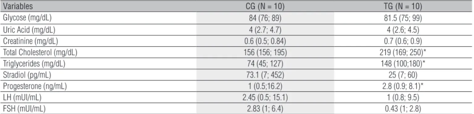
As is shown, in Table 1, that the average values of the blood biochemical exams regarding the plasmatic concentrations of total cholesterol and triglycerides showed statistically signii-cant diferences (p<0.05) and were higher for the TG, and the average values of progesterone for the CG.
It is shown, in Table 2, the ethinylestradiol and prostagen dosages of the OC used by the TG. Prostagens varied between gestoden, or cyproterone acetate, or drospirenone.
he values for potency (Watts), oxygen consumption (VO2) in
mL.kg-1.min-1, carbon dioxide production (VCO2) in L/min, VO2/VCO2
ratio (RER), pulmonary ventilation (PV) in L/min, and HR in bpm, obtained during ET at exercise peak and at anaerobiosis threshold (AT) are shown at Table 3. It is observable that the variables va-lues were similar (p>0.05) in both situations analyzed between the CG and TG.
N Comercial Name Ethinil-estradiol (mg)
Gestoden (Desogestrel) (mg)
Cyproterone acetate (mg)
Drospirenone (mg)
3 Femiane 0.02 0.075 -
-1 Harmonet 0.02 0.075 -
-2 Tâmisa 20 0.02 0.075 -
-1 Selena 0.035 - 2.0
-2 Diane 35 0.035 - 2.0
-1 Yasmin 0.03 - - 3.0
Table 2. Values of low-dose oral contraceptive (OC) obtained by the therapy group subjects.
N=number of oral contraceptive users; mg=milligrams.
Table 3. Values of variables for anaerobic threshold (AT) and at the exercise peak in ergospirometric test (ET) obtained by the control groups (CG) and therapy groups (TG).
ET - Pico Power (W) VO2 (mL.kg-1.min-1) VCO
2 (L/min) RER VE (L/min) FC (bpm)
GC Median 131 25.80 1.78 1.22 56.60 179
Min 108 17.80 1.48 1.10 42.10 168
Max 156 30.40 1.78 1.34 81.10 193
TG Median 115.5 24.20 1.51 1.23 62.20 183
Min 84 18.30 1.13 1.10 41.70 161
Max 167 27.00 2.37 1.34 92.10 198
AT
Median 65.5 12.1 0.67 0.92 19.85 127
CG Min 54 8.6 0.44 0.91 13.00 104
Max 97 18 0.89 0.97 21.90 139
Median 64.5 12.3 0.65 0.94 21.90 133
GT Min 49 8.7 0.39 0.89 13.2 107
Max 86 19 0.97 0.98 31.7 160
Data are reported as medians (minimum; maximum). CG=control group; TG=therapy group; W=Watts; VO2=oxygen uptake; VCO2=carbon dioxide production; RER=ratio of gas exchange VO2/VCO2; PV=pulmonary ventilation; mL.kg-1.min-1= milliliter per kilogram per minute; HR=heart rate; bpm=beat per minutes; min=minutes; ET=ergospyrometric test; Level of significance α=5%.
Data are reported as medians (minimum; maximum). CG=control group; GT=therapy group; N=number of volunteers; mg/dL=milligrams per deciliters; pg/mL=picograms per milliliter; ng/mL=nanograms per milliliter; mUI/mL=international milliunits per milliliters; *p<0.05. Level of α=5%.
Table 1. Median values obtained by biochemical analysis of the blood control group and therapy group subjects.
Variables CG (N = 10) TG (N = 10)
Glycose (mg/dL) 84 (76; 89) 81.5 (75; 99)
Uric Acid (mg/dL) 4 (2.7; 4.7) 4 (2.6; 4.5) Creatinine (mg/dL) 0.6 (0.5; 0.84) 0.7 (0.6; 0.9) Total Cholesterol (mg/dL) 156 (156; 195) 219 (169; 250)* Triglycerides (mg/dL) 74 (45; 127) 148 (100;180)*
Stradiol (pg/mL) 73.1 (7; 452) 25 (7; 60)
Progesterone (ng/mL) 1 (0.5;16.2) 2.8 (0.9; 8.1)*
LH (mUI/mL) 2.45 (0.5; 15.1) 1 (0.8; 9.5)
FSH (mUI/mL) 2.83 (1; 6.4) 0.43 (1; 2.8)
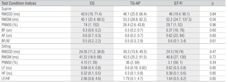
It is observed, in Table 4 that the average values for the time domain indices (RMSSD (ms), RMSM (ms), pNN50 (%)), and frequency domain indices (BF (un), AF (un) and BF/AF ratio) of the HRV, did not demonstrate statistically signiicant diferences (p>0.05) in the supine and seated positions, be-tween the CG and the TG on the active (TG-AP) and inactive (TG-IP) phases.
Discussion
Data are reported as medians (minimum; maximum).ms=milliseconds; %=percent; nu=normalized units; LF=low frequency; HF=high frequency; LF/HF. Level of significance α=5%.
Table 4. Indices values obtained analyses heart rate variability for the control groups (CG) and therapy groups (TG) in the active (TG-AP) and inactive phases (TG-IP) in the supine and sitting positions.
Test Condition Indices CG TG-AP GT-FI p
Supine
RMSSD (ms) 43.9 (19; 71.4) 46.1 (25.9; 66.4) 46 (19.4; 90.1) 0.84 RMSM (ms) 45.1 (22.4; 66.5) 53.3 (28.8; 82.2) 52.2 (24.7; 137.3) 0.56 PNN50 (%) 74 (1; 152) 26.4 (2.6; 43.8) 29.7 (1; 52) 0.96 BF (un) 0.3 (0.6; 0.2) 0.3 (0.2; 0.7) 0.37 (16; 78) 0.60 AF (un) 0.6 (0.7; 0.3) 0.6 (0.2; 0.7) 0.62 (22; 84) 0.60 BF/AF 0.5 (0.2; 2.2) 0.5 (0.3; 2.9) 0.6 (0.1; 3.4) 0.61 Sitting
RMSSD (ms) 24.35 (11.2; 38.8) 30.3 (13.8; 49.3) 24.3 (16;74) 0.47 RMSM (ms) 41.22 (18.9; 68) 43.5 (25.2; 91.5) 40.8 (27; 130) 0.72 PNN50 (%) 4.15 (1; 59) 36 (2; 69) 3.1 (56; 1) 0.34 LF (nu) 0.68 (0.4; 0.8) 0.6 (0.16; 0.82) 0.62 (0.3; 0.8) 0.85 HF (nu) 0.32 (0.1; 0.5) 0.3 (0.1; 0.8) 0.38 (0.1; 0.6) 0.85 LF/HF 2.08 (0.8; 4.6) 1.79 (0.1; 4.7) 1.64 (0.5; 8.2) 0.85
containing high doses, caused collateral efects such as liquid retention, nauseas, headaches, body weight changes, and in-creased risks of thrombo-lythic and ischemic diseases17. With
the reduction of the estradiol and progesterone dosages con-tained in the OC, these collateral efects were only observed in women aged more than 35 years old, or when associated to smoking habits18,19.
The combined single-phase OC utilized by the subjects varied between: ethinilestradiol/gestoden (desogestrel), ethinilestradiol/cyproterone acetate or ethinilestradiol/ drospirenone. These medications have an excellent safety profile, for they belong to the class of modern contracep-tives, with less than 35 micrograms of estrogen (low doses) and the progestogenes belong to the “second and third ge-neration” of OC with similar efficiency and collateral effects among young women20,21.
In the present study, the results from the biochemical exams for total cholesterol and triglycerides of the contra-ceptive therapy for young users, showed higher values in relation to the young women from the CG, which reached the threshold limits regarding the normal band. his occurrence may be attributed to the fact that cholesterol is the elemen-tary block in steroidgenesis22. hese data are in accordance
with the ones by Sherif2 and by Foulon et al.3, which report
that the use of combined OC (estrogen/progesterone) increa-ses the total cholesterol plasmatic levels, which may increase or leave unaltered the triglyceride levels. Foulon et al.3 report
also that this triglyceride increase is found in women who are users of ethinilestradiol/desogestrel combined single-phase OC, similar to the data in this study.
he inluences of the combined OC over the plasmatic lipids will depend on the balance between the estrogen/
progesterone dosages. Koh et al.23, observed in their study
that low estrogen/progesterone dosages of combined contra-ceptives reduce the adverse efects of the plasmatic lipids and lipoproteins. On the other hand, Martins, Gomes and Pasini24,
report that oral contraceptive therapy contributes to the increases of the plasmatic cholesterol levels, with the most evident efects at the age of 30 to 39 years. However, these authors also report that in women at the age of 20 to 29, there seems to be no relationships between the cholesterol levels and the use of OC. However, Godsland et al.25 have observed
in users of low-dose combined OC an increase of 13 to 75% at the triglyceride levels.
his study’s limitations are related to the fact that the ex-perimental design is of a transversal type, and that only the minimal time use was considered, resulting in a considerable dispersal in total oral contraceptive time use. hus, the perfor-mance of lipid proile exams prior to the use of OC is conside-red relevant. It is also important that more detailed exams of the lipid fractions are made both before and during the use of contraceptives, since dyslipidemia is an important risk factor for coronary artery disease.
In the autonomic modulation of the HR from the TD indices (RMSSD, RMSM, and pNN50) and FD (BF,AF and BF/AF), the use of contraceptives was verified and did not influence the results, since no differences were observed between the two groups. Such a fact can be attributed to the pharmacological properties of low doses of estrogen/ progesterone, as well as the maintenance of the integrity of the autonomic modulation of the HR, since the values found were within the normality range26. These findings
corrobo-rated the study by Schueller et al.7, which verified that in
young females, 27 belonging to the CG, and 31 who made
use of single-phase OC, no differences in the autonomic mo-dulation of the HR were observed. This fact was attributed to the duration and the pharmacological properties utilized in the contraceptives. However, the authors report that post-menopause women are more susceptible to the effects of the hormonal repositioning therapy, and demonstrate changes in the autonomic nervous system activity.
Other studies that assessed women before and after OC therapy have observed decreases in aerobic capacity du-ring therapy24. Authors assert that this fact may be related
to the decreases of the autonomic nervous system activity induced by the exogenous estradiol administration, thus increasing the levels of nitric oxide, which is a potent va-sodilator. This mechanism hinders the redistribution of the blood flow to the active musculature, thus limiting its development at peak exercise levels. However, the effects of the contraceptive in the present results during physical exercise (potency (W), VO2 in mL.kg.min-1, VCO
2 (L/min),
RER, VE (L/min) and HR (bpm)) obtained at the peak exercise level during ET, did not demonstrate significant statistical differences (p>0.05) between the CG and TG, suggesting the OC did not influence the aerobic capacity of sedentary young women.
Although the average potency and HR values did not shown any significant differences, a higher reached po-tency value for a lower HR at the peak exercise levels was observed in the CG, in relation to the TG, which suggested a greater cardiorespiratory aptitude. These findings were in accordance to the ones by Giacomoni and Falgairette27,
who found a lower cardiorespiratory aptitude in OC users. It is worth pointing out that the average values obtained for the VO2 during exercise peak (25.80mL/kg/min), are in
accordance to Neves et al.28, who observed average values of
peak VO2=22.6±3.1mL/kg/min in sedentary young women. he breathing and metabolic variables found in the pre-sent study, at the AT level, did not result in statistically signii-cant diferences in the comparisons between the CG and TG. hese data agreed with the study by Redman et al.29, who did
not observe any inluences by the combined single-phase OC in the aerobic capacity at the AT level. Such occurrences in the present study may be due to the fact that the volunteers were using the low dosage OC. hus, more studies regarding this topic are needed, with the inclusion of other ages, and risk factors associated with the use of OC.
Conclusions
he present results suggest that low doses of estrogen/ pro-gesterone did not afect the aerobic capacity and autonomic modulation in this selected age range. However, it does con-tribute to modiications of the lipoprotein metabolism related to increases of the total cholesterol and triglycerides levels. hese are important risk factors in the development of CAD in sedentary women.
Acknowledgements
Genazzani AR, Gadducci A, Gambacciani M; International Menopause 1.
Society Expert Workshop. Controversial issues in climacteric medicine II. Hormone replacement therapy and cancer. Maturitas. 2001;40(2):117-30.
Sherif K. Benefits and risks of oral contraceptives. Am J Obstet Gynecol. 2.
1999;180(6 Pt 2):S343-8.
Foulon T, Payen N, Laporte F, Bijaoui S, Dupont G, Roland F, et al. Effects 3.
of two low-dose oral contraceptives containing ethinylestradiol and either desogestrel or levonorgestrel on serium lipds and lipoproteins with particular regard to LDL size. Contraception. 2001;64(1):11-6.
Notelovitz M, Zauner C, McKenzie L, Suggs Y, Fields C, Kitchens C. 4.
The effect of low-dose oral contraceptives on cardiorespiratory function, coagulation, and lipids in exercising young women: a preliminary report. Am J Obstet Gynecol. 1987;156(3):591-8.
Bonen A, Haynes FW, Graham TE. Substrate and hormonal responses 5.
to exercise in women using oral contraceptives. J Appl Physiol. 1991;70(5):1917-27.
Bush TL, Barrett-Connor E, Cowan LD, Criqui MH, Wallace RB, Suchindran 6.
CM, et al. Cardiovascular mortality and noncontraceptive use of estrogen in women: results from the Lipid Research Clinics Program Follow-up Study. Circulation. 1987;75(6):1102-9.
Schueller PO, Feuring M, Sharkova Y, Grimm W, Christ M. Effects of 7.
synthetic progestagens on autonomic tone, neurohormones and C-reactive protein levels in young healthy females in reproductive age. Int J Cardiol. 2006;111(1):42-8.
Davy KP, DeSouza CA, Jones PP, Seals DR. Elevated heart rate 8.
variability in physically active young and older adult women. Clin Sci. 1998;94:579-84.
Perini R, Fisher N, Veicsteinas A, Pendergast DR. Aerobic training and 9.
cardiovascular responses at rest and during exercise in older men and women. Med Sci Sports Exerc. 2002;34(4):700-8.
Wiegratz I, Jung-Hoffmann C, Gross W, Kuhl H. Effect of two 10.
oral contraceptives containing ethinyl estradiol and gestodene or norgestimate on different lipid and lipoprotein parameters. Contraception. 1998;58(2):83-91.
Gaspard U, Endrikat JP, Desager C, Buicu C, Gerlinger R, Heithecker R. 11.
A randomized study on the influence of oral contraceptives containing ethinylestradiol combined with drospirenone or desogestrel on lipid and lipoprotein metabolism over a period of 13 cycles Contraception. 2004;69(4):271-8.
Bergink EW, Kloosterboer HJ, Lund L, Nummi S. Effects of levonorgestrel 12.
and desogestrel in low-dose oral contraceptive combination on serum lipids, apolipoproteins A-I and B and glycosylated proteins. Contraception. 1984;30(1):61-72.
Silva E, Catai AM, Trevelin LC, Guimarães JO, Silva Jr LP, Silva LMP, et al. 13.
Design of a computerized system to evaluate the cardiac function during dynamic exercise. In: Annals of the World Congress on Medical Physics and Biomedical Engineering. 1994;1:419.
Casazza GA, Suh SH, Miller BF, Navazio FM, Brooks GA. Effects of oral 14.
contraceptives on peak exercise capacity. J Appl Physiol. 2002;93(5):1698-702.
Higa MN, Silva E, Neves VF, Catai AM, Gallo L Jr, Silva de Sá MF. 15.
Comparison of anaerobic threshold determined by visual and mathematical methods in healthy women. Braz J Med Biol Res. 2007;40(4):501-8.
Antila K. Quantitative characterization of heart rate during exercise. Scand 16.
J Clin Lab Invest Suppl. 1979;(153):3-68.
Stadel BV. Oral contraceptives and cardiovascular disease (first of two 17.
parts). N Engl J Med. 1981;305(11):612-8.
Schwingl PJ, Ory HW, Visness CM. Estimates of risk of cardiovascular 18.
death attributable to low-dose oral contraceptives in the United States. Am J Obstet Gynecol. 1999;180(1 Pt 1):241-9.
O’Brien PA. The third generation oral contraceptive controversy. The 19.
evidence shows they are less safe than second generation pills. BMJ. 1999;319(7213):795-6.
Schwingl PJ, Shelton J. Modeled estimates of myocardial infarction and 20.
venous thromboembolic disease in users of second and third generation oral contraceptives. Contraception. 1997;55(3):125-9.
Pinotti JA, Bagnoli VR, Fonseca AM, Halbe HW. Fisiologia menstrual. São 21.
Paulo: Atheneu; 1994.
Teichmann A. Metabolic profile of six oral contraceptives containing 22.
norgestimate, gestodene and desogestrel. Int J Fertil Menopausal Stud. 1995; 40 Suppl 2:98-104.
Koh KK, Shin MS, Sakuma I, Ahn JY, Jin DK, Kim HS, et al. Effects 23.
of conventional or lower doses of hormone replacement therapy in postmenopausal women. Aterioscler Thromb Vasc Biol. 2004;24:1516-21.
Martins IS, Gomes AD, Pasini U. Serum lipids levels and some risk 24.
factors of cardiovascular diseases in a population of the municipality of Sao Paulo, SP (Brazil). Rev Saude Publica. 1989;23(1):26-38.
Godsland IF, Crook D, Devenport M, Wynn V. Relationships between 25.
blood pressure, oral contraceptive use and metabolic risk markers for cardiovascular disease. Contraception. 1995;52:143-9.
Bryner RW, Toffle RC, Ullrich IH, Yeater RA. Effect of low dose oral contraceptives 26.
on exercise performance. Br J Sports Med. 1996;30(1):36-40.
Giacomoni M, Falgairette G. Decreased submaximal oxygen uptake during 27.
short duration oral contraceptive use: a randomized cross-over trial in premenopausal women. Ergonomics. 2000;43(10):1559-70.
Neves VF, Silva de Sá MF, Gallo L Jr, Catai AM, Martins LE, 28.
Crescêncio JC, et al. Autonomic modulation of heart rate of young and postmenopausal women undergoing estrogen therapy. Braz J Med Biol Res. 2007;40(4):491-9.
Redman LM, Scroop GC, Westlander G, Norman RJ. Effect of a synthetic 29.
progestin on the exercise status of sedentary young women. J Clin Endocrinol Metab. 2005;90(7):3830-7.
194