From the Division of Urology, Hospital das Clínicas, Faculty of Medicine, University of São Paulo.
LAPAROSCOPIC TREATMENT OF
RETROPERITONEAL FIBROSIS: REPORT OF TWO
CASES AND REVIEW OF THE LITERATURE
Lísias Nogueira Castilho, Anuar Ibrahim Mitre, Flávio Haruyo Iizuka, Oscar Eduardo Hidetoshi Fugita, José Roberto Colombo Jr. and Sami Arap
RHCFAP/3006 CASTILHO L N et al. - Laparoscopic treatment of retroperitoneal fibrosis: report of two cases and review of the literature.
Rev. Hosp. Clín. Fac. Med. S. Paulo 55(2):69-76, 2000.
SUMMARY: Objectives: We present the results of treatment by laparoscopy of two patients with retroperitoneal fibrosis and
review the literature since 1992, when the first case of this disease that was treated using laparoscopy was published. We also discuss the contemporary alternatives of clinical treatment with corticosteroids and tamoxifen.
Case report: Two female patients, one with idiopathic retroperitoneal fibrosis, and other with retroperitoneal fibrosis associated with Riedel’s thyroiditis, were treated using laparoscopic surgery. Both cases had bilateral pelvic ureteral obstruction and were treated using the same technique: transperitoneal laparoscopy, medial mobilization of both colons, liberation of both ureters from the fibrosis, and intraperitonealisation of the ureters. Double-J catheters were inserted before the operations and removed 3 weeks after the procedures. The first patient underwent intraperitonealisation of both ureters in a single procedure. The other had 2 different surgical procedures because of technical difficulties during the first operation. Both patients were followed for more than 1 year and recovered completely from the renal insufficiency. One of them still has occasional vague lumbar pain. There were no abnormalities in the intravenous pyelography in either case.
Conclusions: Surgical correction of retroperitoneal fibrosis, when indicated, should be attempted using laparoscopy. If possible, bilateral ureterolysis and intraperitonealisation of both ureters should be performed in the same operation.
DESCRIPTORS: Retroperitoneal fibrosis. Ureteral intraperitonealisation. Ureterolysis.
Retroperitoneal fibrosis was first described by French urologist Albarran at the beginning of the century and has received countless other names through time: Ormond’s disease, fibrotic peri-ureteritis, plastic periperi-ureteritis, chronic periureteritis, sclerosing retroperitoneal granuloma, fibrotic retroperitonitis, perianeurismatic retroperitoneal fibro-sis, subclinical chronic periaortitis, and chronic periaortitis1,2. Until recently,
different etiopathogenic conditions were grouped under the name of ret-roperitoneal fibrosis which generated confusion in medical literature. Current practice is to classify retroperitoneal
fi-brosis into 2 groups: idiopathic and secondary1. Idiopathic retroperitoneal
fibrosis (IRF), which comprises to two-thirds of the total cases, is retroperito-neal fibrosis in which no etiology can be defined and that does not associate itself to any other etiopathogenic con-dition (Table 1). Secondary retroperi-toneal fibrosis (SRF) is associated with cancer, drugs, chemical products, in-fections, inflammatory diseases, retro-peritoneal bleeding, or radiotherapy1.
IRF afflicts all races equally, being predominant in the male sex in the pro-portion of 2:1 to 3:1, with its peak of incidence being between 40 and 60 years of age4. There is evidence that the
onset of IRF depends on hereditary predisposition, and may have an etio-logic relation with the arteriosclerotic process of the terminal aorta and the iliac arteries1,5,6. IRF has an initially
90% of the cases; however, this symp-tom is not diagnostic. With the evolu-tion of the disease, manifestaevolu-tions in-dicative of renal insufficiency due to bilateral ureteral compression, such as hypertension, edema and anemia, or even signs caused by the compression of retroperitoneal veins, such as edema of the lower limbs, varicocele, and hy-drocele1,4 may appear. Diagnosis of
IRF in its initial phase is very difficult because the physical examination of the patient is usually normal and the clinical laboratory findings are nonspe-cific, the most constant being the eleva-tion of ESR (erythrocyte sedimentaeleva-tion rate) present in over 80% of the cases.2
The clinical diagnosis depends funda-mentally on the demonstration, by computerized tomography (CT) or, preferably, magnetic nuclear resonance (MRI) 7, of a unilateral, or, far more
frequently, bilateral thickening of the retroperitoneum, which may extend vertically from the renal hilum to the pelvic brim, and laterally from one psoas muscle to the other. In the ma-jority of cases, the fibrotic retroperito-neal thickening is located between the last lumbar vertebrae and the first sac-ral vertebrae, in the region of the
aor-tic bifurcation, sparing the posterior region of the great vessels. The medial deviation of the ureters in their middle portion is typical, however non-pathog-nomonic, as is the approximation of the aorta and the vena cava, in opposi-tion to what occurs in malignant meta-static diseases, in which augmented interaortocaval lymph nodes promote distancing of the great vessels8.
The treatment of IRF is controver-sial, not only because the natural his-tory of the disease is not well known due to its low incidence, estimated by some to be 1/200 000 inhabitants9, but
also because there are reports of spon-taneous regression10 and favorable
re-sponses to several different pharmaceu-tical treatments with corticosteroids, tamoxifen, azatioprin, methotrexate, cyclophosfamide, and penicillamine, used alone or in combination, with or without concomitant use of Double-J ureteral catheters1,11. Unfortunately, all
reports of clinical treatment involve small numbers of patients and are not controlled studies; for this reason, many urologists, claiming good results in the long run, have opted for imme-diate surgical treatment, once the diag-nosis is established1,2,5,12. Surgical
treat-ment consists basically in unilateral or bilateral ureterolysis. Some defend bi-lateral treatment in all cases, even if there are no radiological evidences of involvement of both ureters.1 The
ure-terolysis is the liberation of all the in-carcerated portion of the ureter, which is usually involved by a circular con-centric fibrosis, from its proximal healthy portion to the distal portion, generally free of fibrosis, below the iliac vessels. The ureteral liberation, as an isolated procedure, can lead to re-lapse, hence the preference of the ma-jority of surgeons for wrapping the ure-ters with retroperitoneal fat or greater omentum, or, alternatively, intra-peritonealisation them1,2,9. Whenever
possible, whether to alleviate renal in-sufficiency, reducing the risk of
com-plications, or to facilitate the ureteral identification and dissection, a Double-J catheter is introduced in each of the ureters, days or weeks before surgery, remaining there for 2 to 3 weeks after surgery9.
Open surgery has been traditionally performed through a median trans-peritoneal incision. Some prefer open-ing the retroperitoneum through a me-dian incision in the posterior perito-neum, beginning superiorly between the duodenum and the inferior mesen-teric vein, others opt to incise the line of Toldt, mobilizing the colons medi-ally1,2. Both retroperitoneal approaches
allow ureterolysis, the envelopment of the ureters with fatty tissue followed by intraperitonealisation. The open sur-gery has approximately 9% mortality and 60% morbidity, either because the patients frequently present chronic re-nal insufficiency and poor clinical con-ditions, or because of the extent of the surgery9.
When possible, the underlying dis-ease of SRF is treated. When this is not possible, the clinical or surgical treat-ment for IRF can be performed, with similar results2,9,12.
Since 1992, several cases of laparoscopic ureterolysis and intraperitonealisation have been de-scribed, with low morbidity rates and no deaths reported9,13-18. Elashry, et al
compared the results of 6 laparoscopic surgeries with 7 open surgeries, all uni-lateral ureterolysis cases, and con-cluded that the laparoscopic approach is superior in all considered aspects, with the only exception being mean surgical time: 255 minutes for the laparoscopy versus 232 minutes for the open surgery9. Because of these reports
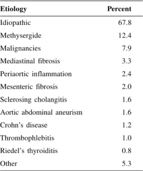
and our previous experience with laparoscopic technique, we decided to evaluate the results of laparoscopic intraperitonealisation in retroperitoneal fibrosis cases and also review the medical information related to the treatment of retroperitoneal fibrosis. Table 1 - Retroperitoneal fibrosis:
etiology in 491 patients*
Etiology Percent
Idiopathic 67.8
Methysergide 12.4
Malignancies 7.9
Mediastinal fibrosis 3.3
Periaortic inflammation 2.4
Mesenteric fibrosis 2.0
Sclerosing cholangitis 1.6 Aortic abdominal aneurism 1.6
Crohn’s disease 1.2
Thrombophlebitis 1.0
Riedel’s thyroiditis 0.8
Other 5.3
CASE REPORTS
Surgical technique
The patient undergoing single-stage bilateral intraperitonealisation of the ureters lies on the operating table in dorsal decubitus, with arms extended alongside the body, under general an-esthesia. Initially, only 4 trocars are used, but more may be inserted de-pending on the difficulty of the surgery and the extent of the ureteral fibrosis. The trocars are inserted in the abdomen based on the extent of the intended dis-section. In bilateral intraperitoneali-sation with ureteral segments incarcer-ated in the sacrolumbar area, a 10 mm trocar is inserted through the umbilical scar (for the optic), a 5 mm trocar is inserted in the hemiclavicular line on each side, 2-3 centimeters below the umbilical scar, and a 10/11mm trocar is inserted in the median line, 2 cm above the pubis. Once the trocars are inserted and the cavity is inspected, the procedure begins.
An incision in the parietal perito-neum is made laterally to the line of Toldt, so as to leave a 2 cm free peri-toneum band connected to the colon, to be later sutured or clipped to the lat-eral border of the peritoneum. The co-lon is mobilized medially (which side first is irrelevant), to expose the Gerota’s fascia and the ureter. The healthy ureteral segment, distal or proximal, is approached first and placed under light traction with a thin Penrose drain to facilitate the dissec-tion. The presence of a Double-J cath-eter inserted prior to surgery greatly diminished the difficulty of this ma-neuver. The incarcerated segment of the ureter is dissected and freed from the fibrosis in its full extent until the normal portion of the ureter is reached (either proximal or distal). Once mo-bilized, the ureteral segment is placed anteriorly to the borders of the incised parietal peritoneum, which are
ap-proximated with sutures or metallic clips, depending on the availability of the material and the surgeon’s prefer-ence. Care is taken not to leave the ure-ter angled or narrowed in the entrances to the retroperitoneum, proximally and distally. The procedure is repeated in the contralateral side, and the surgery is finalized. Both ureteral catheters are removed 3 weeks after the surgery.
Two patients with retroperitoneal fibrosis who had been sent for bilateral intraperitonealisation through laparo-tomy were selected for laparoscopic surgery. After the informed consent of both, the surgeries were performed.
Case 1
G.P.N., white female, 58 years old, born in Piatá (BA), went to the ER of the Hospital das Clinicas da FMUSP in April 1996 with intermittent bilateral lumbar colics, and occasional fever. She had antecedents of idiopathic hy-pothyroidism, arterial hypertension, and diabetes mellitus, adequately
treated. At this occasion, her physical examination was normal. She under-went routine clinical laboratory tests, which were all normal, except for
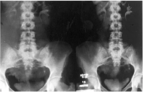
se-rum creatinine (C=2.0). She then took an abdominal echography, revealing bilateral ureterohydronephrosis. Then, under local anesthesia, a retrograde bi-lateral ureteropyelography was per-formed; its result was suggestive of ex-trinsic ureteral obstruction (L4-L5 re-gion), with medial deviation of the ure-ters in their middle portions. In the same procedure, Double-J catheters were introduced bilaterally with no dif-ficulties. After a few weeks, serum creatinine fell to 1.2, and the patient did not present further complaints. The CT revealed a thickening of the retroperitoneum anterior to the great vessels, between L4 and S1, suggest-ing retroperitoneal fibrosis (Fig. 1). On 7-30-1996 the laparoscopic bilateral ureteral intraperitonealisation was per-formed.
Figure 4 - Normal intravenous pyelography 1 year after the operation. Figure 2 - Left retrograde ureteropyelography 4 weeks after the operation.
One can observe pyeloureteral ectasy and an exaggerated ureteral lateralization. On the right side the image was similar.
followed with serum creatinine and in-travenous pyelographies. The creati-nine stabilized at 1.3 and the pyelog-raphies 1 and 2 years after the surgery showed normal kidneys, absence of di-lation, and ureteral lateralization (Figs. 3 and 4). The patient reports light lum-bar pain on the right side, irradiated to the right inferior limb, which ceases with common analgesics.
Case 2
J.A.A., white female, 51 years old, born in Guararapes (SP), presented a sudden increase in the anterior cervi-cal region, with fever and sweating, in October, 1996. Initially treated with antiinflammatories and antibiotics, she underwent clinical laboratory tests until February, 1997, when the diagnosis of hypothyroidism was established and the treatment with T3 and T4 initiated. In 1997, she presented bilateral lumbar pain and dysuria. Her physical exami-nation at the occasion had a single ab-normality: augmented thyroid with pe-trous consistency. The abdominal echography revealed bilateral uretero-hydronephrosis. Serum creatinine was 4.4, and the CT was suggestive of ret-roperitoneal fibrosis. On 9-26-1997, an attempt to pass Double-J catheters bi-laterally was unsuccessful, and two thin ureteral catheters externally con-nected to a Foley catheter were left in place. Two weeks later it was possible to substitute each catheter for a Double-J. On the 10-21-1997, the pa-tient underwent a laparoscopic proce-dure for retroperitoneal biopsies and bilateral ureteral intraperitonealisation. The ureterolysis of the left side was easy, and the intraperitonealisation was completed. However, on the right side, the fibrotic process was far more in-tense and did not allow the identifica-tion of the ureter or of the iliac vessels. Four hours after the start of the surgery, the choice was made for a direct ure-terolysis on another occasion. In the
same anesthetic session, a thyroid bi-opsy was performed, confirming clini-cal suspicion of Riedel’s thyroiditis. The retroperitoneal biopsies revealed only an unspecific inflammatory pro-cess. The patient was discharged on the 10th post-operative day, with no com-plications. There was no need for blood transfusion. The late discharge was due to social problems of the patient; clini-cally, she could have left the hospital on the 2nd or 3rd post-operative day. Due to the extensive fibrosis on the right side, involving not only the ure-ter but also the psoas muscle and the iliac vessels, treatment with prednisone (40 mg/day) and tamoxifen (20 mg/ day) for 3 months was initiated. On the 02-09-98, the patient successfully un-derwent laparoscopic ureterolysis and intraperitonealisation on the right side. During the surgery, it was observed that the fibrosis extended up to the re-nal fossa. The renal pelviswas acciden-tally opened and closed with an intracorporeal suture using chromic catgut 4-0.
In this case, the first surgery took 4 hours; the second, 3 hours and 15 minutes. Bleeding was negligible in both procedures, and the patient was discharged in the 10thand 5th post-op-erative day, respectively. Both ureteral catheters were removed three weeks after the second surgery. After a year of follow-up, the patient had no ab-dominal or lumbar pain, serum creati-nine stabilized at 1.4 and the intrave-nous pyelography was normal.
In none of the 3 procedures was any form of drainage used in the ab-dominal cavity.
DISCUSSION
Due to the relative rarity of retro-peritoneal fibrosis, there are few re-ports in medical literature that present more than a dozen cases originated in the same service, and even then, these
are non-randomized and lack control groups. The reports with the greatest data are usually product of literature reviews and suffer from the same limi-tations4. In face of available published
data, it can be said that not much is known yet about the natural history of retroperitoneal fibrosis, nor about the most appropriate treatment for patients. It is also impossible to determine where clinical treatment attempts should end and in which circumstances should surgical treatment begin. There are those who defend the superiority of clinical treatments11, and those who
choose surgery as the best option5,12;
however, in neither case is there rigor-ous scientific basis. Due to the good results obtained by some with corticoid treatment, in doses (determined empiri-cally) of 20 to 60 mg of prednisone a day during several weeks, or with tamoxifen, 20 mg a day during several months (dosage also determined em-pirically), and eventually used in asso-ciation, it can be argued that the clini-cal treatment should be primarily at-tempted in IRF and in some forms of SRF11. Tamoxifen, used in IRF since
199119, has intense antiestrogenic
ac-tion, and its usefulness in breast can-cer is beyond question. However, even in cases where there are no estrogenic receptors, tamoxifen seems to work, inhibiting the proliferation of fibro-blasts, hence its utilization in fibrotic diseases and mesenchymal tumors20-23.
demon-strates high degree of inflammatory cellularity, or only tamoxifen, when the fibrosis is predominant. As a way to optimize the clinical treatment, system-atic biopsies to ascertain non-malig-nancy and also to determine the degree of inflammatory cellularity has been suggested by some clinicians. Others suggest treatment with corticosteroids in the post-operative period2,5,20,23.
Not all cases respond to clinical treatment; these must treated surgically. Open surgery has significant morbidity and mortality1,9. Since 1992,
laparoscopic technique has been suc-cessfully performed by some groups, with results apparently comparable to those of open surgery, but having lower incidence of complications with no deaths reported due to the surgical pro-cedure13. In the treatment of unilateral
ureteral incarceration, Elashry et al9
con-cluded that laparoscopic surgery was superior to open surgery, despite requir-ing a few more minutes on average to perform. His study compared 6 cases of unilateral fibrosis, only 2 of which hav-ing an IRF diagnosis, with 7 historical cases of fibrosis, also unilateral, 2 of which were associated with aortic aneu-rysms and 1 with a total hysterectomy. His conclusions can be questioned, as they are based on a small data set, non-comparable groups, and a majority of SRF cases. Although there are no groups with large experience in laparoscopically corrected SRF yet, those that have performed it have had the impression, based also on the
expe-rience of successful laparoscopic treat-ment of other retroperitoneal diseases, such as adrenalectomy and nephrec-tomy, that laparoscopy is the method of choice in retroperitoneal fibrosis.
Ureterolysis, performed as an iso-lated procedure, probably has more fre-quent recurrences, which is why most surgeons perform the complimentary ureteral intraperitonealisation, with or without involvement of greater omen-tum, with superior results. Some proceed empirically with pharmaceutical treat-ment after successful surgery as prophy-laxis of recurrence1, 2, 5, 11. Some
investi-gators have also empirically determined that the Double-J catheters must remain for 2 to 3 weeks after surgery.
In the two patients we had the op-portunity to treat and follow, we made no attempt to treat primarily with cor-ticosteroids and/or tamoxifen before prescribing surgery, because these cases were initially considered ad-vanced and had already been selected for open surgery by other colleagues. However, considering the international experience with clinical treatment, to-day the handling of the cases could have possibly been different. After the Double-J catheters had been passed, we would have attempted a clinical treatment with corticosteroids and tamoxifen for 3 or 4 weeks, and then reevaluated. In case of regression of the fibrosis, verified by CT or MRI, we would proceed for a few more weeks and then decide on whether to operate,
based on the results. The empirical clinical treatment introduced in the sec-ond case, after the failed attempt to per-form the ureterolysis on the right side, was not adequately evaluated, making conclusions unfeasible. The subjective intra-operative impression was of a slightly diminished fibrosis, which fa-cilitated the dissection of the pelvic ureter, impossible in the first operation. The association of retroperitoneal fibrosis with Riedel’s thyroiditis, as in our second case, is very rare, and does not require any special therapeutic measure in the approach of retroperi-toneal fibrosis3,24,25. Despite being
etio-logically classified as SRF, the patient was successfully treated as IRF.
The clinical evolution in both cases was excellent, the painful symptoms and the renal insufficiency having dis-appeared rapidly. However, the radio-logical evolution was slower, with pyeloureteral ectasypersisting for sev-eral months (Fig. 3). After a year of evolution, the radiologic images nor-malized completely in both cases, even though signs of retroperitoneal fibrosis persisted (Fig. 4).
In conclusion, although we do not yet know much about retroperitoneal fibrosis, its natural history, or the best treatment alternatives, when there is surgical indication, we understand that the laparoscopic intraperitonealisation is possibly the most appropriate tech-nique for the patient, which may be confirmed in the next few years.
RESUMO RHCFAP/3006
CASTILHO L N e col. - Tratamento laparoscópico de fibrose retroperito-neal: relato de dois casos e revisão da literatura. Rev. Hosp. Clín. Fac. Med. S. Paulo 55(2):69-76, 2000.
Objetivos: Os autores apresentam os resultados de dois pacientes com fibrose retroperitoneal tratados por laparoscopia e fazem a revisão da litera-tura desde 1992, quando o primeiro caso
Relato dos casos: Duas mulheres, uma com fibrose retroperitoneal idiopática, e a outra com fibrose retroperitoneal associada a tireoidite de Riedel, foram operadas por laparoscopia. Ambas apresentavam obstrução ureteral pélvica bilateral e foram operadas por meio da mesma técnica: laparoscopia transperitoneal, mobilização medial de ambos os có-lons, liberação dos ureteres da fibrose e intraperitonização ureteral. Cateteres Duplo-J foram inseridos antes das
ci-rurgias e removidos três semanas de-pois da intraperitonização. A primei-ra paciente teve os dois ureteres intraperitonizados em um único pro-cedimento. A segunda foi submetida a dois procedimentos distintos por di-ficuldades técnicas durante a primei-ra cirurgia. Ambas foprimei-ram acompanha-das por mais de um ano e recupera-ram-se completamente da insuficiên-cia renal. Uma delas ainda tem dor lombar leve ocasionalmente. As urografias excretoras de ambas as
pa-cientes não apresentam mais anorma-lidades.
Conclusões: A correção cirúrgica da fibrose retroperitoneal, quando indicada, deve ser realizada por lapa-roscopia. A ureterolise e a intra-peritonização de ambos os ureteres devem ser realizadas no mesmo ato cirúrgico, sempre que possível.
DESCRITORES: Fibrose retro-peritoneal. Intraperitonização ure-teral. Ureterolise.
REFERENCES
1. HALL MC, VON ESCHENBACH AC & AMES CA - Diseases of the retroperitoneum. In: GILLENWATER JY, GRAYHACK JT, HOWARDS SS et al. (eds.) Adult and Pediatric Urology. St. Louis, Mosby, 1996. p. 1123.
2. RESNICK MI & KURSH ED - Extrinsic obstruction of the ureter. In: WALSH PC, RETIK AB, VAUGHAN JR ED et al. (eds.) Campbell’s Urology. Philadelphia, Saunders, 1996. p. 387.
3. KOEP L & ZUIDEMA GD - The clinical significance of retroperitoneal fibrosis. Surgery1977;81:250-257.
4. LEPOR H & WALSH PC - Idiopathic retroperitoneal fibrosis. J Urol 1979,122:1-6.
5. DE LUCA S, TERRONE C, MANASSERO A et al. - Aetiopathogenesis and treatment of idiopathic retroperitoneal fibrosis. Ann Urol 1998;32:153-159.
6. ZDROJEWSKI Z - Retroperitoneal fibrosis and chronic peri-aortitis: a new hypothesis. Pol Merkuriusz Lek 1998;4:50-53.
7. ENGELKEN JD & ROS PR - Retroperitoneal MR imaging. Magn Reson Imaging Clin N Am 1997;5:165-178.
8. BACHMANN G, BAUER T & RAU WS - MRI and CT in diagnosis and follow-up of idiopathic retroperitoneal fibrosis. Radiology 1995;35:200-207.
9. ELASHRY OM, NAKADA SY, WOLF JR JSI et al. - Ureterolysis for extrinsic ureteral obstruction: a comparison of laparoscopic and open surgical techniques. J Urol 1996;156:1403-1410. 10. TOSCANO C, DI MARZO L, SAPIENZA P et al. - A case of
retroperitoneal fibrosis with spontaneous regression.Minerva Chir 1997;52:1123-1127.
11. OOSTERLINCK W & DERIE A - New data on diagnosis and medical treatment of retroperitoneal fibrosis. Acta Urol Belg. 1997;65:3-6. 12. MARTIN-MARQUINA A, RODRIGUEZ-RUBIO FI, ABAD JI, et al. - Idiopathic retroperitoneal fibrosis. Report of 12 cases. Actas Urol Esp 1995;19:303-306.
13. KAVOUSSI LR, CLAYMAN RV, BRUNT LM et al. - Laparoscopic ureterolysis.J Urol 1992;147:426-429.
14. PUPPO P, CARMIGNANI G, GALLUCI M et al. - Bilateral laparoscopic ureterolysis. Eur Urol1994;25:82-84.
15. KAWABATA G, SHIMOGAKI H & YAMANAKA N - Laparoscopic approach to idiopathic retroperitoneal fibrosis. Nippon Hinyokika Gakkai Zasshi1995;86:1060-1063.
16. BOEKMANN W, WOLFF JM, ADAM G et al. - Laparoscopic bilateral ureterolysis in Ormond’s disease. Urol Int 1996;56:133-136. 17. KAVA BR, RUSSO P & CONLON KC - Laparoscopic diagnosis of
malignant retroperitoneal fibrosis. J Endourol1996;10:535-538. 18. MATTELAER P, BOEKMANN W, BRAUERS A et al. - Laparoscopic ureterolysis in retroperitoneal fibrosis. Acta Urol Belg 1996;64 :15-18.
19. CLARK CP, VANDERPOOL D & PRESKITT JT - The response of retroperitoneal fibrosis to tamoxifen. Surgery1991;109:502-506. 20. OWENS LV, CANCE WG & HUTH JF - Retroperitoneal fibrosis treated
with tamoxifen. Am Surg. 1995;61:842-844.
22. SAVELLI BA, PARSHLEY M & MORGANROTH ML - Successful treatment of sclerosing cervicitis and fibrosing mediastinitis with tamoxifen. Chest 1997;111:1137-1140.
23. ALLENDORFF J, RIEGEL W & KÖHLER H - Regression of retroperitoneal fibrosis by combination therapy with tamoxifen and steroids. Med Klin 1997;92:439-443.
24. MARCHESI M, BIFFONI M, MASTROPIETRO T et al. - Riedel’s thyroiditis: a case report and review of the literature. G Chir 1997;18:820-822.
25. JULIE C, VIEILLEFOND A, DESLIGNERES S et al. - Hashimoto’s thyroiditis associated with Riedel’s thyroiditis and retroperitoneal fibrosis. Pathol Res Pract. 1997;193:573-577.