From the Department of Emergency Medicine and Nutrition Group, Hospital das Clínicas, Faculty of Medicine, University of São Paulo.
ORIGINAL ARTICLES
CHANGES IN BODY FLUID AND ENERGY
COMPARTMENTS DURING PROLONGED HUNGER
STRIKE
Joel Faintuch, Francisco Garcia Soriano, José Paulo Ladeira, Mariano Janiszewski, Irineu Tadeu Velasco and Joaquim J. Gama-Rodrigues
RHCFAP/3002
FAINTUCH J et al. - Changes in body fluid and energy compartments during prolonged hunger strike. Rev. Hosp. Clín. Fac. Med. S. Paulo 55 (2):47-54, 2000.
SUMMARY: Prolonged total food deprivation in non-obese adults is rare, and few studies have documented body composition changes in this setting. In a group of eight hunger strikers who refused alimentation for 43 days, water and energy compartments were estimated, aiming to assess the impact of progressive starvation. Measurements included body mass index (BMI), triceps skinfold (TSF), arm muscle circumference (AMC), and bioimpedance (BIA) determinations of water, fat, lean body mass (LBM), and total resistance. Indirect calorimetry was also performed in one occasion. The age of the group was 43.3±6.2 years (seven males, one female). Only water, intermittent vitamins and electrolytes were ingested, and average weight loss reached 17.9%. On the last two days of the fast (43rd-44th day) rapid intravenous fluid, electrolyte, and vitamin replenishment were provided before proceeding
with realimentation. Body fat decreased approximately 60% (BIA and TSF), whereas BMI reduced only 18%. Initial fat was estimated by BIA as 52.2±5.4% of body weight, and even on the 43rd day it was still measured as 19.7±3.8% of weight. TSF findings were much lower and commensurate with other anthropometric results. Water was comparatively low with high total resistance, and these findings rapidly reversed upon the intravenous rapid hydration. At the end of the starvation period, BMI (21.5±2.6 kg/m2) and most anthropometric determinations were still acceptable, suggesting efficient energy and muscle conservation.
Conclusions: 1) All compartments diminished during fasting, but body fat was by far the most affected; 2) Total water was low and total body resistance comparatively elevated, but these findings rapidly reversed upon rehydration; 3) Exaggerated fat percentage estimates from BIA tests and simultaneous increase in lean body mass estimates suggested that this method was inappropriate for assessing energy compartments in the studied population; 4) Patients were not morphologically malnourished after 43 days of fasting; however, the prognostic impact of other impairments was not considered in this analysis.
DESCRIPTORS: Hunger strike. Acute starvation. Prolonged fasting. Body composition. Nutritional assessment. Bioimpedance analysis.
Body composition changes during long-term hunger strike have not been reported frequently, and no study with bioimpedance analysis in this setting was found in the literature, because this is an uncommon and rarely docu-mented clinical event. Much more is known about food deprivation in ex-perimental animals5, or in other human
nutritional deficiency states13, including
therapeutic starvation for morbid
obe-sity6,20. However, it is debatable
whether such findings should directly apply to long-term hunger strikers7,14,
for the following reasons: 1) hunger strikes are typically undertaken by comparatively healthy adults with
that mortality can be very high, espe-cially as food deprivation reaches or exceeds about two months14.
In the present retrospective study, the results of bioimpedance analysis (BIA) and anthropometric estimations of body composition changes in a group of hunger strikers are reported, with the aim of evaluating the possible contributions of these methods towards a better understanding of the metabolic consequences of long-term total food deprivation.
PATIENTS AND METHODS
The population consisted of eight prisoners serving long sentences who engaged in a difficult legal dispute with government authorities, and within that context started a hunger strike. During the first 11 days they remained within the correctional facility, but afterwards seven of them were transferred to the hospital for clinical observation (the eighth case arrived one week later).
The hospital was requested to do whatever necessary to keep the prison-ers alive, but there obviously were technical, psychological, and legal con-straints. For ethical reasons it was de-cided that, except during emergencies, all diagnostic or therapeutic procedures would be submitted to the prisoners for specific previous approval. As a con-sequence, no investigation protocol or standardized therapeutic approach could be formulated, not only because of the sensitivities and anxieties of the population, but also because it was not possible to forecast the duration of the fasting.
For similar ethical and legal rea-sons, realimentation would only be at-tempted by force if the situation of the patients became critical. Fortunately, by the time it was suspected that their clinical conditions were starting to de-teriorate and intravenous fluids were introduced, their problems with the
au-thorities were adequately solved, and medical advice regarding food intake was therefore spontaneously accepted. The total hunger strike extended for 46 days, but by the 43rd day, a large
in-take of intravenous fluids and electro-lytes was provided to offset hydro-elec-trolytic and vitamin imbalance. From the 43rd day until the 46th day, modest
amounts of hypocaloric parenteral feeding (mean daily intake of 535± 206 kcal and 10.3±2.5 g of amino ac-ids) were supplied to these individuals. Otherwise only water was accepted during the first six weeks, with occa-sional vitamins and electrolytes to cor-rect clinical manifestations or bio-chemical imbalances. The current in-vestigations address the first 43 days of this period.
The age of the population was 43.3±6.2 years (33 – 52), and there were seven males and one female. Ini-tial body mass index (BMI) was
26.1±2.7 kg/m2 (20.8 – 30.1), and no
relevant medical or surgical disease was identified in the clinical history, except for essential arterial hyperten-sion in one male case and urinary in-fection in the female patient. There was no mortality in this series, and after adequate realimentation, all subjects were discharged in good condition.
Anthropometric determinations in-cluded body weight and height, BMI, arm circumference, triceps skinfold (TSF), and arm muscle circumference (AMC). For the last two variables, the adopted normal references were re-spectively 12.5 mm and 25.3 cm for males, and 16.5 mm and 23.2 cm for females2.
Bioimpedance analysis (BIA) was done on a few occasions. The standard tetrapolar technique employing a single current apparatus (50 kHz) was used (Biodynamics model 310, Biodynam-ics, Seattle, Washington, USA). The following variables were documented: body fat, lean body mass (LBM), total body water, resistance, and reactance.
Both absolute values and % of body weight were examined.
Basal energy expenditure (BEE) was calculated according to the Harris Benedict equation (HBE). Whenever corrections or adjustments were intro-duced, the corresponding citation is in-dicated. The opportunity to perform indirect calorimetry occurred only once when a 33-year-old male on the 17th day of food deprivation agreed to
the test. The apparatus was a Deltatrac MB-101, Sensormedics, Yorba Linda, CA, USA, and the measurement was done by a trained technician. Steady state was defined as a 30-minute period during which the coefficient of
varia-tion of oxygen consumpvaria-tion and CO2
production was equal or below 10%9.
Statistical analysis: Values are
pre-sented as mean±SEM or as percentage. For selected variables, linear regression analysis was performed to estimate missing values in the beginning or end of the observation period. In such cir-cumstances, the correlation index (r)
and the (p) value are indicated. Com-parison between initial and final results within the group was done by two-tailed Student’s t test. A significance
level of 5% was adopted.
This study was approved by the Ethical Committee of Hospital das Clinicas and São Paulo University Medical School.
RESULTS
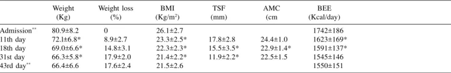
Patients were initially well-nour-ished but lost nearly 18% of their body weight. Reduction for TSF was sub-stantial (from 158.5% to 63.8% of nor-mal, or 94.7% decrease), however AMC was affected by only 15.1%. These results as well as the calculated BEE for various time periods can be seen in Table 1.
findings are displayed in Table 2. Body fat on the 31st day of starvation was 26.8% of body weight. Projecting the rate of daily change to the first day, an unusual proportion of 52.2% of body weight as adipose tissue resulted. To-tal water remained within the low-nor-mal range on the first date of registra-tion, markedly increasing 12 days later, along with the corresponding reduction in resistance.
BEE was routinely obtained from the Harris-Benedict equation (HBE), but the bioimpedance apparatus fur-nishes its own estimate of that equa-tion, which was also taken into ac-count. Because of possible interference of progressive starvation on traditional findings of BEE , as described by sev-eral authors7,14, BEE data was
math-ematically manipulated to assess the probable impact on fuel oxidation and severity of tissue loss, or total en-ergy expenditure. As demonstrated in Table 3, discrepancies concerning BEE were relatively minor; however, much greater differences were unveiled dur-ing the estimation of total energy ex-penditure (TEE).
Indirect calorimetry could not be serially employed due to technical and psychological barriers. It was success-fully measured only once (Table 4). The respiratory quotient was extremely low (0.68), and energy expenditure also remained 13% below the HBE
cal-Table 1 - Anthropometric findings.
Weight Weight loss BMI TSF AMC BEE
(Kg) (%) (Kg/m2) (mm) (cm (Kcal/day)
Admission** 80.9±8.2 0 26.1±2.7 1742±186
11th day 72.l±6.8* 8.9±2.7 23.3±2.5* 17.8±2.8 24.4±1.0 1623±169*
18th day 69.0±6.6* 14.8±3.1 22.3±2.3* 15.5±3.5* 22.9±1.4* 1591±137*
31st day 66.3±5.8* 17.9±2.0 21.4±2.2* 11.9±2.2* 22.5±1.5 1545±146
43rd day** 66.4±6.6 17.6±2.4 21.5±2.6 1550±151
BMI = inicial body mass index TSF= triceps skinfold
AMC= mid arm circumference – 0,314 x TSF BEE= Harris-Benedict equation
*p<0.05 (in comparison with previous values)
**TSF and AMC not available due to technical problems. Estimated average values for TSF (linear regression analysis, r= -0.9985, p=0.035):Admission 20.6 (158.5% of normal) and 43rd day 8.3 (63.8% of normal)
For AMC (r= -0.8423, p=0.362): Admission 25.0 (99.8% of normal) and 43rd day 21.2 (84.7% of normal)
Table 2 - Principal bioimpedance results.
Fat Fat weight H20 H20 volume Resistance
(%) (Kg) (%) (L) (Ohm)
31st day 26.8±6.2% 17.6±3.9 52.l±3.7% 35.l±4.5 639±80
43rd day 19.7±3.8% 12.9±1.8* 57.8±3.2% 38.4±5.2* 550±78*
* p<0.05 (in comparison with initial values)
Table 3 - Estimated basal energy expenditure BEE and total energy expenditure
TEE during period of hunger strike.
Harris-Benedict BEE Apparatus BEE Adjusted BEE TEE** TEE***
Admission 1742±186 2021
31st day 1545±146* 1480±203 1483±80 1414* 1244
43rd day 1550±151 1626±220* 1515±81 1414 1240
BEE and TEE = Kcal/day
Apparatus BEE = Supplied by Biodynamics-BIA model 310
Adjusted BEE = Dulloo & Jacquet,19987 (BMR kJ= 3482 + 52.9 FFM kg +9.7 Fat kg),converted
to Kcal/day
*p<0.05 (in comparison with previous values)
**Leiter & Marliss,198214 (Initial BEE = Harris-Benedict X 1.16; After three weeks: 30%
decrease)
***Dulloo & Jacquet,19987 (Thermogenic economy in moderate starvation:20% below expected
BEE),calculated from Harris- Benedict results
Table 4 - Indirect calorimetry results on the 18th day (33-year-old male, 1.73m,
69.3 kg).
VO2 (ml/min/m2) 217
VCO2 (ml/min/m2) 146
EE (kcal/day) 1440
RQ 0.68
BEE (kcal/day) 1655
EE/BEE 0.87
culation. This case, as well as all oth-ers, had positive urine tests for ketone bodies, not only during the time of the test, but during the entire hunger-strike period inside the hospital.
Spent calories for the complete analyzed period were computed using the straight Harris-Benedict equation, extrapolations based on the BIA read-ings, and modified HBE results as re-ported by several groups14,18. Forecasts
of 59 772 – 70 211 burned kcal were obtained. The conversion of such ex-penditures into actual destruction of fat and non-fat mass produced theoretical contributions of 5 957 – 7 225 g of lipid reserves and 1 046 – 1 808 g of protein and carbohydrate (Table 5).
A similar calculation for probable total energy consumption can be followe in Table 6, but derived from measured tissue losses and their caloric equivalent.Different assumptions were adopted8,14,as appropriate for serial
measurements of body weight as well as of total fat (estimated by three methods).The deducted range of total kcal loss fluctuated widely, from 62311 up to 151704 for the studied period.
DISCUSSION
Uncomplicated starvation differs from traumatic and septic malnutrition in many aspects; one of these is the
contribution of fat toward energy pro-duction. In prolonged hunger, fat is definitely the most consumed fuel, rep-resenting at least 90% of the total ex-penditure7,11,18. Its exhaustion may have
implications for survival5,6,14,20 not
un-like nitrogen (N) loss. In undernour-ished critical patients, N excretion tends to be overwhelming, obfuscating the numerically superior but clinically less worrisome oxidation of fatty acids, and is therefore considered the limit-ing nutrient category.
In trauma, nitrogen balances in the range of -8 g N/ day are common9, and
burn victims exhibit as much as 20 – 25 g of N depletion in some phases of
their hospitalization17.Such damage
Table 5 - Estimated total energy expenditure during 43 days of fasting, as furnished by BIA apparatus and Harris Benedict
equation, and its equivalent in lost body mass (fat and protein oxidation) according to different methods of calculation (Same references as Table 3)*.
Harris-Benedict Apparatus Dulloo&Jacquet7 Adj.Dulloo&Jacquet7 Leiter&Marliss14
0 - 31st day 51 119 49 941 49 987 45 384 53 243
31 - 43rd day 18 636 18 636 17 988 14 388 16 968
Sum total 69 755 68 577 67 975 59 772 70 211
Weight equiv.** 1796+6952 1766+6835 1750+6775 1539+5957 1808+6998
(8748 ) (8601 ) (8525 ) (7496 ) (8806 )
Weight equiv.*** 1221+7208 1200+7086 1190+7024 1046+6176 1229+7225
(8429) (8286 ) (8214 ) (7222 ) (8484 )
Real weight lossf
---14500---*Daily energy expenditure calculated as mean between initial and final values for the period; Initial energy expenditure always based on Harris-Benedict equation except when the apparatus measurements are mentioned ;Energy in kcal, weight in g; caloric value of fat: 9 kcal/g; caloric value of carbohydrate or protein:4 kcal/g
**Leiter & Marliss,198214: 10.3% of calories supplied by protein+carbohydrate, remainder by fat
***Owen et al, 199818: 7.0% of calories furnished by protein+carbohydrate,remainder by fat
fAverage weight loss for the studied patients during the analyzed period, including fluids and electrolytes (Initial weight, 80.9±8.2 kg, weight on 43rd
day, 66.4±6.6 kg)
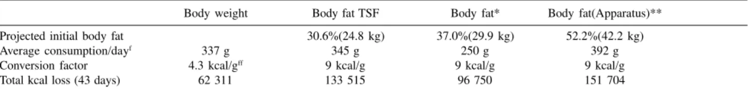
Table 6 - Energy expenditure estimated from measured loss of body weight and body fat.
Body weight Body fat TSF Body fat* Body fat(Apparatus)**
Projected initial body fat 30.6%(24.8 kg) 37.0%(29.9 kg) 52.2%(42.2 kg)
Average consumption/dayf 337 g 345 g 250 g 392 g
Conversion factor 4.3 kcal/gff 9 kcal/g 9 kcal/g 9 kcal/g
Total kcal loss (43 days) 62 311 133 515 96 750 151 704
TSF = Initial triceps skinfold estimated as 158.5% of normal (linear regression analysis, r= -0.99, p= 0.035); ideal body fat deducted from Heitmann equation10 (% Fat=0.988 BMI+0.242 Wt +0.094 A-30.18=19.3% body weight), and corrected for the measured excess adiposity, to 30.6%
*Valhalla equation (Apud Elia & Lunn,19978): Fat% = 9.07 + 0.603 weight (kg) - 0.581 height2 (m)/resistance (ohm), projected for first day
**Biodynamics model 310
fConsidering first and 43rd day findings
compounds the prevailing tendency to-wards body mass erosion, even when food intake is present. Before the mod-ern era of clinical nutrition, adults suf-fering from burns exceeding 40% of body surface lost 22% of body weight within two months, even though they were receiving standard complete hos-pital diets17.
In the hypothetical situation of a healthy 70-kg adult male with 13 kg of body protein (2080 g of nitrogen) and 160 000 kcal of fat (17.8 kg)2, who
ex-periences a loss of 60 g of protein (9.6 g N) and 150 g of fat per day19,
pro-tein would be exhausted in 217 days and adipose tissue in 119 days. Given the fact that in acute conditions, sur-vival is unlikely when more than one
third of body protein is degraded13,
available life span would not exceed 72 days. If a negative nitrogen balance of -20 g/day is assumed, for instance be-cause of trauma or sepsis, then that theoretical deadline would recede to only 35 days. As emphasized previ-ously, the limiting substrate is more of-ten protein, but fat deficit may become critical in circumstances of uncompli-cated but prolonged starvation, when massive consumption of body fat stores are prone to occur4,14.
Hunger strike is not counted among the metabolically stressful conditions, and catabolism is subdued in this set-ting. Consequently, survival for 2.0 – 2.5 months is usually expected4,23,
simi-larly to the theoretical example that was described previously. This assump-tion must be reassessed within the con-text of therapeutic fasting for morbid obesity, a rather similar situation but where lipid reserves are much more plentiful, and in which total food dep-rivation for up to one year has been well tolerated6,7,18,20.
The population here investigated was not obese, with the possible excep-tion of one subject with a BMI of 30.1 kg/m2, but their initial BMI was
some-what above the usual range (Table 1).
Therefore, they probably had more en-ergetic tissues to burn than leaner sub-jects, and anthropometric and BIA de-terminations tend to confirm this sus-picion. In such circumstances, daily fat oxidation should not markedly change, but the end result could be a healthier profile of body composition than pre-viously described for long-term fasting individuals7,14,18.
Table 1 reveals that weight loss was precipitous but far from dramatic (ap-proximately 18% change), and arm muscle circumference, a variable re-lated to muscle mass, was even less af-fected during the studied period. Tri-ceps skinfold thickness reduced at a higher magnitude, and extrapolation for the total period would suggest a de-crease of almost 60% during the period of food deprivation.
Assuming the figures on Table 5 with regard to total fat oxidation, if 60% of fat reduction corresponds to approxi-mately 7 kg of burned fat, then initial body fat should be in the range of 11.7 kg, or 14.5% of body weight (80.9±8.2 kg). Probably actual fat before fasting was appreciably higher, close to 30% of body weight, as suggested by body weight and triceps skinfold determina-tions (Table 6). However, propordetermina-tions of 37% or 52%, as foreseen by a theoreti-cal equation or bioimpedance analysis, are hardly compatible with other anthro-pometric and clinical findings. If one accepts the figure of 30%, which seems reasonable, it must be concluded that fat oxidation did not correspond to 60% of body reserves, because that would amount to more than 14 kg of con-sumed fat, or nearly 100% of real weight loss. According to the reported data, water alone usually represents roughly 50% of the weight decrease in such circumstances15,21,23, which is still
less than the 74% lost by surgical pa-tients.12 The more logical hypothesis is
that 7 kg of adipose tissue were lost, representing about 30% of available stores.
In this study, nitrogen elimination could not be fairly estimated by BIA, which varies directly with fat-free mass (FFM) and varies inversely with body fat. Since anthropometric assessments indicated a greater loss in fat grams than in protein grams, the BIA deter-minations were influenced most heavily by the fat loss, confounding in-terpretation with regard to nitrogen elimination. Therefore, the BIA find-ings suggesting that FFM (and LBM, which is a related variable) increased as fasting advanced must be rejected. Shifts of intracellular water would per-haps furnish more useful indications regarding nitrogen elimination, but this compartment could not be investigated in the present situation. Standard an-thropometric assessment in the form of arm muscle circumference is also rela-tively insensitive for estimating nitro-gen elimination because of the possible interference of fluids and bone on the result. In any case, AMC was affected in the proportion of 18% for the entire fasting period.
If one assumes 13 kg of total muscle for a healthy young male2, the
loss should be equivalent to 2.3 kg of muscle or approximately 20% of that value in protein (460 g)2,15,19. As
dem-onstrated in Table 5, even the most conservative prediction of protein ero-sion was above 1 kg, thus suggesting an underestimation of real changes by AMC .
Indirect calorimetry was only per-formed once in a single patient. Nev-ertheless, the results fully correspond to the predicted values, thus lending credence to the assumptions of Table 3. It is interesting to note that the res-piratory quotient was under 0.7, which is theoretically improbable, since 0.7 corresponds to exclusive fat oxidation. Yet such results seem to be entirely typical of prolonged food deprivation and could be explained by fatty acid desaturation18.
shifts during severe long-term volun-tary wasting can be retrieved from the literature. During chronic malnutrition, especially within the context of surgi-cal catabolism, but also in kwashiorkor cases of third-world countries, it is ap-parent that water accumulates every-where, especially in the extracellular space8,13,15,23. In contrast, initial
abun-dant diuresis is usually observed in obese subjects that engage in total fast-ing, tapering down after a couple of days, but a relative state of dehydration is maintained6,20,23.Although fluid
bal-ance was not available in this experi-ence, there is indirect evidence that the reported patients followed the obese paradigm, because of the compara-tively low readings of total body wa-ter, and appropriately elevated determi-nations of total resistance on the 31st day of hunger strike (Table 2).
The phase of conservation of the remaining water and sodium after the initial depletion is the rule in healthy fasting human beings, but interestingly rats proceed straight to fluid retention when denied food, without any previ-ous negative fluid balance.3 The next
step, coinciding with refeeding after prolonged starvation, may display some tendency toward peripheral edema, as apparent during the first days or weeks of realimentation of both chronically malnourished and starved obese subjects6,23. In the current
series, body water restoration with some questionable degree of over-shooting was noted immediately upon intensive hydration, as BIA-estimated water rapidly expanded and body resis-tance diminished in the same propor-tion (Table 2). Nevertheless, clinical edema was not asignificant problem in any of the patients.
The metabolic profile of the fasting subjects after adaptation that best fits the subjects in this study consists of a total daily energy expenditure of about 1500 kcal/day (roughly 25% below comparable non-starving subjects, and
close to the suggestion of Leiter &
Marliss14); a probable consumption
pattern of at least 90% lipid calories versus 7% – 10% generated by pro-tein18; and a total oxidation (43 days)
in the range of 60 000 – 70 000 kcal, divided as fat (6.0 – 7.2 kg ) and pro-tein + carbohydrate (1.0 –1.8 kg ). If one subtracts these numbers from the average total weight loss (14.5 kg), a balance of 6.0-7.5 kg of water and electrolytes will appear (rather close to the suggested 50%15,21,23).
Based on these premises, a very proper physiologic response was thus elicited in the subjects in this study, with marked reduction of energy ex-penditure, adequate defense of vital muscle stores, and massive but not pro-hibitive burning of adipose tissue. In-deed, if total body glycogen is ac-cepted as 600 g in the adult,2,12,19 and
total protein + carbohydrate loss was 1.0-1.8 kg, then nitrogen excretion was fairly inconspicuous for the circum-stances.
Given the fact that the patients ap-parently started with sufficient organic reserves at the beginning of the hunger strike, their body composition at the end of 43 days was still nominally more acceptable than what is observed in many hospitalized patients, with a
BMI of 21.5±2.6 kg/m2.
One is tempted to speculate that the subjects could have fasted considerably longer, similarly to morbidly obese in-dividuals6,20. However, this may be a
premature impression, since total food deprivation in normal adults is some-times accompanied by pathophysi-ologic derangements quite different from what is seen in severe obesity or in chronic partial starvation. It cannot be forgotten that morphologic assess-ments such as those performed here are comparatively weak prognostic indices
regarding morbidity and mortality.16
Vital functions may become compro-mised during rapid weight loss, tissue deproteinization may lead to damage
and rupture of myocardial fibres,22 and
certain crucial micronutrients are more quickly exhausted in some circum-stances than in others. Kinney12,
re-viewing previous investigations by Keys et al11 and Benedict1, emphasizes
the fact that intake of as little as 20% of the daily calorie requirements may extend the average period before le-thality of total fasting from two months or even less to about six months.
Many details of the natural history of human absolute starvation are still obscure, and most were originally documented 50 years ago or earlier. Professional fasting specialists (who fasted in exchange for payment) as used in the beginning of the century by
Benedict1, cannot be used in modern
scientific protocols. Experimental ani-mals do provide many insights, but biological differences with patients cer-tainly exist. Similarly, information ac-cumulated in various modalities of hu-man primary and secondary malnutri-tion are very useful for understanding fuel consumption and body composi-tion changes during starvacomposi-tion16.
Nev-ertheless, actual clinical observations of hunger strikers as here analyzed should be collected whenever avail-able, feasible, and ethical, in order to advance knowledge about this infre-quent modality of self-inflicted injury and to provide insights for nutritional assistance and patient care.
CONCLUSIONS
1) All body compartments dimin-ished with fasting, but adipose tissue was by far the most depleted, thus con-firming the overwhelming participation of body lipids in energy balance dur-ing uncomplicated prolonged starva-tion.
3) Exaggerated fat percentage esti-mated from BIA tests and simulta-neous estimation of increased lean body mass suggested that BIA was in-appropriate for estimating energy com-partments in the studied population.
4) Anthropometry yielded a body profile more commensurate with
clini-cal findings, although some shortcom-ings concerning muscle mass estima-tion could be identified as well.
5) According to calculations of both fat and fat-free tissue consump-tion, patients were not initially mal-nourished and remained apparently free from high nutritional risk after 43
days of fasting; however, the impact of functional impairments, tissue damage, and micronutrient depletion was not considered in this analysis, and could have severely affected their prognosis if fasting had been prolonged beyond 43 days.
RESUMO RHCFAP/3002
FAINTUCH J e col. - Alterações nos compartimentos hídricos e energéticos do organismo durante a
greve de fome. Rev Hosp Clin
Fac Med S Paulo 55 (2):47-54,
2000.
A privação total e prolongada de alimentos em adultos não-obesos é ra-ramente vista, e poucos estudos docu-mentaram as modificações da compo-sição corpórea neste contexto.Num grupo de oito casos de greve de fome durante 43 dias, procedeu-se à estima-tiva dos compartimentos hídricos e energéticos, visando averiguar a influ-ência sobre os mesmos da desnutrição progressiva.Os métodos incluiram ín-dice de massa corporal (IMC), prega cutânea do tríceps (PCT), circunferên-cia muscular do braço, e determinação através da bioimpedância (BIA) da água, massa gorda, massa magra e re-sistência corpórea total..A calorimetria indireta foi realizada em uma ocasião apenas.A idade do grupo era de 43,3± 6,2 anos (sete homens, uma mulher),
somente água e ocasionais eletrólitos e vitaminas foram ingeridos no jejum, e a perda de peso média foi de 17,9%.
Por volta do 43º dia da greve ini-ciou-se a reposição venosa rápida de fluidos, vitaminas e eletrólitos,antes de se prosseguir com a realimentação.A gordura corporal diminuiu em aproxi-madamente 60% (BIA e PCT), ao pas-so que o IMC caiu apenas 18%.A esti-mativa da gordura total inicial por BIA foi de 52,2± 5,4% do peso corporal, e mesmo no 43º dia do evento o valor cal-culado era de 19,7± 3,8% do peso.Os valores correspondentes deduzidos da PCT mostraram-se substancialmente in-feriores, e mais compatíveis com os de-mais índices antropométricos.
A água corporal revelou-se inicial-mente contraída, com resistência elevada,sendo que estes achados se re-verteram rapidamente por ocasião da hidratação venosa rápida.Quando do término da greve de fome o IMC
(21,5± 2,6 kg/m2) e outras variáveis
antropométricas revelavam-se numeri-camente aceitáveis, sugerindo
eficien-te conservação de musculatura e ener-gia na fase de dieta zero.Conclui-se que: 1) Todos os compartimentos orgâ-nicos se contrairam na greve de fome, porém o tecido adiposo foi de longe o mais afetado; 2) A água corporal mostrou-se reduzida com elevada resis-tência total, mas estes achados inverte-ram-se prontamente mediante hidratação parenteral; 3) O encontro de gordura total excessiva e de aumento da massa magra com o avançar do je-jum sugerem que as leituras de BIA são inapropriadas para esta população e fornecem resultados incoerentes; 4) Com base nos parâmetros expostos os doentes não estavam morfologicamente desnutridos ao cabo de 43 dias, toda-via não foram aqui avaliados outros transtornos de considerável importân-cia prognóstica.
DESCRITORES: Greve de fome.
REFERENCES
1. BENEDICT FG - A study of prolonged fasting. Washington Publ 1915; 203.
2. BLACKBURN GL, BISTRIAN BR, MAINI BS et al. - Nutritional and metabolic assessment of the hospitalized patient. J Parent Ent Nutr 1977;1:11-22.
3. BOIM MA & SCHOR N - Renal sodium conservation during starvation in rats. Braz J Med Biol Res 1992; 25:1209-1213.
4. CAHILL Jr GF - Survival in starvation. Am J Clin Nutr 1998; 68:1-2.
5. CUENDET GS, LOTEN EG, CAMERON DP et al. - Hormone-substrate responses to total fasting in lean and obese mice. Amer J Physiol1975; 228:276-2831.
6. DRENICK EJ, SWENDSEID ME, BLAHD WH et al. - Prolonged starvation as treatment for severe obesity. J Amer Med Assoc 1964; 187:100-105.
7. DULLOO AG & JACQUET J - Adaptive reduction in basal metabolic rate in response to food deprivation in humans: a role for feedback signals from fat stores. Am J Clin Nutr 1998; 68:599-606. 8. ELIA M & LUNN PG - Biological markers of protein-energy
malnutrition. Clin Nutr 1997; 16 (Suppl 1):11-17.
9. FRANKENFIELD DC, SMITH JS & COONEY RN - Accelerated nitrogen loss after trauma is not attenuated by achievement of nitrogen balance. J Parent Ent Nutr 1997; 21:324-329. 10. HEITMANN BL - Evaluation of body fat estimated from body mass
index, skinfolds and impedance. A comparative study. Eur J Clin Nutr 1990; 44:831-837.
11. KEYS A, BROZEK J, HENSCHEL A et al. - The biology of human starvation. Minneapolis USA, Univ of Minnesota Press, 1950. 12. KINNEY JM - The tissue composition of surgical weight loss. In:
JOHNSTON I. DA (ed) - Advances in parenteral nutrition. Lancaster, MTP Press, 1978. p 511-518.
13. KOTLER DP, TIERNEY AR, WANG I et al. - Magnitude of body cell mass depletion and the timing of death from waisting in AIDS. Am J Clin Nutr 1989; 50:444-447.
14. LEITER LA & MARLISS EB - Survival during fasting may depend on fat as well as protein stores. J Amer Med Assoc 1982; 248 :2301-2307.
15. MOORE FD & BOYDEN CM - Body cell mass and limits of hydration of the fat-free body. Their relation to estimated skeletal weight. Ann NY Acad Sci 1963; 110:62-71.
16. MORAIS AAC, COMARELLA AO, PITANGA KC et al. - Interest of conventional clinical, biochemical, and bioimpedance measurements as indicators of mortality risk in critical patients. Rev Hosp Clin Fac Med S Paulo 1998; 53:176-180.
17. NEWSOME TW, MASON Jr AD & PRUITT Jr BA - Weight loss following thermal injury. Ann Surg 1973; 178:215-217. 18. OWEN OE, SMALLEY KJ, D’ALESSIO DA et al. - Protein, fat and
carbohydrate anaplerosis and cataplerosis. Am J Clin Nutr 1998; 68:12-34.
19. PASSMORE R & ROBSON JS. - A companion to medical studies. Oxford, UK, Blackwell, 1974. v. 3.
20. RUNCIE J & MILLER V - Treatment of obesity by total fasting for up to 249 days. Lancet 1966;992-996.
21. SMITH R & DRENICK EJ - Changes in body water and sodium during prolonged starvation for extreme obesity. Clin Sci 1966; 31;437-441.
22. SOURS HE, FRATALLI VP, BRAND CD et al. - Sudden death associated with very low calorie weight reductions regimens. Amer J Clin Nutr 1981; 34:453-46.
23. WEINSIER RL - Fasting-A review with emphasis on the electrolytes. Amer J Med. 1971; 50:233-240.