65
REV. HOSP. CLÍN. FAC. MED. S. PAULO 55(2):65-68, 2000 MARCH-APRIL
From the Department of Medical Clinic, Triangulo Mineiro University, School of Medicine and the Cancer Combact Association of Brazil.
PARASITIC THYROID NODULE IN A PATIENT WITH
HASHIMOTO’S CHRONIC THYROIDITIS
Vitorino Modesto dos Santos, Marcus Aurelho de Lima, Eurípedes Oliveira Marinho, Marco Aurélio de Oliveira Marinho, Lister Arruda Modesto dos Santos and Cristiane Mendes Raphael
RHCFAP/3005
SANTOS V M dos et al. - Parasitic thyroid nodule in a patient with hashimoto’s chronic thyroiditis. Rev. Hosp. Clín. Fac. Med.
S. Paulo 55(2):65-68, 2000.
SUMMARY: A case of parasitic thyroid nodule is presented. The patient was a non symptomatic 53-year-old white woman, on irregular course of L-thyroxine to treat hypothyroidism due to Hashimoto’s thyroiditis. Without a history of thyroid trauma or surgery, she presented a 1.6 x 0.7 x 0.5cm right pre-laryngeal lymph node–like mass which, on ultrasonography, appeared distinct from the gland. TSH, thyroid peroxidase antibody and thyroglobulin antibody serum levels were elevated and T4-free level was normal. Thyroid and total body 99mTc isonitrile scintiscan showed a topic thyroid without radionuclide uptake in the nodule.
Fine-needle aspiration of the nodule showed epithelial cells with nuclear atypia and oncocytic changes plus intense lymphoid infiltration and germinative center formation, simulating lymph node metastasis of papillary thyroid carcinoma. Conventional biopsy revealed a parasitic thyroid nodule with Hashimoto’s chronic thyroiditis. Parasitic thyroid nodule must always be remembered so that unnecessary surgical assessment and undesirable sequels may be avoided.
DESCRIPTORS: Thyroid. Parasitic nodule. Sequestered nodule. Accessory nodule. Hashimoto’s chronic thyroiditis.
Ectopic thyroid tissue is usually de-scribed as being situated in the medial or lateral cervical region, between the base of the tongue and the gland’s nor-mal site, due to thyroid migration dis-orders during embryogenesis 1. Besides that condition, portions of thyroid tis-sue may be mechanically separated from the gland and develop as acces-sory nodules.
These nodules, also termed para-sitic or sequestered, do not show any lymph node architectural vestiges and may or may not be connected to the thyroid by a pedicle 1-5. This benign condition, considered rare, may consti-tute a serious diagnostic dilemma when one suspects lymph node metastasis from an occult thyroid carcinoma 1, 2, 4, 6. Parasitic nodules have been
de-scribed in association with Graves dis-ease and colloid goiter 1, 3.
Recently, we examined a case of parasitic thyroid nodule in a female with Hashimoto’s chronic thyroiditis. The needle aspiration biopsy sample resembled papillary thyroid carcinoma lymph node metastasis.
CLINICAL CASE
A 53-year-old non symptomatic woman, body mass index 25.0kg/m2, with no history of trauma or thyroid
hetero-66
REV. HOSP. CLÍN. FAC. MED. S. PAULO 55(2):65-68, 2000 MARCH-APRIL
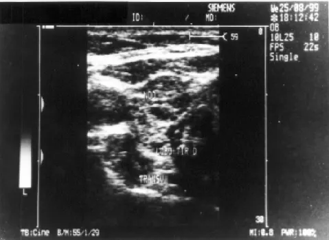
geneous aspect, suggestive of chronic thyroiditis and a 0.8 x 0.7cm nodule without connection with the upper por-tion of the gland but in the same fascial plane (Fig. 1). 99mTc isonitrile (MIBI) scintiscan of the thyroid and total body showed a normal topic thyroid with no radionuclide uptake in the separated nodule (Fig. 2).
Fine-needle aspiration biopsies of the thyroid and of the nodule were per-formed. Nodule aspiration revealed groups of epithelial cells with irregu-lar nuclei, nuclear grooving and/or in-tranuclear cytoplasmic inclusions, sur-rounded by abundant mature lympho-cytes. Lymph node metastasis of pap-illary thyroid carcinoma was suspected (Fig. 3). Thyroid aspiration revealed intense lymphocyte infiltrates, occa-sional plasmocytes, and epithelial cells with oncocytic changes, compatible with Hashimoto’s chronic thyroiditis. Patient was submitted to a nodule re-section which appeared as a chestnut colored, smooth and brilliant 1.6 x 0.7 x 0.5cm mass. Microscopically, thyroid parenchyma represented by small and medium sized follicles surrounded by cells with irregular and voluminous nuclei presenting some nuclear groov-ing and cytoplasmic inclusions was observed. Cytoplasma were either scanty or abundant and granular, with an oncocytic appearance. Thyroid fol-licles were permeated and surrounded by an intense inflammatory lymphoid infiltrate forming germinative centers (Fig. 4). The absence of marginal si-nuses in all the silver staining sections dismissed the possibility of thyroid tis-sue in the lymph node. Diagnosis: parasitic thyroid nodule with Hashimoto’s chronic thyroiditis.
DISCUSSION
Besides embryogenic disorders, trauma or surgery on the gland, the presence of sequestered thyroid tissue
has been related to the mechanical ac-tion of neck muscles in cases of nodu-lar colloid goiter. Projecting portions of the goiter, extending through the fas-cia, may be severed by muscular
pres-sure and separated from the thyroid. In such case, the appearance is that of nodular goiter, without any evidence of malignancy both in the gland and the nodule 3, 5.
Figure 1 - Thyroid ultrasonography revealed a diffuse heterogeneous aspect, suggestive of chronic thyroiditis, and a nodule, 0.8 x 0.7cm, in the same fascial plane of the gland and without contact with the upper limit of the right lobe.
Figure 2 - 99mTc MIBI scintiscan of thyroid and of total body showed a normal topic thyroid with no
67
REV. HOSP. CLÍN. FAC. MED. S. PAULO 55(2):65-68, 2000 MARCH-APRIL
The diagnosis of thyroid parasitic nodule requires that the tissue be placed in the same fascial plane as the thyroid, be unassociated with the lymph node and exhibit a similar his-tological aspect as the gland. The ana-tomical relationship between the nod-ule and the thyroid may be lost or missed, as occurs in cases of parasitic uterine leyomioma 4.
Our patient had Hashimoto’s chronic thyroiditis and, on physical ex-amination, she presented a topic thyroid and an accessory thyroid nodule in the anterior cervical region, separated from the gland. In such cases, besides lymph node metastasis, one must consider the possibility of benign conditions such as implants of thyroid tissue after trauma or surgery and thyroid inclusion in cer-vical lymph nodes7.
In the present case, the diagnosis of a parasitic nodule was confirmed by the absence of any lymph node archi-tectural association; fact which dis-missed the possibility of thyroid tissue inclusion in the lymph node. The lack of trauma or surgery in the gland dis-counted the possibility of mechanical implants. Although the presence of nuclear atypia and cytoplasmic changes associated with lymphoid fol-licles were suggestive of papillary lymph node carcinoma metastasis, this diagnosis was also discounted by the absence of marginal sinuses in all the silver staining sections examined 5.
In Hashimoto’s thyroiditis, abun-dant lymphocytic inflammatory infil-trate and germinative centers forma-tion, may simulate lymph node me-tastasis of papillary thyroid carcinoma, especially the follicular variant 1, 6. If there was metastasis from papillary thyroid carcinoma in this patient, total thyroidectomy and group VI lymph node dissection should be performed. Therefore, parasitic thyroid nodule must always be remembered so that unnecessary surgical assessment and undesirable sequels may be avoided. Figure 4a - Fotomicrography of the thyroid nodule showing Hashimoto’s chronic
thyroiditis (HE 40X).
Figure 4b - In detail, Hashimoto’s chronic thyroiditis characterized by thyroid follicles with nuclear atypia and oncocytic changes (arrow), in addition to intense lymphocytic inflammatory infiltrate (HE 400X).
68
REV. HOSP. CLÍN. FAC. MED. S. PAULO 55(2):65-68, 2000 MARCH-APRIL
RESUMO RHCFAP/3005
SANTOS V M dos e col. - Nódulo tireoidiano parasítico em paciente com tireoidite crônica de Hashimoto. Rev. Hosp. Clín. Fac.
Med. S. Paulo 55(2):65-68, 2000.
Relata-se caso de nódulo tireoidiano parasítico em mulher bran-ca, 53 anos, assintomátibran-ca, sem ante-cedentes de trauma ou cirurgia, usan-do irregularmente L-tiroxina para hipotireoidismo decorrente de tireoidite de Hashimoto. O nódulo, situado na região pré-laríngea direita, media 1,6 x 0,7 x 0,5cm, estando separado da
tireóide, conforme ultra-sonografia. Os níveis de TSH, anticorpos antitireóide peroxidase e antitiroglobulina estavam elevados, com T4-livre normal. Cintilografia da tireóide e corpo total com 99mTc isonitrila mostrou glândula tópica, sem captação no nódulo. Pun-ção biópsia aspirativa com agulha fina demonstrou atipias nucleares e altera-ções oncocíticas nas células epiteliais e acentuado infiltrado linfocitário for-mando centros germinativos, simulan-do metástase linfática de carcinoma papilífero. Biópsia convencional reve-lou nódulo parasítico com tireoidite
crônica de Hashimoto. Tratando-se de metástase de carcinoma papilífero de tireóide, tireoidectomia total e dissec-ção de linfonodos do grupo VI estari-am indicadas. Para evitar procedimen-tos cirúrgicos desnecessários e seqüe-las indesejáveis, deve-se sempre con-siderar a possibilidade de nódulo tireoidiano parasítico.
DESCRITORES: Tireóide. Nódulo
parasítico. Nódulo seqüestrado. Nódulo acessório. Tireoidite crônica de Hashimoto.
REFERENCES
1. SHIMIZU M, HIROKAWA M & MANABE T - Parasitic nodule of the thyroid in a patient with Graves’ disease. Virchows Arch 1999; 434:241-244.
2. ASSI A, SIRONI M, DI BELLA C et al. - Parasitic nodule of the right carotid triangle. Arch Otolaryngol Head Neck Surg 1996; 122:1409-1411.
3. LIU RS, YEN TC, YEH SH et al. - Scintigraphic demonstration of sequestered nodular goiter. A lateral aberrant thyroid rest. Clin Nucl Med 1992; 17:402-403.
4. ROSAI J - Thiroid gland. In: ROSAI J - Ackerman’s Surgical Pathology. 8th ed. St Louis, Mosby, 1996. p. 493-567.
5. SISSON JC, SCHMIDT RW & BEIERWALTES WH - Sequestered nodular goiter. N Engl J Med 1964; 270:927-932.
6. HOMAN MR, GHARIB H & GOELLNER JR - Metastatic papillary cancer of the neck. A diagnostic dilemma. Head Neck 1992; 14:113-118.
7. MEYER JS & STEINBERG LS - Microscopically benign thyroid follicles in cervical lymph nodes. Serial section study of lymph node inclusions and entire thyroid gland in 5 cases. Cancer 1969; 24:302-311.