Article
Printed in Brazil - ©2016 Sociedade Brasileira de Química0103 - 5053 $6.00+0.00
*e-mail: [email protected]
Application of Enzymes in Sunflower Oil Extraction: Antioxidant Capacity and
Lipophilic Bioactive Composition
Suellen A. O. Ribeiro,a Antonio E. Nicacio,a Ana B. Zanqui,a Polyana B. F. Biondo,a
Benício A. de Abreu-Filho,b Jesui V. Visentainer,a Sandra T. M. Gomesa and
Makoto Matsushita*,a
aDepartmento de Química and bDepartmento de Biologia, Universidade Estadual de Maringá,
87020-900 Maringa-PR, Brazil
A 23 factorial design with center point in triplicate was used to evaluate the application of
different enzymes in extraction of sunflower oil, comparing its quality with the oil obtained by conventional methods (solvent and pressing) in relation to antioxidant capacity, phytosterol and tocopherol contents, and fatty acid composition. Three enzymes were used; Pectinex Ultra SPL, Celluclast 1.5 L and Alcalase 2.4 L; and three factors were evaluated: enzyme concentration, sample/water ratio and extraction time. Enzymatic aqueous extraction produced oil with better quality in relation to some of the evaluated parameters: highest antioxidant capacity against peroxyl radicals (371 µmol Trolox g−1 of oil), total phytosterols (183 mg 100 g−1 of oil) and omega-3 fatty
acid content, as well the lowest content of saturated fatty acids. The application of enzymes in sunflower oil extraction represents an environmentally friendly methodology, free of toxic solvent residues and providing a final product of high quality.
Keywords: oilseed, clean method, enzymes, lipids, antioxidant capacity
Introduction
The sunflower grain (Helianthus annuus L.) is an oilseed
that has wide adaptability to different soil and climatic
conditions.1 Currently, it is grown mainly in the Russian
Federation, Ukraine, Argentina, China and France, which are
the top five global producers.2 One of the main destinations of
the production of sunflower grain is the edible oil industry,1
since it has high oil content in its composition.
In industries, the oil extraction is performed mainly through techniques using solvents or by pressing, and both techniques are considered as conventional methods. The extraction process using solvents presents some disadvantages, such as the elimination of the solvent at the end of extraction, the use of non-renewable resources, the need for special care in its handling (high volatility and flammability of liquids), the toxicity of the solvents, the need for treatment of the residues, and alteration of the original product quality in relation to some important compounds, such as antioxidants and fat-soluble vitamins, which is especially caused by the high temperature reached
in just a few steps.3
Current trends in food production have demonstrated interest in alternative approaches for extraction of vegetable oils by clean techniques that are more environmentally
friendly,4,5 since the use of solvents must be replaced
by more sustainable technological processes, to meet the requirements of bodies ranging from government to
environmental protection agencies.3
Enzymatic aqueous extraction is a green method, which consists of employing different enzymes which hydrolyze
the cell wall and membranes of oleosomes,6 where the lipids
are stored. Some studies have shown that the application of enzymes in the extraction of vegetable oils has improved
the oils’ quality.7-9
One of the main problems with enzymatic extraction is the extraction yield and the cost of the enzymes. However, several studies have reported the optimization of this process using different enzymes, the development of demulsification steps to increase the percentage of free oil and the production of enzymes through solid state fermentation on a large scale using different low-cost agro-industrial residues as the substrate, which is very attractive, since it adds value by decreasing the cost of enzyme production and reducing the quantity of solid
This study aimed to evaluate the application of different enzymes (pectinase, cellulase and protease) in extraction of sunflower oil, comparing its quality with the oil obtained by conventional methods (solvents and pressing) in relation to antioxidant capacity, phytosterol and tocopherol contents, and fatty acid composition.
Experimental
Sample preparation
The sunflower grains were purchased in local market in Maringá-PR, Brazil. The grains were ground in a Wiley mill (Thomas Scientific, Swedesbroro, NJ, USA) to obtain a fine flour that was then sieved, using the fraction that passed through a 16 mesh Tyler series sieve (WS Tyler, Mentor, OH, USA). Pectinex Ultra SPL, Celluclast 1.5 L and Alcalase 2.4 L enzymes were obtained from Sigma-Aldrich (St. Louis, MO, USA).
Enzymatic aqueous extraction
The enzymatic aqueous extraction of sunflower oil
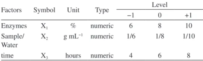
was conducted from a 23 factorial design with center point
in triplicate (Table 1) using the Design Expert software,
version 7.1.3.15 The extraction yield response was evaluated
by the influence of different concentrations of Pectinex Ultra SPL, Celluclast 1.5 L and Alcalase 2.4 L enzymes, sample/water ratio and extraction time.
The extraction experiments were performed according
to Santos and Ferrari16 with adaptations. Initially, the
samples were subjected to heat treatment at 105 °C for 45 min. In the first step, 5.0 g of sample was weighed and mixed with distilled water at a ratio of 1/6, 1/8 or
1/10 (g mL−1). The pH of the mixture was adjusted to
4.5 with 1.0 mol L−1 aqueous HCl solution and then
added Pectinex Ultra SPL and Celluclast 1.5 L enzymes in concentrations of 6, 8 and 10% (v/v) of each single enzyme. Afterwards, the mixture was maintained at 50 °C for 4, 6 and 8 h, with shaking at 100 rpm in an incubator shaker (CT 712, Cientec, Belo Horizonte, MG, Brazil). In the second step, the pH was adjusted to
7.0 through the addition of 1.0 mol L−1 aqueous NaOH
solution followed by the addition of the Alcalase 2.4 L enzyme in concentrations of 6, 8 and 10% (v/v). Then, the sample was incubated at 55 °C under the same conditions mentioned in the first step. After, the mixture was heated at 60 °C for 15 min and the extract was centrifuged for 15 min. The free oil was collected with a micropipette and weighed to determine the extraction yield.
Solvent extraction
The sample was submitted to a lipid extraction process with a mixture of chloroform-methanol-water
(2:2:1.8, v/v/v) according to Bligh and Dyer.17
Extraction by pressing
For oil extraction by pressing, 100 g of sunflower grains ground and sieved (16 mesh), previously dried in a fan oven at 50 °C for about 14 h, was placed in a stainless steel hydraulic cylinder (PHP Model 30 tons, Metal PEM Metallurgical, Maringa, PR, Brazil) under a pressure of 10 tons for 5 h.
Fatty acid composition
Fatty acid methyl esters (FAME) of the sunflower oil
were prepared by the methylation of lipids.18 The FAME
were separated by gas chromatography (Trace Ultra 3300, Thermo Fisher Scientific, Waltham, MA, USA) equipped with a flame ionization detector (FID) and a cyanopropyl
capillary column (100 m × 0.25 i.d., 0.25 µm film
thickness, CP 7420 Varian). The injector, detector and gases conditions, and the main operational parameters were
performed according to Sargi et al.19
Quantification of fatty acids was performed using tricosanoic acid methyl ester (Sigma-Aldrich, St. Louis,
MO, USA) as an internal standard (23:0).20 Theoretical
FID (flame ionization detector) correction factor values
were used to obtain concentration values.21
Phytosterols and tocopherols
Phytosterols and tocopherols were simultaneously evaluated by gas chromatography with mass spectrometer
(GC-MS).22 The extracted oils were previously derivatized23
and the analysis was performed in a gas chromatograph (Thermo Focus GC, Thermo Finnigan, Milan, Italy) equipped with a capillary column DB-5 (5% phenyl, 95% methylpolysiloxane) fused silica, 30 m, 0.25 mm i.d and 0.25 µm thick film stationary phase (J&W Scientific, Table 1. Factors and levels for the 23 factorial design with center point
Factors Symbol Unit Type Level
−1 0 +1
Enzymes X1 % numeric 6 8 10
Sample/ Water
X2 g mL−1 numeric 1/6 1/8 1/10
Folsom, CA, USA) coupled to a mass spectrometer (DSQ II, Thermo-Finnigan, Milan, Italy) equipped with an electron ionization (EI) source. The conditions for
analysis by GC-MS were described by Zanqui et al.24
Quantification was carried out in relation to the internal
standard 5-α-cholestane (Sigma-Aldrich, St. Louis, MO,
USA).25
Antioxidant capacity
DPPH radical assay
The 2,2-diphenyl-1-picrylhydrazyl (DPPH) radical scavenging activity assay was performed as described in
literature26,27 including modifications.28 The absorbance of
the solutions was measured at 517 nm using a ultraviolet-visible (UV-Vis) spectrophotometer (Genesys 10S, Thermo Fisher Scientific, Waltham, MA, USA).
Lipophilic-oxygen radical absorbance capacity (L-ORAC)
The lipophilic antioxidant capacity was determined
using the L-ORAC assay,29 with randomly methylated
β-cyclodextrin (RMCD) as a solubility enhancer.30 The
decay of the fluorescence spectra was obtained with a spectrofluorimeter (Victor X4, PerkinElmer, Waltham, MA, USA) at an excitation wavelength of 485 nm and an emission wavelength of 520 nm.
The calibration curve for both assays was performed using Trolox (Sigma-Aldrich, St. Louis, MO, USA) as a reference standard and the results were expressed in Trolox
equivalent antioxidant capacity per gram of oil.
Statistical Analysis
Data were expressed as mean values ± standard deviations of the analytical error propagation. The results were submitted to analysis of variance (ANOVA) and mean values were compared by Tukey’s test, using the
Statistica software, version 8.0,31 at 5% level of significance
(p < 0.05) for rejection of the null hypothesis. The principal
and interaction effects resulted of factorial design were calculated and the variance analysis was used to evaluate the effect of independent variables on the response using the mathematical model expressed by equation 1:
Y = β0 + β1X1 + β2X2 + β3X3 + β12X1X2 + β13X1X3 +
β23X2X3 + β123X1X2X3 + ε (1)
where Y is the expected response, X1 is the enzyme
concentration, X2 is the sample/water ratio, X3 is the
extraction time, and the other term refers to interactions effects.
Results and Discussion
Considering that the plant cell walls are mainly composed of proteins and polysaccharides, such as
cellulose, hemicelluloses and pectins,32 the enzymatic
extracts should contain enzymes with different activities. Therefore, Pectinex Ultra SPL (pectinase), Celluclast 1.5 L (cellulase) and Alcalase 2.4 L (protease) enzymes were chosen. Some parameters, such as the pH and optimal temperature of the enzymes, sample/water ratio and extraction time were also considered, since these conditions could affect the extraction yield of this process.
The data obtained, with a 23 factorial design with
center point in triplicate for sunflower oil extraction using enzymes, are shown in Table 2. Table 3 shows the ANOVA for the extraction yield of sunflower oil.
Table 3. Analysis of variance (ANOVA) for the response enzymatic aqueous extraction yield of sunflower oil
Source Degrees of freedom
Sum of squares
Mean
square F-value p-value
X1a 1 1.78 1.78 5.60 0.0643
X2b 1 79.70 79.70 251.02 < 0.0001
X3c 1 87.98 87.98 277.11 < 0.0001
X1X2 1 2.57 2.57 8.08 0.0361
Curvature 1 5.15 5.15 16.22 0.0100
Residual 5 1.59 0.32 − −
Lack of fit 3 1.51 0.50 13.29 0.0708
Pure error 2 0.08 0.04 − −
Total 10 178.75 − − −
aEnzyme concentration; bsample/water ratio; cextraction time.
Table 2. Factorial design 23 with center point and enzymatic aqueous
extraction yield of sunflower oil
Run
Coded variable Actual variable
X1a X2b X3c X1a / %
X2b /
(g mL–1) X3c / h Yd / %
1 −1 −1 −1 6 1/6 4 27.4
2 +1 −1 −1 10 1/6 4 29.7
3 −1 +1 −1 6 1/10 4 22.3
4 +1 +1 −1 10 1/10 4 23.1
5 −1 −1 +1 6 1/6 8 34.7
6 +1 −1 +1 10 1/6 8 36.6
7 −1 +1 +1 6 1/10 8 29.4
8 +1 +1 +1 10 1/10 8 28.3
9 0 0 0 8 1/8 6 27.6
10 0 0 0 8 1/8 6 27.3
11 0 0 0 8 1/8 6 27.3
aEnzyme concentration; bsample/water ratio; cextraction time; denzymatic
The highest yield obtained from the extraction of sunflower oil from the factorial design was 36.6% under these conditions: 10% of enzymes, a sample/water ratio of
1/6 (g mL−1) and eight hours of extraction.
The ANOVA for the enzymatic aqueous extraction of sunflower oil showed that the three terms in the study were significant for the model. The analysis of the effects showed that the extraction time and the sample/water ratio were the most significant factors of the extraction process with contributions of 49.22 and 44.58%, respectively. The curvature of the model was significant (measured by difference between the average of the center points and the average of the factorial points) in the design space. The lack of fit was not significant and the mathematical equation and the regression coefficient obtained for this model are reported in equation 2. The interaction effects which were not significant were removed from the model equation. The response surface is shown in Figure 1.
Y = 28.95 + 0.47X1 – 3.16X2 + 3.32X3 – 0.57X1X2 (2)
R2 = 0.991
where Y is the extraction yield, X1 is the enzyme
concentration, X2 is the sample/water ratio and X3 is the
extraction time.
Table 4 shows the yield obtained by different methods used in the extraction of sunflower oil.
No significant difference between the pressing and enzymatic techniques was observed, showing that enzymatic method under the evaluated conditions could extract the same amount of oil as conventional methodology.
Several studies claim that it is not possible to compare the antioxidant capacity quantitatively among different techniques, because of differences between the methodologies (solvents, types of radicals and different targets of oxidation). Therefore, it is recommended to perform at least two or, if possible, all of the most widely used assays, as DPPH, ferric reducing antioxidant power (FRAP), ORAC and 2,2’-azino-bis(3-ethylbenzothiazoline-6-sulphonic acid (ABTS), for vegetable oils to obtain more complete information about
the antioxidant capacity of the oils.33,34
To evaluate the antioxidant capacity of the sunflower oil extracted, either enzymatically or via conventional methods, two assays were performed: DPPH radical scavenging activity and antioxidant activity by inhibiting oxidation induced by peroxyl radical using the L-ORAC assay. Table 5 shows the results obtained from these tests.
The sunflower oil showed the highest antioxidant capacity against the DPPH radical when extracted by a pressing process. In relation to solvents and enzymatic methods, the values reported were considered statistically equal by Tukey’s test at 95% confidence. For the L-ORAC assay, the oil obtained by enzymatic aqueous extraction showed the highest antioxidant capacity against the peroxyl radical, being 1.6 times higher than the antioxidant capacity of the oil extracted using solvents and about twice as high as that obtained by pressing.
The sunflower oil that presents the greatest antioxidant capacity brings more health benefits, since the natural antioxidants have the potential effect in preventing chronic diseases, being capable of protecting biological systems against the action of reactive oxygen species and nitrogen, which are responsible for oxidative damage to lipids,
proteins and nucleic acids.35,36 Furthermore, it shows higher
protection against the action of free radicals that initiate and Figure 1 Response surface for the enzymatic aqueous extraction of
sunflower oil.
Table 5. Antioxidant capacity of sunflower oil extracted by different methods
Method Assay / (µmol Trolox g
−1 of oil)
DPPH L-ORAC
Solvent 86.71b ± 3.54 225.71b ± 2.68
Pressing 106.18a ± 3.89 181.94c ± 11.11
Enzymatic 87.98b ± 3.89 371.53a ± 12.65
DPPH: 2,2-diphenyl-1-picrylhydrazyl; L-ORAC: lipophilic-oxygen radical absorbance capacity. Mean (n = 3) ± standard deviation. Different letters in the same column indicate significant difference by Tukey’s test (p < 0.05).
Table 4. Extraction processes yield of sunflower oil obtained by different methods
Method Yield / %
Solvent 55.1a ± 0.7
Pressing 36.8b ± 0.5
Enzymatica 36.6b ± 0.6
aMaximum extraction yield obtained from the factorial design. Mean
perpetuate lipid peroxidation, which consists in the main form of degradation of vegetable oils and an important
source of losses to the food industry.37
The α-tocopherol and the three major phytosterols
(campesterol, stigmasterol and sitosterol) were identified in sunflower oil. Table 6 shows the quantification of these compounds in the oil sample.
In relation to the α-tocopherol in the sunflower oil,
the pressing and solvent methods were able to extract
the largest amount of this compound. Tuberoso et al.38
identified the α-tocopherol using high-performance liquid
chromatography (HPLC) with fluorescence detector in sunflower oil obtained by pressing grains and obtained 49.42 mg of this compound in 100 g of oil, a value very close to that determined in this work using GC-MS.
The phytosterols present in sunflower oil, evaluated separately, did not show significant differences between the extraction methods, solvents, pressing and enzymatic,
except for β-sitosterol, in which the oil extracted with
enzymes showed the highest amount. Phillips et al.39
evaluated the composition of sterols in several edible oils and analyzed the main phytosterols in refined sunflower oil. The results obtained in the present study for all extraction methodologies were higher in sunflower oil without a refining process. The authors affirm that the refining stage could cause a reduction in the amount of total and free sterols in edible oils.
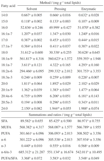
Table 7 shows the results of the fatty acid quantification of the sunflower oil extraction using solvent, pressing and enzymatic aqueous extraction methods.
The statistical analysis indicated that there was significant variation in some fatty acids among the different methodologies for the oilseed under study. The sunflower oil composition was mainly the fatty acids 16:0, 18:0, 18:1n-9 and 18:2n-6, showing differences among the methods only for the 16:0 fatty acid content.
Nimet et al.,40 who evaluated the extraction of sunflower
oil by supercritical and subcritical (CO2 and propane), and
Corsini and Jorge,41 who studied changes in the profile of
fatty acids of sunflower oil used in frying, also reported the same fatty acids as those mentioned above.
The presence of the linoleic (18:2n-6) and α-linolenic
(18:3n-3) fatty acids was identified in sunflower oil. These fatty acids are extremely important in the human diet in the synthesis of other long chain fatty acid routes, such as 22:6n-3 and 22:5n-3, which play an important role in
preventing inflammation and fostering immunity.42,43
Table 7. Fatty acid quantification of sunflower oil
Fatty acid Method / (mg g
−1 total lipids)
Solvent Pressing Enzymatic
14:0 0.667a ± 0.005 0.666a ± 0.016 0.632a ± 0.028
15:0 0.118b ± 0.002 0.133a ± 0.003 0.107c ± 0.009
16:0 52.836a ± 0.246 49.999c ± 0.427 51.070b ± 0.382
16:1n-7 1.207b ± 0.037 1.347a ± 0.030 1.240b ± 0.016
17:0 0.387b ± 0.002 0.453a ± 0.033 0.444a ± 0.015
17:1n-7 0.384a ± 0.014 0.411a ± 0.037 0.387a ± 0.022
18:0 31.612a ± 0.600 30.338a ± 0.255 30.628a ± 0.645
18:1n-9 561.817a ± 4.316 560.623a ± 1.572 559.393a ± 1.948
18:1n-7 3.611b ± 0.121 4.322a ± 0.165 4.293a ± 0.160
18:2n-6 294.466a ± 6.095 299.332a ± 2.012 301.753a ± 3.353
18:3n-3 0.246a ± 0.009 0.259a ± 0.009 0.226b ± 0.007
20:0 1.811b ± 0.064 1.892b ± 0.029 2.102a ± 0.029
20:1n-9 1.362a ± 0.039 1.383a ± 0.047 1.477a ± 0.064
20:4n-6 6.755a ± 0.099 6.206b ± 0.051 6.181b ± 0.143
20:5n-3 0.194c ± 0.008 0.296b ± 0.015 0.343a ± 0.011
24:0 2.150a ± 0.082 1.944ab ± 0.055 1.988b ± 0.074
Summations and ratios / (mg g−1 total lipids)
SFA 89.582a ± 0.655 85.425b ± 0.500 86.971b ± 0.755
MUFA 568.382a ± 4.317 568.087a ± 1.577 566.789a ± 1.955
PUFA 301.661a ± 6.096 306.093a ± 2.013 308.502a ± 3.356
n-6 301.222a ± 6.096 305.538a ± 2.013 307.934a ± 3.356
n-3 0.440b ± 0.010 0.555a ± 0.016 0.568a ± 0.009
n-6/n-3 685.312a ± 21.287 551.134b ± 16.674 542.011b ± 10.495
PUFA/SFA 3.368b ± 0.072 3.583a ± 0.032 3.548a ± 0.049
SFA: total saturated fatty acids; MUFA: total monounsaturated fatty acids; PUFA: total polyunsaturated fatty acids; n-6: total omega-6 fatty acids; n-3: total of omega-3 fatty acids. Mean (n = 3) ± standard deviation. Different letters in the same column indicate significant difference by Tukey’s test (p < 0.05).
Table 6. Quantification of phytosterols and tocopherols of sunflower oil
Method α-Tocopherol /
(mg 100 g−1)
Phytosterol / (mg 100 g−1)
Total phytosterol / (mg 100 g−1)
Campesterol Stigmasterol Sitosterol
Solvent 48.46a ± 2.38 17.17a ± 2.16 19.75a ± 1.27 130.03b ± 3.75 166.95b ± 3.65
Pressing 48.69a ± 5.43 17.29a ± 3.31 17.16a ± 2.06 121.06b ± 1.29 155.52b ± 4.01
Enzymatic 40.84b ± 0.17 19.85a ± 0.99 19.34a ± 1.30 143.81a ± 6.70 183.00a ± 6.90
The sunflower oil extracted using enzymes showed the
highest amount of the 20:5n-3 fatty acid (0.568 mg g−1),
reflected in the omega-3 summation. A diet that contains omega-3 and polyunsaturated fatty acids brings health benefits, such as reducing the risk of coronary artery disease, increase of high density lipoprotein (HDL), stabilization of heartbeat, hypertension control, prevention of cancers, lower effects of autoimmune diseases, relief of depression, help maintain under normal conditions, cell membranes, brain function, nerve impulse transmission, and participate in the transfer of atmospheric oxygen to the blood plasma, synthesis of hemoglobin and cell division.42,44,45
Conclusions
The experimental design used for the enzymatic aqueous extraction proved adequate to evaluate the influence of the independent variables in the enzymatic process. The terms that most influenced the response were the sample/water ratio and the extraction time, whereby the reduction in the sample/water ratio and the increase in time favored higher extraction yields.
The sunflower oil extracted using enzymes and non-toxic solvents showed the highest antioxidant capacity against peroxyl radicals, total phytosterols and omega-3 fatty acids contents, as well as the lowest content of saturated fatty acids, when compared with the oil obtained by conventional methods. Therefore, the application of enzymes for sunflower oil extraction could indeed represent a promising alternative that is environmentally friendly and free of toxic solvent residues, and provides a final product of high quality.
Acknowledgments
The authors thank Coordination for the Improvement of Higher Education Personnel (CAPES) and the National Council for Scientific and Technological Development (CNPq) for the financial support.
References
1. http://www.embrapa.br/soja/cultivos/girassol accessed in November 2015.
2. http://faostat3.fao.org/browse/Q/QC/E accessed in November 2015.
3. Parmentier, M.; Guillemin, S.; Barbar, R.; Linder, M.; Fanni, J.; Ol., Corps Gras, Lipides2004, 11, 377.
4. Gaia, Q.-Y.; Jiao, J.; Mua, P.-S.; Wanga, W.; Luo, M.; Li, C.-Y.; Zu, Y.-G.; Wei, F.-Y.; Fu, Y.-J.; Ind. Crops Prod. 2013, 45, 303.
5. Rai, A.; Mohanty, B.; Bhargava, R.; Food Chem. 2016, 192, 647.
6. http://www.botaneco.com/index-10.html accessed in November 2015.
7. Ribeiro, S. A. O.; Nicacio, A. E.; Zanqui, A. B.; Biondo, P. B. F.; Abreu-Filho, B. A.; Visentainer, J. V.; Gomes, S. T. M.; Matsushita, M.; LWT -- Food Sci. Technol.2016, 65, 464. 8. Li, J.; Zu, Y.-G.; Luo, M.; Gu, C-B.; Zhao, C-J.; Efferth, T.; Fu,
Y.-J.; Food Chem. 2013, 138, 2152.
9. Teixeira, C. B.; Macedo, G. A.; Macedo, J. A.; Silva, L. H. M.; Rodrigues, A. M. C.; Bioresour. Technol.2013, 129, 575. 10. Bansal, N.; Tewari, R.; Soni, R.; Soni, S. K.; Waste Manage.
2012, 32, 1341.
11. Thomas, L.; Larroche, C.; Pandey, A.; Biochem. Eng. J.2013, 81, 146.
12. Hansen, G. H.; Lübeck, M.; Frisvada, J. C.; Lübeck, P. S.; Andersen, B.; Process Biochem. (Oxford, U. K.)2015, 50, 1327. 13. Zhang, Y.-L.; Li, S.; Yin, C.-P.; Jiang, D.-H.; Yan, F.-F.; Xu, T.;
Food Chem.2012, 135, 304.
14. Zhang, S. B.; Liu, X. J.; Lu, Q. Y.; Wang, Z. W.; Zhao, X.; J. Am. Oil Chem. Soc. 2013, 90, 1261.
15. Stat-Ease, Inc; Design Expert Software version 7.1.3; Minneapolis, USA, 2008.
16. Santos, R. D.; Ferrari, R. A.; Food Sci. Technol. 2005, 25, 132. 17. Bligh, E. G.; Dyer, W. J.; Can. J. Biochem. Physiol.1959, 37,
911.
18. Hartman, L.; Lago, R. C. A.; Lab. Pract. 1973, 22, 475. 19. Sargi, S. C.; Silva, B. C.; Santos, H. M. C.; Montanher, P. F.;
Boeing, J. S.; Santos-Júnior, O. O.; Souza, N. E.; Visentainer, J. V.; Food Sci. Technol. 2013, 33, 541.
20. Joseph, J. D.; Ackman, R. G.; J. AOAC Int.1992, 75, 488. 21. Visentainer, J. V.; Quim. Nova2012, 35, 274.
22. Du, M.; Ahn, D. U.; J. Food Sci.2002, 67, 1696.
23. Beveridge, T. H. J.; Li, T. S. C.; Drover, J. C. G.; J. Agric. Food Chem.2002, 50, 744.
24. Zanqui, A. B.; Moraes, D. R.; Silva, C. M.; Santos, J. M.; Gomes, S. T. M.; Visentainer, J. V.; Eberlin, M. N.; Cardozo-Filho, L.; Matsushita, M.; Food Chem. 2015, 188, 452.
25. Li, T. S. C.; Beveridge, T. H. J.; Drover, J. C. G.; Food Chem.
2007, 101, 1633.
26. Brand-Williams, W.; Cuvelier, M. E.; Berset, C.; Food Sci. Technol.1995, 28, 25.
27. Masuda, T.; Yonemori, S.; Oyama, Y.; Takeda, Y.; Tanaka, T.; Andoh, T.; J. Agric. Food Chem.1999, 47, 1749.
28. Ma, X.; Wu, H.; Liu, L.; Yao, Q.; Wang, S.; Zhan, R.; Xing, S.; Zhou, Y.; Sci. Horticult. (Amsterdam, Neth)2011, 129, 102. 29. Prior, R. L.; Hoang, H.; Gu, L.; Wu, X.; Bacchiocca, M.;
Howard, L.; Hampsch-Woodill, M.; Huang, D.; Ou, B.; Jacob, R.; J. Agric. Food Chem.2003, 51, 3273.
31. StatSoft, Inc.; Statistica: Data Analysis Software System, version 8.0; Tulsa, USA, 2007.
32. Taiz, L.; Zeiger, E.; Plant Physiology, 3rd ed.; Sinauer
Associates: Massachusetts, 2002.
33. Alves, C. Q.; David, J. M.; David, J. P.; Bahia, M. V.; Aguiar, R. M.; Quim. Nova2010, 33, 2202.
34. Castelo-Branco, V. N.; Torres, A. G.; Rev. Nutr.2011, 24, 173. 35. Szydlowska-Czerniak, A.; Karlovits, G.; Dianoczki, C.;
Recseg, K.; Szlyk, E.; J. Am. Oil Chem. Soc.2008, 85, 141. 36. Ramadan, M. F.; Moersel, J. T.; J. Food Compos. Anal. 2006,
19, 838.
37. Chaiyasit, W.; Elias, R. J.; McClements, D. J.; Decker, E. A.; Crit. Rev. Food Sci. Nutr. 2007,47, 299.
38. Tuberoso, C. I. G.; Kowalczyk, A.; Sarritzu, E.; Cabras, P.; Food Chem. 2007, 103, 1494.
39. Phillips, K. M.; Ruggio, D. M.; Toivo, J. I.; Swank, M. A.; Simpkins, A. H.; J. Food Compos. Anal.2002, 15, 123.
40. Nimet, G.; Silva, E. A.; Palú, F.; Dariva, C.; Freitas, L. S.; Medina-Neto, A.; Cardozo-Filho, L.; Chem. Eng. J. (Amsterdam, Neth.)2011, 168, 262.
41. Corsini, M. S.; Jorge, N.; Quim. Nova2008, 31, 956. 42. Pratt, S.; Matthews, K.; Super Alimentos: os Incríveis Efeitos
de uma Comida que Pode Mudar Sua Vida, 1ª ed.; Prestígio Editorial: São Paulo, 2005.
43. Simopoulos, A. P.; Food Rev. Int.2004, 20, 77.
44. Yehuda, S.; Rabinovitz, S.; Carasso, R. L.; Mostofsky, D. I.; Neurobiol. Aging2002, 23, 843.
45. Youdim, K. A.; Martin, A.; Joseph, J. A.; Int. J. Dev. Neurosci.
2000, 18, 383.
Submitted: September 23, 2015