Article
J. Braz. Chem. Soc., Vol. 25, No. 7, 1246-1252, 2014. Printed in Brazil - ©2014 Sociedade Brasileira de Química 0103 - 5053 $6.00+0.00
A
*e-mail: [email protected]
A New Spectrophotometric Method for Determining the Enzymatic Activity of
endo-
β
-mannanase in Seeds
Gevany P. Pinho,*,a Juliana R. M. Matoso,a Flaviano O. Silvério,a Welha C. Mota,a
Paulo Sérgio N. Lopesa and Leonardo M. Ribeirob
aInstitute of Agricultural Sciences, Federal University of Minas Gerais,
39404-547 Montes Claros- MG, Brazil
bDepartment of General Biology, State University of Montes Claros,
Campus Professor Darcy Ribeiro, 39401-089 Montes Claros-MG, Brazil
O objetivo deste trabalho foi desenvolver um novo método espectrofotométrico, de elevada sensibilidade, eficiente e de fácil execução para determinar a atividade enzimática da endo-β -mananase em sementes. O método espectrofotométrico foi otimizado e aplicado em amostras de sementes de Butia capitata para determinar a localização da atividade enzimática em diferentes partes das sementes. O método consiste em extrair a enzima da semente e utilizá-la para hidrolisar o galactomanano Locust Bean Gum. Os açúcares redutores formados reagem com a hidrazida ácida p-hidroxibenzóico e são quantificados por espectrofotometria na região do visível. O método otimizado e validado apresentou baixos limites de quantificação para o açúcar redutor formado (0,6 µg de manose) e atividade da endo-β-mananase (1 mU). Apenas 5 mg de sementes em germinação são necessárias para executar o método.
The objective of this study was to develop a new, highly sensitive, efficient and easy touse spectrophotometric method to determine the enzymatic activity of endo-β-mannanase in seeds. The method was optimized and applied to determine the site of enzyme activity in different parts of seeds of Butia capitata. It consists in using the enzyme extracted from seeds to hydrolyze Locust bean gum galactomannan. The reducing sugars react with p-hydroxybenzoic acid hydrazide and are quantified by spectrophotometry in the visible region. The optimized and validated method showed low limits of quantification for the reducing sugars (0.6 µg mannose) and endo-β-mannanase activity (1 mU). Only 5 mg of germinating seeds are needed to perform the method.
Keywords: galactomannan, germination, reducing sugar
Introduction
The difficulty of seed germination may be related, among other factors, to the chemical composition of the
cell wall. In some species, such as Coffea arabica, Phoenix
dactilifera, Annona crassiflora and Arabidopsis thaliana, the endosperm has thick cell walls, consisting of insoluble carbohydrates in the form of galactomannans, which act as a physical barrier restricting embryo growth and hampering
radicle protrusion.1-3
During germination, galactomannans are hydrolyzed
by the enzymes endo-β-mannanase (EC 3.2.1.78),
β-mannosidase (EC 3.2.1.25) and β-galactosidase
(EC 3.2.1.22).4 Endo-β-mannanase is the main degrading
enzyme, acting specifically in the hydrolysis of β (1→4)
linkages in the galactomannan present in seed cell walls.5
The synthesis of this enzyme occurs especially during
germination,6 growth or senescence of the seed.7
The enzymatic activity of endo-β-mannanase in
seeds has been determined by the modified
Nelson-Somogyimethod8-10 and the dinitrosalicylic acid (DNS)
method of Miller.11,12 Both methods are based on reducing
sugar production and colorimetric determination. However, endogenous components present in the samples (glucose, fructose, mannose, etc.) have been shown to interfere with the results.
Currently, the main method used to determine the mananase activity is by gel diffusion using the
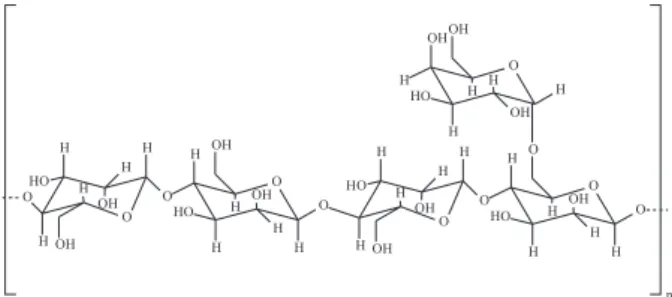
galactomannan locust bean gum (LBG) as substrate.1,13
β(1→4)-D-mannopyranosyl units randomly linked with
α(1→6)-D-galactopyranosyl units occurring as side
branches (Figure 1).
LBG has a mannose:galactose ratio of 4:1 and the
endo-β-mannanase hydrolyses only the glycosidic linkages
involving mannose to form the reducing oligosaccharides manotetrose, manotriose and manobiose as hydrolysis
products.14 Although the method has shown satisfactory
results, it is a visual, laborious and very time consuming method, taking more than 16 h to be accomplished.
Another method that has also been used to determine
the enzymatic activity of endo-β-mannanase is based on
spectrophotometric analysis.6,15 It consists in extracting the
enzyme from seed and putting it in contact with the synthetic polysaccharide AZCL-galactomannan that has carboxylate groups, zirconium and ammonium linked to the structure of the galactomannan, forming a colored compound. The hydrolysis of the synthetic substrate shows decreased absorbance at 590 nm. However, data from the calibration curve to determine the enzymatic activity are obtained from the hydrolysis of LBG by mananase. The reducing sugars are quantified by the spectrophotometric method proposed
by Lever et al..16 Furthermore, AZCL-galactomannan is
expensive.
Therefore, the aim of this work was to develop a new, highly sensitive, efficient and easy touse spectrophotometric
method to determine the enzymatic activity of endo-β
-mannanase in seeds.
Experimental
Standard solutions
A standard solution of galactomannan locust bean gum (LBG) (Sigma, USA) 0.25% (w/v) was prepared
in 0.1 mol L-1 sodium acetate buffer, pH 4.7. A standard
solution of p-hydroxybenzoic acid hydrazide (PHBAH) (Sigma, USA) 5% (w/v) was prepared in acid medium
(HCl 0.5 mol L-1) and diluted at the moment of analysis
to 0.5% (w/v) in alkaline medium (NaOH 0.5 mol L-1).
Standard solutions of endo-β-mannanase (Megazyme,
Ireland) were prepared by diluting the enzyme standard to 500U (U = unit enzyme activity) in suspension of
ammonium sulfate 3.2 mol L-1. The standard solution of the
monosaccharide D-mannose (Sigma, USA) was prepared at different concentrations in distilled water.
Seed samples
F r u i t s o f B . c a p i t a t a w e r e c o l l e c t e d f r o m the experimental orchard located in the Institute of Agricultural Sciences, Federal University of Minas Gerais (16º40’53.8’’/43º50’22.7’’). The fruits were depulped using a blender at low speed and the pyrenes (seed + endocarp) were dried in the shade. Seeds were extracted from endocarps using a bench vice.
Equipment
The samples were homogenized by vortexing (AP-56, Phoenix, Brazil). A centrifuge (Vision Scientific, Korea) was used in the extraction procedure of the enzyme. A reciprocating shaker water bath (Dubnoff 304 ID, Tecnal, Brazil) was used in the enzymatic hydrolysis of galactomannan. A spectrophotometer (Agilent Cary 60 UV/ Visible, Australia) was used to quantify the reducing sugar.
Quantification of reducing sugar
To determine the amount of reducing sugar, mannose standard solutions were prepared at concentrations of 1,
15, 20, 25, 30, 35, 40, 50 and 60 mg L-1 in distilled water.
In test tubes, 600 µL of each solution was mixed with 3 mL of PHBAH 0.5% (w/v) in basic medium and heated to 95 °C in water bath for 5 min. The sample was cooled and immediately read in the UV-Visible spectrophotometer (398 nm). Assays were performed in triplicate. A calibration curve was obtained by linear regression of the absorbance readings versus concentration of reducing sugar.
Enzyme activity of the endo-β-mannanase standard
Six test tubes were filled with 900 µL of LBG solution 0.25% (w/v). Each tube was added with 225 µL of mananase standard solution with the following enzymatic activities: 1, 3, 5, 7, 9 and 11 mU (nmol of reducing sugar
min-1). The tubes were incubated at 40 oC in a reciprocating
shaker water bath for 3 h. In test tubes, 600 µL of the hydrolyzate was mixed with 3 mL of PHBAH 0.5% (m/v) in basic medium and heated to 95 °C in a water bath for 5 min. The sample was cooled and immediately read in
the UV-Visible spectrophotometer (398 nm). Assays were performed in triplicate. A calibration curve was obtained
by linear regression of the absorbance readings versus
enzymatic activity of mannanase.
Optimization of enzymatic activity method
The method for determining the activity of endo-β
-mannanase was based on previous studies.6,15 Preliminary
experiments were conducted to optimize the main factors
affecting the extraction of endo-β-mannanase from seeds,
the enzymatic hydrolysis of the galactomannan standard and quantification of reducing sugars. The results of optimization parameters were assessed by t-test, calculated with SPSS 19 Statistics Software.
Samples from endosperms of B. capitata were
macerated in a porcelain mortar. Different masses of the sample (Table 1) were measured on an analytical scale (0.0010 g accuracy) in Eppendorf tubes in triplicate and
mixed with an aliquot of sodium acetate buffer 1 mol L-1,
pH 4.7. The tubes were vortexed for 1 min and centrifuged at 16000 g at 4 °C for 45 min for enzyme extraction.
In test tubes, 225 µL of the endo-β-mannanase extract
were mixed with 900 µL of LBG galactomannan 0.25% (w/v). The samples were incubated in a shaker water bath with controlled temperature (Table 1) for 3 h for galactomannan hydrolysis. The same procedure was carried out by adding the extract enzyme to sodium acetate buffer (pH 4.7) without LBG to be used as a blank sample.
An aliquot of the hydrolyzate (Table 1) previously obtained, either with LBG or without LBG, was mixed with 3 mL of PHBAH 0.5% (w/v) in a basic medium in a test tube and heated at 95 °C in water bath for determined periods of time (Table 1). The sample was cooled and then read in a UV-Visible spectrophotometer (398 nm).
Validation of the method
After optimization of the method, the kinetic assay was carried out under steady state conditions and validated. The main figures of merit of validation were selectivity, limit of quantification (LOQ), linearity and accuracy as suggested
by the National Institute of Metrology, Standardization and
Quality (INMETRO).17 Data were examined by statistical
analysis and coefficient of variation (CV) were calculated using software Excel.
Site of enzyme activity
Seeds of Butia capitata (200 units) were surface
sterilized in ethanol 70% (v/v) for 1 min and washed 3 times with distilled water. Seeds were then immersed in a sodium hypochlorite 6% (w/v) for 15 min and washed 3 times in distilled water. After disinfection, the opercular integument of 100 seeds was removed using a cutter knife. Seeds with and without operculum were placed in gerboxes containing sand moistened to 60% of field capacity. The seeds were kept in a growth chamber at 25 °C for 11 days and were considered germinated when the cotyledonary
petiole elongated 2 mm.18
The endosperm, embryo, operculum and region adjacent to the embryo of each seed were separated (Figure 2). The optimized and validated method was used to determine enzyme activity in each of the four parts. The results of enzymatic activity assays were assessed by t-test, calculated with SPSS 19 Statistics Software.
Results and Discussion
Spectrophotometric analysis
The proposed method aims to extract endo-β
-mannanase from seeds and use the enzyme to hydrolyze the standard solution of LBG galactomannan. The hydrolyzate consists of reducing sugars, which react with PHBAH to form compounds of yellow color which can be quantified by spectrophotometry in the visible region. The maximum absorbance wavelength at 398 nm was determined from the reaction between the D-mannose standard solution and PHBAH (Figure 3).
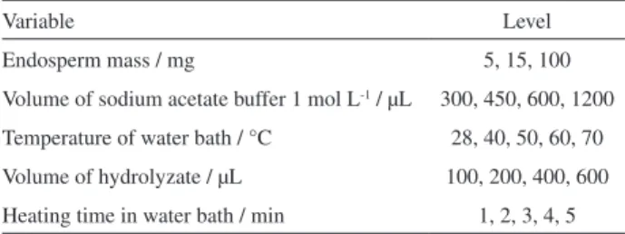
Table 1.Optimization parameters for the enzyme activity method
Variable Level
Endosperm mass / mg 5, 15, 100
Volume of sodium acetate buffer 1 mol L-1 / µL 300, 450, 600, 1200
Temperature of water bath / °C 28, 40, 50, 60, 70
Volume of hydrolyzate / µL 100, 200, 400, 600
Heating time in water bath / min 1, 2, 3, 4, 5
Figure 2. Diagram of the longitudinal section of Butia capitata seed
The reaction between the reducing sugar mannose and the reagent PHBAH was linear in the concentration
range of 1.0-60.0 mg L-1 and calibration data obtained
via spectrophotometric analysis were y = 0.014x + 0.093 (y = absorbance and x = concentration of reducing sugar) with coefficient of determination greater than 0.99. To
determine the enzymatic activity of endo-β-mannanase, the
standard solution (500 U) was diluted to the range 1-11 mU for response linearity in the spectrophotometric analysis (398 nm). The calibration data were y = 0.15x – 0.099 (y = absorbance and x = enzymatic activity) with a coefficient of determination greater than 0.99.
Optimization of the enzymatic activity method
The methods described in the literature use sample masses ranging from 50 to 300 mg. These values are considered high to study the enzymatic activity of
endo-β-mannanase in seeds,1,6 making it difficult to investigate
the activity of this enzyme in structures with small masses, such as operculum (± 0.45 mg) and embryo (± 1.17 mg). In addition, the reported methods are laborious and endogenous seed components significantly interfere with the results. Therefore, to remove matrix interferents, seed samples were also used as blank in the development of the method.
The optimization step of the method was carried out using endosperms of non-germinating seeds with low
enzymatic activity of endo-β-mannanase. In Figure 4
there was significant difference between the results of enzymatic activity for the samples 100 mg / 1200 µL and 15 mg / 1200 µL by the p value from the t-test (p = 0.025). A greater amount of enzyme is obtained in 100 mg, due to the concentration of 0.08 mg sample / mL extractor versus 0.01 mg sample / mL (15 mg). Another important aspect of the results was the significant difference in the
enzyme activities of the samples 15 mg / 1200 L and 15 mg / 300 µL (p = 0.020), which can be explained also by the difference in the concentration of mannanase in the extracts. We chose to work with only 15 mg of endosperm, since the enzyme activity was satisfactory with low volumes extractor.
However, with 300 µL of extract, the removal of the aliquot after centrifugation is difficult because seed particles are also pipetted, interfering with the spectrophotometer readings in later steps. Thus, 350 µL of sodium acetate buffer was used with satisfactory results. By contrast, it was not possible to determine the enzymatic activity using only 5 mg of endosperm samples of non-germinating seeds.
Previous studies showed that enzyme activity is higher
at pH near 4, we used the buffer solution with pH 4.7.6
Besides the pH, the mannanase enzyme activity is also dependent on the hydrolysis temperature and heating time. Fixing the hydrolysis time in 3 h, the best result was obtained at 40 °C (Figure 5). Similar results were reported
by Blibech et al..14
Figure 3. Absorption spectrum in the visible region: A) PHBAH standard
solution using distilled water as blank, B) Product formed from the reaction of D-mannose with PHBAH using PHBAH solution as blank.
Figure 4. Enzymatic activity using different masses of endosperm and
extractor. *p value = 0.025, **p value = 0.020, p value were calculated using t-test.
Figure 5. Effect of temperature on the enzymatic activity of
Reducing sugars do not react with PHBAH using basic medium in ambient conditions. Only at high temperatures (95 °C) it is possible to detect the reaction products (yellow color). Figure 6 shows that the reaction between PHBAH and reducing sugars occurs satisfactorily with a 5 min heating time. In addition, heating at 95 °C also stops the galactomannan hydrolysis by denaturation of
the mananase.14
Samples of galactomannan standard were also subjected to heating with PHBAH to evaluate the hydrolysis of the polysaccharide in basic medium. However, no changes in galactomannan occurred with 5, 10 and 15 min of
heating at 95 oC. These results were expected because the
acetal-ether bond formed at the glycosidic linkage is not
cleaved in basic medium.19 In this sense the basic hydrolysis
of galactomannan does not affect the results.
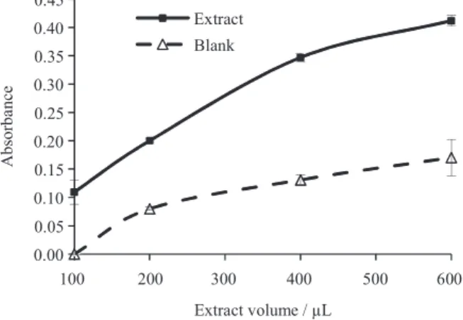
In addition to heating time, the amount of hydrolyzate that is subjected to reaction with PHBAH also enhances detection of the colored compound in the spectrophotometric analysis. The addition of increasing amounts of hydrolyzate significantly increases the absorbance of the extracts (Figure 7). The results in Figure 7 also show that the endosperm has endogenous interferents, because compounds naturally present in seed extracts react with PHBAH increasing the absorbance of the blank with the increase in extract volume. Previous studies have shown that monosaccharides such as glucose, fructose, galactose, etc., or disaccharides such as maltose, potentially present in the
seed, can react with PHBAH interfering with the results.16
Thus, the extract without the galactomannan standard must be used as blank in the spectrophotometric analysis.
Method Validation
The selectivity of the method was obtained when endogenous interferents present in the matrix were removed using a blank in the spectrophotometric analysis. The blank
has the same compounds as the sample except the LBG substrate. However, this procedure does not eliminate
the actions of β-mannosidase and β-galactosidase. These
two enzymes hydrolyze the byproducts of galactomannan
after the activity of endo-β-mannanase,20 increasing the
amount of reducing sugars. Although the proposed method
overestimates the enzymatic activity of endo-β-mannanase,
it can be satisfactorily used to study the degradation of galactomannan in different conditions of seed germination.
The limit of quantification was considered when the absorbance in the spectrophotometric analyzes were higher than 0.1, corresponding to the lower limit of linearity of the instrument. At this absorbance, the extract showed 0.6 µg
of mannose. A similar result was found by Lever16 in the
quantification of glucose by reaction with PHBAH and spectrophotometric analysis which was 0.1 µg. The limit of quantification of enzyme activity was 1 mU (nmol of
reducing sugar per minute) because of the limit of linearity
of the spectrophotometer, which was similar to that of mannose. Lower enzyme activities were considered merely
as detection of endo-β-mannanase activity in the sample.
The qualitative analysis of the endo-β-mannanase activity
using the gel diffusion method required an enzyme standard
solution of 0.2 U (200 mU) for positive control.21 However,
Still et al.22 found a detection limit for endo-β-mannanase
activity of 0.500 mU by the method of gel diffusion (0.5 nmol
of reducing sugar per minute) similar to that of this work.
The precision of the method was evaluated in terms of repeatability on the same day. Seven identical repetitions of the proposed method were performed and the coefficients of variation (CV) of enzymatic activities were less than 10% (Table 2). The same analyst evaluated the intermediate precision of the method on three different days (1, 7 and
Figure 6. Heating time required for formation of the colored product from
the reaction between reducing sugars and PHBAH.
Figure 7. Absorption as a function of the volume of extract subjected
30 days). Furthermore, the method was repeated by a different analyst after 45 days. The results are shown in Table 2.
Site of enzyme activity
The optimized and validated method was applied to
different parts of the seed of B. capitata. These seeds have
a high degree of dormancy what cause slow germination
rate.23 These characteristics may be related to the difficult
degradation of galactomannan in cell walls by hydrolases
such as endo-β-mannanase.
Although the optimized method required 15 mg of
samples to quantify the endo-β-mannanase activity, during
germination only 5 mg of samples were sufficient for application of the method. After 11 days of germination,
the enzymatic activity of endo-β-mannanase was detected
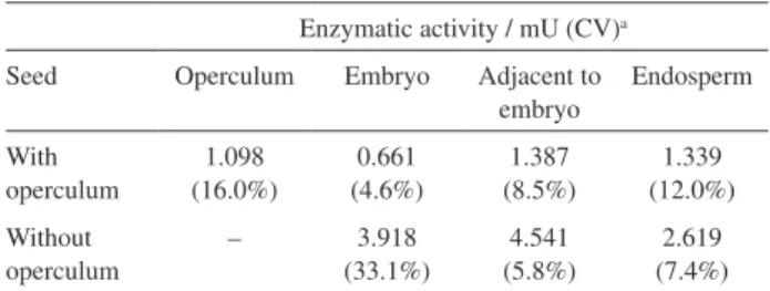
in seeds with and without operculum in all parts studied (Table 3). In seeds with operculum, the highest enzyme activity was detected in the region adjacent to embryo and the lowest activity in the embryo (Table 3). There were significant differences between these results (p < 0.01). It was also found that the enzymatic activity in the operculum was higher than in the embryo. The operculum in seeds serves as a mechanical barrier that hinders germination, thus its higher enzyme activity could be related to the weakening of this structure to allow embryo elongation.
In seeds without operculum, there were significant differences between the results of enzyme activity for region adjacent to embryo and in the endosperm (p < 0.01). The highest enzyme activity was detected near the embryo and the lowest activity in the endosperm (Table 3). Seeds that had the operculum removed showed high levels of enzyme activity in the parts studied, which allowed seeds to overcome dormancy and led to germination and
consequently reserve mobilization.18
Conclusion
The method developed was proven to be simple, accurate and rapid, with analysis time of less than 8 h. It is
also affordable because of the amount and cost of reagents used. The main advantage of the method is the use of only 5 mg samples to quantify the enzymatic activity of
endo-β-mannanase in germinated seeds. The application of the
method to Butia capitata showed that seeds germinated
without the operculum have higher enzyme activity than seeds with the operculum. Differences in enzyme activities were also detected between the seed parts operculum, endosperm, region adjacent to the embryo and embryo.
Acknowledgements
To the National Council of Technological and Scientific Development (CNPq) and the Foundation for Research Support of the State of Minas Gerais (FAPEMIG) for the undergraduate research scholarship and the financial support, and the Federal University of Minas Gerais (UFMG) for the financial support and the facilities.
References
1. Da Silva, E. A.; Toorop, P. E.; Van Aelst, A. C.; Hilhorst, H. W.;
Planta 2004, 220, 251.
2. Gong, X.; Bassel, G. W.; Wang, A.; Greenwood, J. S.; Bewley, J. D.; Ann. Bot. 2005, 96, 1165.
3. Da Silva, E. A. A.; De Melo, D. L. B.; David, A. C.; De Bode, N.; Abreu, G. B.; Faria, J. M. R.; Hilhorst, H. W. M.; Ann. Bot. 2007, 99, 823.
4. Tonini, P. P.; Lisboa, C. G. S.; Freschi, L.; Mercier, H.; Mazzoni-Viveiros, S. C.; Buckeridge, M. S.; Trees2006, 20, 669. 5. Dotsenko, G. S.; Semenova, M. V.; Sinitsyna, O. A.; Hinz,
S. W. A.; Wery, J.; Zorov, I. N.; Kondratieva, E. G.; Sinitsyn, A. P.; Biochemistry (Moscow) 2012, 77, 1303.
6. Iglesias-Fernández, R.; Rodríguez-Gacio, M. C.; Barrero-Sicilia, C.; Carbonero, P.; Matilla, A.; Planta 2010, 233, 25. 7. Schroder, R.; Ross, G. A.; Redgwell, J. R.; Ann. Bot. 2009, 104,
197.
8. Nelson, N. J.; Biol. Chem. 1944, 153, 375. 9. Somogyi, M.; Biol. Chem. 1952, 195, 19.
Table 2. Precision of the proposed method in terms of repeatability and
intermediate precision
Method accuracy Enzyme activity / mU (CV)
1 daya 1.83 (1.94%)
7 daysb 1.66 (8.89%)
30 daysa 1.24 (1.70%)
Different analysta 1.58 (9.73%) athree replicates; bseven replicates; CV: coefficient of variation.
Table 3. Enzymatic activity of endo-β-mannanase in different parts of
Butia capitata seeds after 11 days of germination
Enzymatic activity / mU (CV)a
Seed Operculum Embryo Adjacent to embryo
Endosperm
With operculum
1.098 (16.0%)
0.661 (4.6%)
1.387 (8.5%)
1.339 (12.0%)
Without operculum
– 3.918
(33.1%)
4.541 (5.8%)
2.619 (7.4%)
10. Klyosov, A. A.; Dotsenko, G. S.; Hinz, S. W.; Sinitsyn, A. P.;
Carbohydr. Res. 2012, 352, 65. 11. Miller, G. L.; Anal. Chem.1959, 31, 426.
12. Katrolia, P.; Zhoua, P.; Zhanga, P.; Yanb, Q.; Li, Y.; Jianga, Z.; Xua, H.; Carbohydr. Polym. 2012, 87, 480.
13. Downie, B.; Hilhorst, H. W. M.; Bewley, J. D. A.; Phytochemistry 1994, 36, 829.
14. Blibech, M.; Chaari, F.; Bhiri, F.; Dammaka, L.; Ghorbel, R. E.; Chaabouni, S. E.; J. Mol. Catal. 2011, 73, 111.
15. Iglesias-Fernández, R.; Matilla, A.; J. Exp. Bot. 2009, 60, 1645. 16. Lever, M. A.; Anal. Biochem.1972, 47, 273.
17. National Institute of Metrology, Quality and Technology (INMETRO). Guidance on validation of analytical methods, DOQ-CGCRE-008, 2010.
18. Fior, C. S.; Rodrigues, L. R.; Leonhardt, C.; Schwarz, S. F.;
Cienc. Rural. 2011, 41, 115.
19. Barbosa, L. C. A. Introdução à Química Orgânica. 2th ed.;
Pearson Prentice Hall: São Paulo, Brazil, 2011.
20. Moreira, L. R. S.; Filho, E. X. F.; Appl. Microbiol. Biotechnol. 2008, 79, 165.
21. Lin, J.; Pantalone, V. R.; Li, G.; Chen, F.; J. Agric. Food Chem. 2011, 59, 4622.
22. Still, D. W.; Dahal, P.; Bradford, K. J.; Plant Physiol. 1997, 11, 13.
23. Lopes, P. S. N.; Aquino, C. F.; Magalhães, H. M.; Silva, B. J.; Brandão Junior, D. S.; Pesq. Agropec. Trop. 2011, 41, 120.
Submitted: November 15, 2013