Experimental study of the
tissue reaction caused by the
presence of cellulose produced
Summary
Wander Lopes Amorim1, Henrique Olival Costa2,
Flávia Coelho de Souza3, Marilia Germanos de
Castro4, Leonardo da Silva5
1 Master’s degree student, Santa casa de Sao Paulo. Otorhinolaryngologist.
2 Otorhinolaryngologist, head & neck surgeon. Doctor in Otorhinolaryngology, adjunct professor of the Otorhinolaryngology Department, Santa Casa de Sao Paulo,
coordinator of the graduate program in Otorhinolaryngology, Santa Casa de Sao Paulo.
3 Master’s degree in veterinary science, doctoral degree student, Santa Casa de Sao Paulo. 4 Professor (instructor), Santa Casa de Sao Paulo, pathologist.
5 Doctor in Otorhinolaryngology, professor (instructor), Santa Casa de Sao Paulo.
Faculdade de Ciencias Médicas da Santa Casa de Sao Paulo.
Address for correspondence: Rua Prof. Arthur Ramos 183 3o. andar 01454-011 São Paulo Brasil.
This paper was submitted to the RBORL-SGP (Publishing Manager System) on 5 November 2007. Code 4944. The article was accepted on 1 February 2008.
S
everal materials have been proposed for nasal reconstruction. There is no consensus on which is the best. The cellulose blanket produced by bacteria may be a possible cartilaginous addition element to the nose. Aim: to study tissue reaction to cellulose in the dorsal nose of rabbits.Materials and Methods: 22 New Zealand rabbits were used. In 20 a cellulose blanket was implanted in the nasal dorsum and 2 served as controls. They were followed up through a period of three and six months, after which their nostrils and nasal dorsums were removed and histological studies were carried out on them, considering defined parameters of inflammation such as vascular congestion, intensity of the inflammatory process and presence of purulent exudate.
Results: The inflammatory process remained stable, showing its relationship with the surgical procedure and not with the presence of the cellulose blanket. There were no statistical differences in the other parameters. Conclusion: The cellulose blanket produced by Acetobacter xylinum presented good biocompatibility, remained stable during the entire study period, and could be considered a good material for elevating the nasal dorsum.
Keywords: biocompatibility, cellulose, rabbits, nose. ORIGINAL ARTICLE
INTRODUCTION
Humankind has always been interested in searching for an esthetic ideal and improving bodily contours. The nose is in a strategic central position on the face, and is thus more susceptible to trauma, which may result in facial deformity and lead to social stigma and prejudice of many sorts.
Although its main function is in breathing, the nose is also important esthetically; its central anatomical position reveals the genetic burden in facial contours.
Reconstruction of the nose has been a concern in medicine since antiquity. In the Roman Empire, prisoners of war had their noses amputated as punishment. In Ancient India, adultery and theft were punished by nasal amputation. Thus, the first reports of nasal reconstruction
of deformed or mutilated noses date from these times.1
Modern rhinoplasty started in 1860, when the scientific community began to think about the structure of the nose. Success in the use of the maxillary and frontal bones to recompose the nasal structure led researchers to
try using the ulna, tibia and ribs.2
Based on the first efforts in rhinoplasty, nasal re-construction techniques have developed continuously, and currently provide excellent results. There are many options for nasal reconstruction, due to the development of stable, non-reactive and easily available alloplastic
ma-terials, among other factors.3
Preferred materials for supporting the dorsum of the nose should provide adequate resistance, volume and shape persistence, easy insertion and coating, and suffi-cient availability and ability to mimic the natural contour
of the nasal dorsum.1,2,4-7
Many materials have been proposed for nasal reconstruction. There is, however, no consensus about which is superior. Further studies are needed to seek new materials or substances that have never been used in this application, or that have been used in other parts of the human body, and that might become feasible solu-tions for reasons of practicality, economy and decreased comorbidity.
In 1984, Luis Fernando Xavier Farah, a microbio-logist, developed an economically feasible industrial pro-cess for producing cellulose based on the fermentation of bacteria of the Acetobacter genus. After processing, the resulting membrane has selective permeability; it is permeable to water vapor but not to microorganisms. It is homogeneous, its mean thickness is 0.05 mm, and it contains no adhesives or additives. It consists basically of cellulose, which is inert, resistant and insoluble in all organic solvents. Additional specific physical features in-clude: defined permeability to liquids and gases, tensile and traction resistance, and characteristic and stable mo-lecular weight and structure. Cellulose membranes have
been tested in a variety of applications, from artificial skin to bullet-proof vests, computer screens and paper for
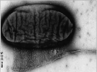
preserving historical documents.8 (Fig. 1)
Figure 1. Microscopy of Acetobacter producing cellulose
In searching for alternatives to reconstruct the dorsum of the nose, we decided to study healing when applying a bacterial (Acetobacter xylinum) cellulose spon-ge produced by Bionext® for raising the nasal dorsum. We wished to increase the volume of the dorsum without changing the usual texture and esthetic consistency and to avoid the disadvantages or harvesting and tolerability of a graft material.
The Bionext® cellulose sponge (ANVISA N 80255120001) is produced by Bionext Produtos Biologi-cos. It consists of a flexible, semitransparent, yellowish membrane composed of polysaccharides synthesized by bacteria of the genus Acetobacter. It is biodegradable, non-toxic, non-pyrogenic, and can be sterilized; it has been
used successfully as a temporary skin graft.9-11
OBJECTIVE
The purpose of this study was to assess the tissue response in rabbits to the presence of cellulose produced from bacteria (Acetobacter xylinun) as a material for ele-vating the dorsum of the nose.
MATERIAL AND METHOD
Surgical procedures were done according to the Ethics Committee guidelines for the Experimental Surgery Unit (Unidade de Técnica Cirúrgica Experimental) of the Santa Casa de Sao Paulo, the norms in the Federal Law number 6 638 (8 May 1979) and the ethical principles for experiments of the Brazilian Code for Animal Experiments (Codigo Brasileiro de Experimentacao em Animais or COBEA).
Sample Size and Selection
Twenty-two male New Zealand rabbits aged 6 mon-ths were studied during three to six monmon-ths. Study groups were established randomly according to the follow-up time. The choice of this animal was based on the facts that they are easy to handle, monitor and assess.
Animal groups were chosen as follows:
a - 2 rabbits for control purposes of the surgical procedure;
b - 20 rabbits for the study group; 10 were assessed 3 months after surgery, and another 10 were assessed 6 months after surgery;
c - the choice of animals for the follow-up groups was made by a random draw on the day of euthanasia.
Preparation of the Material
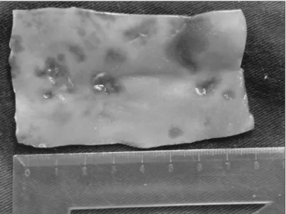
Before surgery, the cellulose sponge was sterilized in a glutaraldehyde solution for 10 minutes and washed in saline. A strip measuring 4 cm long and 1 cm wide was cut, prepared, folded and molded to become an elevation ele-ment to be inserted in the dorsum of the nose (Fig. 2).
40 mg/kg and xylazine hydrochloride 10 mg/kg intrape-ritoneally, and were ventilated mechanically throughout the procedure.
The fur was removed from the skin over the dorsum of the nose to standardize the conditions for photography, to note volume expansion and to evaluate the clinical status of the cellulose graft.
A 1 cm incision was made horizontally on the frontal area and dissection was done subperiotally from the interorbitary incision towards the tip of the nose to attain a 7 cm x 1 cm insertion tunnel. The same procedure was done in the controls, without inserting the cellulose sponge, to note the response to the procedure.
Placing the Cellulose in the Dorsum of the Nose
A strip measuring 4 cm length by 1 cm width, which had been previously designed and cut, was placed in the tunnel made over the dorsum of the nose.
After insertion of the cellulose sponge in the tunnel, 3.0 mononylon sutures were done to close the frontal inci-sion; at this point the procedure was ended. The animals were given 1 ml intramuscular benzilpenicillin benzatin and 0.2 ml dipyrone.
Figure 2. Cellulose membrane before folding for insertion
Surgical Procedure
General anesthesia was done following a 4-hour fasting period. All animals were anesthetizes with ketamine
Figure 3. Sagittal section of specimen showing cellulose included in the dorsum of the nose (arrow)
Euthanasia
Specimens were stored in a 10% formaldehyde solution for anatomy and pathology studies. A similar procedure was done after a 6-month follow-up period.
Histopathology
The anatomical specimens were frozen and decal-cified, after which serial coronal sections were made at 5 mm from the tip of the nose to the frontal suture. In-flammation, the graft thickness and the graft-host relation were studied.
An otorhinolaryngologist and a pathologist standar-dized the parameters that were to be observed. One patho-logist analyzed the slides without knowing to which group each slide belonged. The sections were hematoxyllin-eosin (HE) stained for histology.
An optical microscope was used. The following parameters for defining inflammation were evaluated and graded:
Vascular congestion:
0 - mild 1 - moderate 2 - intense
Intensity of inflammation:
0 - absent 1 - mild 2 - moderate 3 - intense
Pus:
0 - absent 1 - present
Status of the cellulose sponge:
0 - absent 1 - intact
2 - partially fragmented 3 - fragmented
Digital photography was used to record images of all slides (Fig. 4).
Macroscopic Evaluation
A macroscopic parameter was used in assessing the elevation of the dorsum of the nose after placing the cellulose sponge; this parameter consisted of observing the profile of the animal 3 and 6 months after surgery.
Elevation of the dorsum: 1 - Flat dorsum
2 - Elevated dorsum
Statistical Analysis
Results of the anatomical and pathologic
examina-tions were tabulated as categorical variables and compared using the Mann-Whitney non-parametric test (independent samples) to assess the intervention pairs.
The Mann-Whitney test is a non-parametric test similar to the T-test for independent samples. It is used when the distribution of results is not normal and cannot be distributed using a logarithmic transformation. The test combines and classifies the results of two samples and calculates the statistical difference among the sum of rankings. A 5% value was considered statistically signifi-cant (P<0.05).
RESULTS
Tables 1 and 2 show the histological findings of inflammation, as represented by the preestablished pa-rameters (vascular congestion, pus, acute inflammation and status of the cellulose sponge), and the macroscopic findings of the elevated nasal dorsa.
The macroscopic assessment of the elevated dorsa showed that all animals had an elevated dorsum by the end of three months; the skin was partially necrotic in two of 10 animals. Similarly, all animals had an elevated dorsum by the end of six months; in this case, the skin was partially necrotic in one animal.
Based on the preestablished parameters, micros-copy revealed that the cellulose sponge was intact in five animals by the end of three months; the material was partially fragmented in another five animals. There was partial fragmentation of the cellulose sponge by the end of six months, but this finding was not statistically signi-ficant (p = 0.065).
Table 1. Macroscopic and microscopic parameters assessing in-flammation and the status of the cellulose sponge after 3 months follow-up.
Rabbit External aspect
of dorsum Status of cellulose VC P AI
1 Elevated Intact 0 0 1
2 Elevated Intact 1 0 1
3
Elevated with partially necrotic
skin
Partially
fragmented 0 1 3
4 Elevated Intact 2 0 2
5 Elevated Partially
fragmented 1 0 2
6 Elevated Intact 0 0 1
7 Elevated Partially
fragmented 1 0 1
8
Elevated with partially necrotic
skin
Partially
fragmented 2 2 3
9 Elevated Intact 0 0 1
10 Elevated Partially f
ragmented 2 0 3
11* - - - -
-12** - - - -
-VC - vascular congestion; 0: mild; 1: moderate; 2: intense. P - pus 0: absent; 1: present.
AI - inflammation 0: absent; 1: mild; 2: moderate; 3: intense. * this animal died in laboratory a few days after the procedure. ** this animal was sacrificed, the specimen was sent to pathology but was lost.
Table 2. Microscopic and macroscopic parameters for assessing inflammation and the status of the implanted cellulose sponge after 6 months.
Rabbit External aspect of dor-sum
Status of
cellulose VC P AI
1 Elevated Partially
fragmented 2 1 2
2 Elevated Partially
fragmented 1 0 1
3 Elevated Partially
fragmented 0 0 2
4 Elevated Partially
fragmented 0 0 1
5 Elevated Partially
fragmented 0 0 2
6 Elevated Partially
fragmented 0 0 2
7 Elevated with partially necrotic skin
Partially
fragmented 0 0 1
8 Elevated Partially
fragmented 0 0 2
9 Elevated Partially
fragmented 0 0 0
10 Elevated Partially
fragmented 0 0 0
11* - - - -
-VC - vascular congestion 0:mild, 1:moderate, 2: intense P - pus 0: absent, 1: present
AI - inflammation 0: absent, 1: mild, 2: moderate, 3: intense *** animal died in laboratory a few days after the procedure.
The Mann-Whitney test for independent samples
Vascular Congestion
Mean rank of the 90-day group 12,00
Mean rank of the 180-day group 9,00
p-value 0,279
The test results indicated that there was no signifi-cant difference (p-value > 0.05) in vascular congestion at 90 and 180 days.
Pus
Mean rank of the 90-day group 11,05
Mean rank of the 180-day group 9,95
Valor-p 0,684
The test results indicated that there was no signi-ficant difference (p-value > 0.05) in pus at 90 and 180 days.
Inflammation
Mean rank of the 90-day group 11,75
Mean rank of the 180-day group 9,25
Valor-p 0,317
The test results indicated that there was no signi-ficant difference (p-value > 0.05) in inflammation at 90 and 180 days.
significant difference in the presence of pus between the 3-month and 6-month group (p = 0.684).
DISCUSSION
Otorhinolaryngological studies on the feasibility of using bacterial cellulose in mucosal tissues, such as the nasal septum, the turbinates and as substitutes for tympanic membranes have been undertaken and appear promising. Material with such properties has not been duly investigated in patients undergoing nasal remodeling. As animal tests have not included an evaluation of this material in the region of interest, we found that it was necessary to ascertain the healing conditions and efficacy of cellulose sponges as a material for elevating the nasal dorsum of rabbits.
The surgical technique for implanting cellulose in the nasal dorsum of rabbits was feasible and easily done. A frontal interorbitary incision with subperiosteal detach-ment to the tip of the nose made it possible to construct a tunnel for easily placing the implant material. Increased sensitivity was noted on the tip of the nose in some ani-mals, which did not impede constructing the tunnel for inserting a cellulose sponge.
The main concerns in this study were to establish whether the material was biocompatible - if the inflam-matory response would be tolerable - and to assess if the elevated dorsum would remain unaltered with time.
In the literature we found a variety of methods for assessing graft biocompatibility in a host. Such methods
are: tissue and cell culture, histochemical analysis,12
bioche-mical studies,13,14 histological studies, and perfusion studies
of a whole organ. There are also measures of weight,
stiffness, elasticity, elongation, mechanical breakages, and surface changes revealed by electron microscopy. More recently, radiological exams - such as computed tomo-graphy and magnetic resonance imaging with or without specific radioactive markers - have been used in assessing tissue responses to metallic implants that may dissolve
and cause inflammation.14-17 However, the most common
method used in experimental studies is histological analysis
of hematoxilin-eosin stained specimens.18-28
We chose the histological analysis because it is a simpler method and provides general information about tissue responses to implanted materials.
The fact that cellulose did not exacerbate or pro-long the inflammatory process may be explained by its biocompatibility, which results in a more favorable tissue reaction that does not perpetuate inflammation, as shown
in various published reports.13,14,29,30
Biocompatibility was investigated by observing local inflammation. Vascular congestion and pus were rarely present.
The inflammation encountered in the third month of follow-up did not change significantly by the sixth month; it was thus related with the surgery rather than the presence of the cellulose sponge. Inflammation invol-ves participation of cells in tissue repair. There were no signs of an increased inflammatory response in operated animals with grafts compared to those in which cellulo-se was not placed. In animals with the cellulocellulo-se graft, a polymorphonuclear inflammation with the presence of multinucleated giant cells was observed. There was not chronic inflammation with macrophages and lymphocytes, neither granuloma formation after the appearance of giant cells, which would be typical of an immune response and macrophage sequestration.
An assessment of the final quality of the graft as a material for elevating the nasal dorsum showed that the-re was a marked immediate postoperative change in the structure of the nasal dorsum that persisted until the end of follow-up. All animals in both groups had an elevated dorsum (p=1).
Histology of the graft revealed that there was a tendency for fragmentation to occur with time; there was, however, no statistical difference between the three-month and six-three-month groups (p=0.065). Fragmentation was not accompanied by macrophagia; thus, there was no cellulose resorption within the follow-up period. Loss of properties of the implanted material could suggest lack of biocompatibility; the tissue response, however, did not appear unsatisfactory to be considered as such. Cellulose fragmentation may also be seen as a positive sign that the material is being incorporated into tissues with no excessive or pathological inflammation.
Status of cellulose
Mean rank of the 90-day group 7,75
Mean rank of the 180-day group 12,50
Valor-p 0,065
The test results indicated that there was no signifi-cant difference (p-value > 0.05) in cellulose status at 90 and 180 days..
Elevation of the dorsum
Mean rank of the 90-day group 10,50
Mean rank of the 180-day group 10,50
Valor-p 1
Material biocompatibility studies generally require assessments in different periods, given the pathophysiolo-gy of tissues responses to foreign bodies. The progression time was important for an evaluation to be made of the cellulose sponge, local inflammation, and the status of the nasal dorsum within the follow-up period. As there are no other published papers in the literature about the use of this material for the abovementioned end, there is no pos-sibility at the moment for a comparative analysis. Possibly further research with longer follow-up times may provide additional information about fragmentation - which was seen in some samples - and whether the elevation of the nasal dorsum might be affected eventually.
In this study we were able to see that the cellulose sponge was adequately malleable and easily handled. Once inserted in a subcutaneous pouch, it provided a natural consistency to the nasal dorsum and an excellent elevation of the nasal profile. Its physical features and biocompatibility, and the fact that this material is easily placed and may probably be modeled during insertion, make of this product a possible candidate for treatments that required an addition of cartilage and/or bone.
CONCLUSION
The cellulose sponge made by Acetobacter xylinum was biocompatible and remained stable as the study pro-gressed. It may be considered as a good material for use in nasal dorsum elevation in rabbits.
REFERENCES
1. Maniglia AJ. Reconstrutive rhinoplasty. Laryngoscope. 1989;99:865-7.
2. Joseph J. Joseph’s Rhinoplasty and Facial Plastic Surgery with a Supplement on Mammaplasty and Other Operations in the Field of Plastic Surgery of the Body. An Atlas and Textbook. A Limited First Edition en English. Phoenix: Columella Press; 1987.p.:213-20.
3. Min S, Ahn, M.D, Nathan Monhian, M.D., Corey S. Maas, M.D., F.A.C.S., and Nadim B. Bikhazi, M.D. Facial plastic surgery. 1998; 14(2):145-50.
4. Millard DR. Total reconstructive rhinoplasty and a missing link. Plast Reconstr Surg. 1966: 37:167-83.
5. Jackson IT, Smith J, Mixter RC. Nasal bone grafting using split skull grafts. Ann Plast Surg. 1983;11:533-40.
6. Frodel JL Jr., et al. Calvarial bone graft harvest. Arch Otolaryngol Head Neck Surg. 1993;119:17-23.
7. Cheney ML, Glicklich RE. The use of calvarial bone in nasal re-construction. Arch Otolaryngol Head Neck Surg. 1995;643-8. 8. Abrantes ACS. Do avião à urna eletrônica. Fonte: Ministério da
Ciência & Tecnologia (jovem). Disponível em <http://ctjovem. mct.gov.br> (03/11/2006).
9. De Paola DQ, Souza MGPP. Membrana celulósica. Novo curativo biológico para melhoria do leito receptor de enxertia cutânea. Rev Bras Cir. 1987;77(3):135-8.
10. Rebello C, Almeida DA, Lima Júnior EM, Dornelas MP. Bio-fill, um novo substituto de pele:nossa experiência. Rev Bras Cir. 1987;77(6):407-14.
11. Peixoto RS, Santos DLN. Biofill: uso e avaliação clínica de uma membrana celulósica em lesões cutâneas. Rev Bras Cir. 1998;78(2):141-5.
12. Schadel A, Thun G, Stork L, Metzler R. Immunodiffusion and immuno-histochemical investigations on the reactivity of oxide ceramic middle-ear implants. ORL. 1993;55:216-21.
13. Sevastjanova NA, Mansurova LE, Dombrovska LE, Slutskii LI. Biochemical characterization of connective tissue reaction to synthetic polymer implants. Biomaterials. 1987;8(4):242-7. 14. Merchant SN, Nadol Junior JB. Histophology of ossicular
im-plants. Otolaryngol Clin North Am. 1994;27(4):813-33. 15. Vince DG, Hunt JA, Williams DF. Quantitative assessment of
the tissue response to implanted biomaterials. Biomaterials. 1991;12:731-6.
16. Dormer KJ, Bryce GE, Hough JVD. Selection of biomaterials for middle and inner ear implants. Otolaryngol Clin North Am. 1995;28(1):17-27.
17. Uo M, Watari F, Yokoyama A, Matsuno H, Kawasaki T. Tissue reaction around metal implants observed by X-ray scanning analytical microscopy. Biomaterials. 2001;22:677-85.
18. Högset O, Bredberg G. Plaster of Paris: thermal properties and biocompatibility. Acta Otolaryngol. 1986;101:445-52.
19. Williams KR, Blayney AW. Tissue response of several poly-meric materials implanted in the rat middle ear. Biomaterials. 1987;8:254-8.
20. Bonzon N, Carrat X, Deminière C, Daculsi G, Lefebvre F, Rabaud M. New artificial connective matrix made of fibrin monomers, elastin peptides and type I III collagens: structural study, biocompatibility and use as tympanic membranes in rabbit. Biomaterials. 1995;16(11):881-5.
21. Schwager K, Geyer G. Titanium and glass-ionomer cement as ossicular replacement materials: biocompatibility results after implantation in the rabbit. ORL .1998;60:322-8.
22. Ye Q, Ohsaki K, II K, Li DJ, Zhu CS, Yamashita Y et al. Subcu-taneous inflammatory reaction to a synthetic auditory ossicle (Bioceram®) in rats. Acta Otolaryngol. 1999;119:83-8. 23. Laidlaw DW, Costantino PD, Govindaraj S, Hiltzik DH,
Cata-lano PJ.Tympanic membrane repair with a dermal allograft. Laryngoscope. 2001;111(4 Pt 1):702-7.
24. Meijer AGW, Segenhout HM, Albers FWJ, van de Want HJL. Histopathology of biocompatible hydroxylapatite-polyethylene composite in ossiculoplasty. ORL. 2002;64:173-9.
25. Hoffmann KK, Kuhn JJ, Strasnick B. Bone Cements as Adjuvant Techniques for Ossicular Chain Reconstruction. Otol Neurotol. 2003;24:24-8.
26. Trabandt N, Brandes G, Wintermantel E, Lenarz T, Stieve M. Limitations of Titanium Dioxide and Aluminum Oxide as Os-sicular Replacement Materials: An Evaluation of the Effects of Porosity on Ceramic Prostheses. Otol Neurotol. 2004;25:682-93.
27. Spiegel JH, Kessler JL. Tympanic membrane perforation repair with acellular porcine submucosa. Otol Neurotol. 2005;26:563-6.
29. Cullen B, Watt PW, Lundgyst C, Silcock D, Schmidt RJ, Bogan D, Light ND. The role of oxidised regenerated cellulose/colla-gen in chronic wound repair and its potential mechanism of action. Int J Biochem Cell Biol. 2002; 34(12):1544-56.