ARQ. BRAS. OFTAL. 63(3), JUNHO/2000 - 223
Analysis of the intraocular pressure in diabetic,
hyper-tensive and normal patients (Glaucoma Project)
Purpose: To correlate the values of intraocular pressure (IOP) obtained by applanation tonometer (Haag-Streit, Bern, Switzerland) in patients with diabetes mellitus (with or without diabetic retinopa-thy), hypertension (with or without hypertensive diabetic retinopathy) and the normal population.
Patients and Methods: Subjects participating in the Glaucoma Project (n = 924, 40 or more years old) were examined according to standard protocols including IOP measurement, fundoscopy, blood pressure and glucose determination. Patients were divided into seven groups: diabetic patients without diabetic retinopathy, hypertensive patients without hypertensive retinopathy, diabetic hypertensive patients without retinopathy, diabetic patients with diabetic retino-pathy, hypertensive patients with hypertensive retinoretino-pathy, diabetic hypertensive patients with retinopathy and population-based control. Results: Applying the one-way ANOVA test to a normal distribution at 5% significance, it was observed that IOP values were higher in the groups of hypertensive patients without retinopathy (mean = 16.10), hypertensive patients with hypertensive retinopathy (mean = 16.33) and diabetic hypertensive patients with retinopathy (mean = 16.95) when compared with the other groups.
Conclusion: The mean IOP value increases progressively with the evolution of systemic hypertensive disease and when it is associated with diabetes.
Estudo do comportamento da pressão intra-ocular em pacientes diabéticos, hipertensos
e normais (Projeto glaucoma)
This study was carried out on the basis of data of the Glaucoma Project University Extension Project -UFPR. The Glaucoma Project has the support of the Rotary Club - Cristo Rei, Allergan-Frumtost Labora-tories, town councillor Rui Hara, and Extension and Cultural Sub-Rectory - UFPR.
(1) Responsável pelo Setor de Glaucoma do Departamen-to de Oftalmo-ODepartamen-torrinolaringologia do Hospital das Clínicas – UFPR.
(2) Residentes do 2º ano de Oftalmologia do Hospital das Clínicas – UFPR.
(3) Residente do 1º ano de Oftalmologia Hospital das Clínicas – UFPR.
(4) Acadêmicos do 4º ano do curso de Medicina – UFPR. The authors have no proprietary interest in the deve-lopment of marketing of the instruments or medications referred to in the text.
Address for correspondence: Kenji Sakata. R. Itupava, 1428, Alto da Rua XV. Curitiba (PR) CEP 80040-000. e-mail: [email protected]
Kenji Sakata (1)
Maurício Maia (2)
Leonardo Matsumoto (2)
Emerson K. Oyamaguchi (3)
Ana Cristina A. Carvalho (4)
Northon Knoblauch (4)
Adilson Gil de Oliveira Filho (4)
SUMMARY
Keywords: Diabetes; Hypertension; Intraocular pressure; Glaucoma.
INTRODUCTION
Intraocular pressure constitutes the most important risk factor for the emergence of glaucoma 1,2, a pathology often associated with systemic
arterial hypertension and diabetes mellitus. According to several studies, no significant relationship between systemic blood pressure and glaucoma has been observed. There is a report on the coexistence of hypertensive disease and glaucoma with normal IOP and glaucoma with high IOP 3.
Diabetes mellitus is an important ocular risk factor 4,5 with the
occur-rence of retinopathy, certain types of lens opacification (cataract), intrao-cular pressure increase, rubeosis iridis and possibly open angle glaucoma during its course 5. Many studies have suggested an increase in the relative
224 - ARQ. BRAS. OFTAL. 63(3), JUNHO/2000
Analysis of the intraocular pressure in diabetic, hypertensive and normal patients (Glaucoma Project)
the clinical course of the disease favoring the emergence of open angle glaucoma 5.
The purpose of this study is to observe the intraocular pressure behavior in arterial hypertension and diabetes mel-litus which are clinical entities proven to be related to a higher risk for glaucoma development by verifying if there is a significant difference between IOP values in patients with hypertension, diabetes and the control population.
PATIENTS AND METHODS
Nine hundred and twenty-four eyes from 428 patients with age equal to or over 40 years (mean = 56.7 years), 140 (29.04%) being males and 324 (70.95%) females, residents of the city of Curitiba - PR, were studied (data from 11 patients, 21 eyes, were excluded because did could not be included in any of the analyzed groups). These patients were divided into 7 groups as shown in Table 1.
Sequential examination of arterial blood pressure, blood glucose values, fundoscopy and tonometry were performed.
Patients with a systolic blood pressure (SP) ≥160 mmHg and/or diastolic blood pressure (DP) ≥100 mmHg, assessed by the authors, as well as those with a previous history of arterial hypertension, either under treatment or indicated for, as confir-med during the examination, were considered hypertensives.
Patients were considered diabetics when fasting glucose was above 120 mg/ml or postprandial glucose ≥150 mg/dl, by the Dextrostix method, as well as those with a previous diag-nosis of diabetes mellitus, under treatment, as confirmed during the examination.
IOP measurements were always performed between 8.00 a.m. and 12.00 p.m. by Goldmann applanation tonometry.
Direct and indirect fundoscopies were performed with the aim to detect lesions due to diabetes mellitus and/or arterial hypertension. In the case of diabetic patients, lesions compa-tible with proliferative and nonproliferative diabetic retinopa-thy, such as macular exudate, microaneurysms, dilated and tortuous retinal vessels, retinal hemorrhage, ischemia, macu-lar edema and neovascumacu-larization, were searched. In hyper-tensive patients, the purpose of fundoscopy was to observe alterations in retinal arterioles, cotton-wool spots, exudates, hemorrhage, extensive microvascular alterations and
papille-dema, according to the classification by Wagener and Keith of 1939 for patients with hypertensive retinopathy.
Eventual findings of other lesions, not characteristic of diabetes mellitus or arterial hypertension, were excluded from this study.
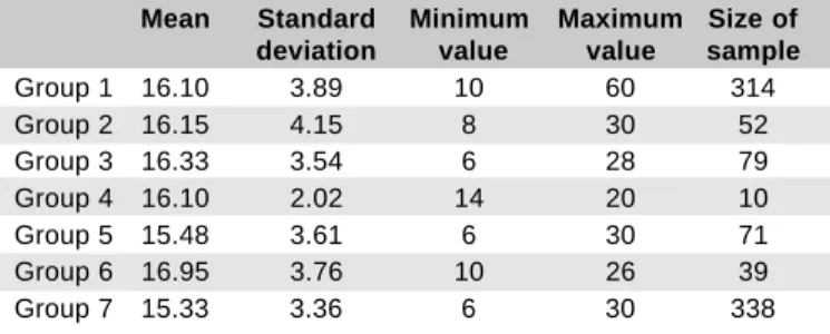
Descriptive analyses showed the values for means, stan-dard deviations, minimum and maximum IOP in all patient groups, in addition to the differences of values in each group. The one-way ANOVA test, with normal distribution and 5% significance was used to compare the mean IOP values between the studied patient groups.
RESULTS
The IOP values of each of the 7 studied groups, their means, standard deviations, minimum and maximum values as well as the number of eyes in each group (total of 914 eyes from 428 patients) are shown in Table 2.
The one-way ANOVA test, by which each group is separa-tely compared with the control group, showed significant values for IOP increase in groups 1, 3 and 6. On the basis of the 5% level of significance, groups 2, 4 and 5 did not show significant results with p = 0.11398, p = 0.47419 and p = 0.74505, respectively.
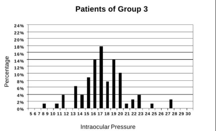
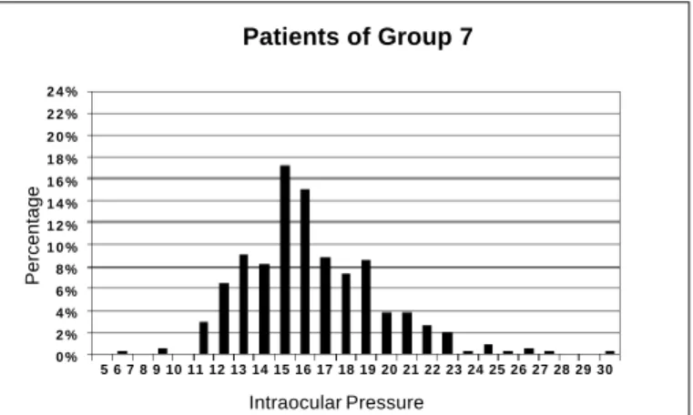
IOP variations found for each group of this study may be seen in Figures 1-7, which show the absolute value of this variable and respective percentage in each group.
Of the 314 hypertensive patients without retinopathy, 51 (16.24%) showed IOP≥19 mmHg, with one value of 60 mmHg, the highest among all groups.
Of the 79 hypertensive patients with hypertensive retino-pathy, 17 (21.52%) presented IOP ≥ 19 mmHg.
Of the total of diabetic and hypertensive patients with some retinopathy, either diabetic or hypertensive, 11 patients (28.20%) showed IOP ≥ 19 mmHg. In this group, p = 0.00526 shown by the one-way ANOVA test was the most significant among the correlated groups.
DISCUSSION
As already mentioned in the Result section, the IOP values
Table 1. Classification of the studied population according to the clinical ophthalmologic diagnosis
Group 1 Hypertensive patients without retinopathy
Group 2 Diabetic patients without retinopathy
Group 3 Hypertensive patients with hypertensive retinopathy
Group 4 Diabetic patients with diabetic retinopathy
Group 5 Hypertensive and diabetic patients without retinopathy
Group 6 Hypertensive and diabetic
patients with some retinopathy
Group 7 Control group - without hypertension,
diabetes and respective retinopathies
Table 2. IOP values of each group of the studied population
Mean Standard Minimum Maximum Size of deviation value value sample
Group 1 16.10 3.89 10 60 314
Group 2 16.15 4.15 8 30 52
Group 3 16.33 3.54 6 28 79
Group 4 16.10 2.02 14 20 10
Group 5 15.48 3.61 6 30 71
Group 6 16.95 3.76 10 26 39
ARQ. BRAS. OFTAL. 63(3), JUNHO/2000 - 225
Analysis of the intraocular pressure in diabetic, hypertensive and normal patients (Glaucoma Project)
increased progressively from the hypertensive patients without retinopathy, to the hypertensive with retinopathy and diabetic hypertensive with retinopathy. This shows the relati-onship between ocular hypertension and evolution of
systemic hypertensive disease and association with diabetes mellitus.
In this study no increased IOP values could be observed when considering only diabetic patients, which disagrees
Patients of Group 3
Percentage
Intraocular Pressure
Fig. 3 - Absolute values of the intraocualr pressure and respective percentages in Group 3.
Patients of Group 6
Percentage
Intraocular Pressure
Fig. 6 - Absolute values of the intraocular pressure and respective percentages in Group 6.
Patients of Group 1
Percentage
Intraocular Pressure
Fig. 1 - Absolute values of the intraocular pressure and respective percentages in Group 1. One IOP value = 60 was excluded for better visualization of the graph.
5 6 7 8 9 10 11 12 13 14 15 16 17 18 19 20 21 22 23 24 25 26 27 28 29 30 2 4 %
2 2 % 2 0 % 1 8 % 1 6 % 1 4 % 1 2 % 1 0 % 8 % 6 % 4 % 2 % 0 %
Patients of Group 2
Percentage
Intraocular Pressure
Fig. 2 - Absolute values of the intraocular pressure and respective percentages in Group 2.
5 6 7 8 9 10 11 12 13 14 15 16 17 18 19 20 21 22 23 24 25 26 27 28 29 30 2 4 %
2 2 % 2 0 % 1 8 % 1 6 % 1 4 % 1 2 % 1 0 % 8 % 6 % 4 % 2 % 0 %
Patients of Group 5
Percentage
Intraocular Pressure
Fig. 5 - Absolute values of the intraocular pressure and respective percentages in Group 5.
5 6 7 8 9 10 11 12 13 14 15 16 17 18 19 20 21 22 23 24 25 26 27 28 29 30 2 4 %
2 2 % 2 0 % 1 8 % 1 6 % 1 4 % 1 2 % 1 0 % 8 % 6 % 4 % 2 % 0 %
5 6 7 8 9 10 11 12 13 14 15 16 17 18 19 20 21 22 23 24 25 26 27 28 29 30 2 4 %
2 2 % 2 0 % 1 8 % 1 6 % 1 4 % 1 2 % 1 0 % 8 % 6 % 4 % 2 % 0 %
5 6 7 8 9 10 11 12 13 14 15 16 17 18 19 20 21 22 23 24 25 26 27 28 29 30 2 4 %
2 2 % 2 0 % 1 8 % 1 6 % 1 4 % 1 2 % 1 0 % 8 % 6 % 4 % 2 % 0 %
Patients of Group 4
Percentage
Intraocular Pressure
Fig 4 - Absolute values of the intraocular pressure and respective percentages in Group 4.
5 6 7 8 9 10 11 12 13 14 15 16 17 18 19 20 21 22 23 24 25 26 27 28 29 30 30%
226 - ARQ. BRAS. OFTAL. 63(3), JUNHO/2000
Analysis of the intraocular pressure in diabetic, hypertensive and normal patients (Glaucoma Project)
with the Australian “The Blue Mountains Eye Study” 6.
Con-trariwise, according to the “Baltimore Eye Survey” 7, there is
an association of diabetes mellitus with IOP increase; however, this not very expressive increase is not associated with evolution towards open angle glaucoma.
The results of the “Barbados Eye Study” 8 showed that IOP
increase is related to systemic arterial blood pressure, a fact that supports the present study. In addition, that study reports a relationship between IOP increase and diabetes mellitus, a fact that could not be proved in the present study, but supports the “The Blue Mountains Eye Study” and the “Baltimore Eye Survey”.
Although there is a tendency of patients with long-standing diabetes to develop diabetic retinopathy and progress to amaurosis, the relationship between IOP increase, glaucoma and diabetes is still a matter of controversy7.
RESUMO
Objetivo: Analisar os valores da pressão intra-ocular (PIO) entre diferentes grupos de pacientes (diabéticos, hipertensos com ou sem retinopatia) e a população normal.
Métodos: Realizou-se a aferição da pressão intra-ocular (PIO) em um total de 924 olhos de 482 pacientes com idade
igual ou maior que 40 anos (x=56,70; dp=11,89) examina-dos segundo um protocolo de estudo que incluiu medida da PIO, pressão arterial e glicemia, além da fundoscopia. A determinação da PIO foi obtida pelo tonômetro de aplanação de Goldmann e a glicemia foi aferida por meio de tiras reativas (Dextrostix – Bayer). Em seguida, os pacientes foram divididos em 7 (sete) grupos: hipertensos, hiper-tensos com retinopatia, diabéticos, diabéticos com retino-patia, hipertensos e diabéticos, hipertensos e diabéticos com retinopatia e a população controle.
Resultados: Pelo teste de ANOVA-uma via, numa distribuição normal no nível de significância de 5%, observou-se que o valor médio da PIO é significativamente maior no grupo de pacientes hipertensos sem retinopatia (média PIO = 16,10), no grupo dos hipertensos com retinopatia (média PIO = 16,33) e no grupo de diabéticos e hipertensos com retinopatia (média PIO = 16,95).
Conclusões: Observou-se que o valor médio da PIO é progressivamente mais elevado conforme o tempo de evolução da doença hipertensiva e também quando esta se encontra associada a diabetes mellitus.
Palavras-chave: Diabetes; Hipertensão; Pressão
intra-ocular; Glaucoma.
REFERENCES
1.Leske CM, Connel AMS, Wu SY, Hyman LG, Schachat AP. Risk factors for open-angle glaucoma. Arch Ophthalmol 1995;113:918-24.
2. Nicolela, MT. Fluxo sanguíneo ocular em glaucoma: métodos de avaliação e importância. Arq Bras Oftalmol 1997;60:639-49.
3. Dielemans I, Vingerling JR, Algra D, Hofman A, Grobbee DE, Jong PTVM. Primary open-angle glaucoma, intraocular pressure, and systemic blood pres-sure in the general elderly population. Ophthalmology 1995;102:54-60. 4. Quigley HA, Enger C, Katz J, Sommer A, Scott R, Gilbert D. Risk factors for
the development of glaucomatous visual field loss in ocular hypertension. Arch Ophthalmol 1994;112:644-9.
5. Klein BEK, Klein R, Moss SE. Incidence of self reported glaucoma in people with diabetes mellitus. Br J Ophthalmol 1997;81:743-7.
6. Mitchell P, Smith W, Chey T, Healey PR. Open-angle glaucoma and diabetes. Ophthalmology 1997;104:712-8.
7. Tielsch JM, Katz J, Quigley HA, Javitt JC, Sommer A. Diabetes, intraocular pressure, and primary open-angle glaucoma in the Baltimore eye survey. Ophthalmology 1995;102:48-53.
8. Wu SY, Leske C. Associations with intraocular pressure in the Barbados eye study. Arch Ophthalmol 1997;115:1572-6.
Patients of Group 7
Percentage
Fig. 7 - Absolute values of the intraocular pressure and respective percentages in Group 7.
Intraocular Pressure
5 6 7 8 9 10 11 12 13 14 15 16 17 18 19 20 21 22 23 24 25 26 27 28 29 30 2 4 %
2 2 % 2 0 % 1 8 % 1 6 % 1 4 % 1 2 % 1 0 % 8 % 6 % 4 % 2 % 0 %
Novidades na Internet!!!
Agora no site CBO você tem disponível todas as informações na íntegra dos Arquivos Brasileiros de Oftalmologia