Cerebral biopsy
Comparison between frame-based stereotaxy
and neuronavigation in an oncology center
Carlos Augusto Ferreira Lobão
1, Janio Nogueira
1,
Antonio Aversa Dutra do Souto
2, José Antonio de Oliveira
1abstract – Treatment of intracranial tumoral lesions is related to its correct histological diagnostic. We present a retrospective analysis of 32 patients submitted to 36 cerebral biopsies using neuronavigation and 44 patients using frame-based stereotaxy. Mean age was 46.6 and 49.3 years old respectively. Sex distribution in both groups was 50% for each. Most of lesions were lobar in both groups. Diagnostic yielding was 91.7% and 83.4%, respectively (p=0.26). We found in the postoperative CT scans intracranial hemorrhages in 13.8% cases of the first group and 9.8% cases in the second. Most of them were mild post-operative hemorrages in the biopsy site. There was one death related to the procedure in each group. Astrocytomas and metastatic adenocarcinomas were the most frequent diagnosis. Diagnostic yielding and the number of postoperative hemorrhage and death were similar on both groups and the same found in the literature.
KEY WORDS: brain tumor, neuronavigation, stereotaxy.
biópsia cerebral: comparação entre estereotaxia com arco e neuronavegação em um centro de oncologia resumo – O manejo das lesões intracranianas tumorais está relacionado ao seu diagnóstico histológico adequado. Foi realizado estudo retrospectivo com 32 pacientes submetidos a 36 biópsias cerebrais por neuronavegação e 44 pacientes por estereotaxia com arco. A idade média foi 46,6 e 49,3 anos respectivamente. Nos dois grupos a distribuição por sexo foi 50% para cada. A maioria das lesões biopsiadas eram lobares nos dois grupos. A positividade diagnóstica foi 91,7% para neuronavegação e 83,4% para a estereotaxia com arco, respectivamente (p=0,26). Identificou-se hemorragia intracraniana na TC pós-operatória em 13,8% dos casos no primeiro grupo e em 9,8% no segundo, a maioria de pequena monta sem provocar piora neurológica. Ocorreu uma morte relacionada ao procedimento em cada grupo. Os diagnósticos mais freqüentes foram astrocitomas e adenocarcinomas metastáticos. A positividade diagnóstica, taxas de hemorragia pós-operatória e de mortalidade foram equiparáveis estatisticamente entre os dois métodos e se assemelham com as descritas na literatura. PALAVRAS-CHAVE: tumor cerebral, neuronavegação, estereotaxia.
1Neurocirurgião, Instituto Nacional de Câncer (INCA), Rio de Janeiro RJ, Brazil; 2Neurocirurgião, Instituto Nacional de Câncer (INCA) e Universidade
Federal do Rio de Janeiro (UFRJ), Rio de Janeiro RJ, Brazil.
Received 18 March 2009, received in inal form 8 July 2009. Accepted 29 July 2009.
Dr. Carlos Augusto Ferreira Lobão – Vila Dona Maria Leopoldina 236 - 66060-180 Belém PA - Brasil. E-mail: calobao@yahoo.com.br
Treatment of intracranial mass lesions is highly reliant in its correct histological diagnosis. Treatment based only on clinical and radiological aspects is unsuccessful in one third of cases even using modern diagnostic techniques1,2.
Advances on stereotactic techniques and more recent-ly on image guided surgery or neuronavigation have left empirical treatment of intracranial tumors, without his-tological conirmation, to be exceptions3. Nowadays the
concept of minimally invasive surgery is becoming a
dai-ly reality in modern neurosurgery4. Among the
advantag-es of this kind of surgery we can list smaller and more pre-cise approaches, lower surgical time, less damage to elo-quent structures, and, as a consequence, smaller morbid-ity and infection rates, and hospital staying5,6. Navigation
means, by deinition, orientation in space. On medical set-tings we consider it as the orientation on a given anatom-ical volume7. Image guided surgery is the use of
sur-gical guidance during surgery8. It enables the surgeon to
make 3D localization of a lesion, program surgical trajec-tory, and localize anatomical structures and surgical in-struments at real time9. The technique evolved during the
80´s permits a more accurate preoperative planning with smaller and more precise approaches10. Knowing the
ex-act location of important and eloquent intracranial struc-tures makes its preservation more likely and permits the surgeon a better resection control11. Many evidences
sug-gest that neuronavigation reduces costs when compared it to standard localization procedures6,12. On the other hand,
frame-based stereotactic biopsy was the gold standard for acquiring intracranial samples for a long time13-15. The
morbidity rate associated to frameless stereotaxy is 0 to 27%13,16,17, mortality rate ranges from 0 to 9%18, and the
di-agnostic yielding is 79 to 100%16,19.
With the advent of neuronavigation the use of frame-based biopsy is becoming smaller because image guided biopsies offers many advantages, like: (1) more comfort to the patient without the using of the stereotactic frame20;
(2) good diagnostic yielding and few complications11,21; (3)
the surgical instruments used on neuronavigation occu-pies less place related to the stereotactic frame applied to the patient head22; (4) the trajectory of the biopsy
nee-dle can be changed any time during the surgery without new calculations23,24; (5) quicker surgical times23. Taking
these advantages in consideration it is easy to realize why neuronavigation reached such a great importance in actu-al neurosurgery. Chiely in neurooncology services, neuro-navigation became a very useful tool with many applica-tions in the treatment of intracranial neoplasm.
The objective of this study is to present the experi-ence of the Oncologic Neurosurgery Service of the Na-tional Institute of Cancer in Brazil with frameless cerebral biopsies and to make a comparison with the frame-based method analyzing diagnostic yielding, postoperative hem-orrhage, and mortality, related to the two procedures.
Method Patients selection
All patients were treated at the Neurosurgery Service of the National Institute of Cancer in Bazil from 2004 to 2007. We did a retrospective analysis of two groups: one group of patients was submitted to frameless cerebral biopsies (neuronavigation), and in the other group frame-based cerebral biopsies were used. The irst group was composed of 32 patients with the mean age of 46.6 years-old, ranging from 8 to 80 years-old, were 36 biopsies were performed on. The second group of 44 patients with the mean age of 49.3 years-old, (3–77 years-old) had 51 frame-based procedures carried out. Half of the patients in each group was composed of male patients.
Surgery was performed by the members of neurosurgery staff of the service for getting the correct diagnosis of varied
in-tracranial mass lesions. Hospital staying considered the time nec-essary for any procedures related to treatment, not only the bi-opsies. Immediate pos-operative CT scans were performed in the 32 patients (88,8%). Criteria for performing the immediate post-operative CT scans were not strictly adopted and were consid-ered at an individual basis. The main reasons to indicate the CT scans were a small bleeding through the biopsy needle occurred, or patients exhibiting post-operative neurological deterioration.
Patients provided informed consent agreeing with data pub-lication and the study was accepted by the Ethic Committee of the National Institute of Cancer - Brazil.
Frameless cerebral biopsy
Roughly seventy 1.5 Tesla T1-weighted gadolinium enhanced axial MRI images were transferred to the neuronavigation work-station preoperatively and the software Cranial Planning v. 1.2 (BrainLAB AG, Heimstetten, Alemanha) was used. This interface helped to establish the exact surgical target, entry point, and surgical trajectory, avoiding vital structures and through the saf-er way. Choosing the surgical target was based in many factors mainly as the most enhancing point of the lesion or the center of a hipointense mass. Proximity of vascular structures and very eloquent areas were avoided. After that planning, the data were transferred to the BrainLab VectorVision Compact neuronaviga-tor (BrainLAB AG, Heimstetten, Alemanha).
Depending on the pre-operative patient consciousness lev-el we have chosen among general or local anesthesia with seda-tion. Head was ixed with the three-points Mayield head-holder in which we adapted a device with infrared relexive spheres for recognition with the neuronavigator. It was used a Sedan-Nash-old biopsy needle with relexive spheres attached to it. Forehead and facial surface anatomical landmarks were obtained with the laser pointer scanner. Minimal accepted registration error was 2 mm. Whenever a bigger estimated error was considered, a new registration process was performed. Biopsy needle was calibrat-ed and recognizcalibrat-ed by the neuronavigation software in relation to the head position and inserted based on three-planar imag-es generated by the neuronavigator (Fig 1A,B). The number of the samples of tissue obtained and the decision to switch to a new target were considered according to the per-operative neu-ropathologist smear hystological analysis of the samples. The software used for neuronavigation was Cranial Navigation v. 6.0 (BrainLAB AG, Heimstetten, Alemanha).
Frame-based cerebral biopsy
Frame-based cerebral biopsy is a well-known and standard-ized technique7,10. Moments before the surgery an ETM03-B
tar-get was the most enhancing point of a lesion on CT-scan or the center of a hipodense mass. A new target point was calculated, and we chose a different trajectory in the case of inconclusive in-print analysis by the pathologist.
Statistical analysis
We performed non-parametric χ2-test (qui-square test),
con-sidering signiicant p<0.05.
results
Table 1 shows data relative to both studied groups: group I, frameless cerebral biopsy, and group II, frame-based cerebral biopsy. We observed an increasing trend for using frameless biopsy throughout the time period studied. Both groups had a preponderance of lobar le-sions biopsies, 68.8% and 70.5%, respectively. There was no signiicant difference between lesion localization on
Fig 1. [A e B] Neuronavigation antenna pointed to the star-shaped tool ixed to three points Mayield head-holder. There most be no obstacles between the antenna and relexive spheres attached to sur-gical instruments.
Table 1. Frequencies, percentiles, and χ2-test (qui-square).
Neuronavigation Frame-base stereotaxy
χ2-test
n % n %
Year studied 2004 2005 2006 2007
1 7 14 14
3.0 19.0 39.0 39.0
12 14 24 1
23.5 27.0 47.5 2.0
p=0.001
Gender Male Female
16 16
50.0 50.0
22 22
50.0 50.0
–
Lesion side Left Right Midline
20 9 3
62.5 28.1 9.4
21 20 3
47.7 45.5 6.8
p=0.31
Localization Lobar Basal nucleus Dien/Mes Pineal
22 5 3 2
68.8 15.6 9.4 6.2
31 9 3 1
70.5 20.4 6.8 2.3
p=0.25
each hemisphere or in the midline considering two groups (p=0.31).
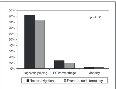
Diagnostic yielding for frameless cerebral biopsy was 91.7%, and 83.4% for frame-based biopsies. This difference was not statistically signiicant (p=0.26, Fig 2). Considering non-diagnostic frame-based biopsies, seven were repeat-ed using the same method, one was repeatrepeat-ed with frame-less stereotaxy, one patient was submitted to craniotomy for surgical tumor resection and open biopsy, and one pa-tient had empirical corticoid treatment with lesion disap-pearance. Five out of seven non-diagnostic cases repeat-ed with frame-basrepeat-ed stereotaxy were diagnostic on sec-ond procedure, one case was considered and treated as a demializating disease, and one patient died before a new diagnosis attempt. Only four of the frameless biopsy pro-cedures were not diagnostic. These cases were submitted to a second frameless procedure with positive diagnosis.
Mean hospital staying was 11.6 days for frame-based biopsies, and 15.9 days for frameless procedures. There was no signiicant difference between then (p>0.05).
We observed ive post-operative intracranial hemor-rhages on frameless biopsies group (13.8%) in the
post-operative CT-scans. Three small volume cerebral hemor-rhages on biopsy site, and one case of small third ventri-cle hemorrhage, all of then without clinical signiicance. Nevertheless there was one case of surgical treated cere-bral hemorrhage that evolved to death.
Five cases of frame-based cerebral biopsies developed small post-operative cerebral hemorrhage at biopsy site (9.8%) observed in the post-operative CT-scans. One pa-tient submitted to frame-based biopsy presented impor-tant clinical deterioration at PO day 1. CT-scan showed tr-anstentorial herniation, sub-falcine herniation, herniation of the left uncus, and intense supratentorial edema at the lesion site. This patient was submitted to decompressive craniotomy immediately after diagnosis but evolved to death at PO day 4 (Table 2, Fig 2). There is no statistically signiicant difference among symptomatic and non-symp-tomatic post-operative intracranial hemorrhage and mor-tality comparing both groups (p>0.05).
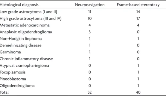
Table 3 presents histological diagnosis at both groups studied. There is a clear predominance of low-grade and high-grade gliomas on both of then. There are four cases of frame-based biopsies not presented on this table: one
Table 2. Post-operative hemorrhage and mortality related to stereotactic cerebral biopsies.
Neuronavigation Frame–based stereotaxy
n % n %
PO hemorrhage 5 13.8 5 9.8
Small ICH 3 8.3 5 9.8
Small IVH 1 2.7 – –
Huge ICH 1 2.7 – –
Hypertensive oedema – – 1 1.9
Mortality 1 2.7 1 1.9
PO: post-operative; ICH: intra-cerebral hemorrhage; IVH: intra-ventricular hemorrhage.
Table 3. Histological diagnosis on stereotactic cerebral biopsies.
Histological diagnosis Neuronavigation Frame-based stereotaxy Low grade astrocytoma (I and II) 11 14
High grade astrocytoma (III and IV) 10 17 Metastatic adenocarcinoma 4 4 Anaplasic oligodendroglioma 3 0
Non-Hodgkin linphoma 1 1
Demielinizating disease 1 0
Germinoma 1 0
Chronic inlammatory disease 1 0 Atypical craniopharingioma 0 1
Toxoplasmosis 0 1
Pineoblastoma 0 1
Oligodendroglioma 0 1
was treated with corticoid without deined diagnosis, a second non-diagnosed case was diagnosed by frameless biopsy, the third one needed craniotomy for tumoral re-section, and the latter died before a second biopsy.
disCussion
According to a meta-analysis of 7471 frameless biop-sies the method achieves a diagnostic yielding of 91%, morbidity of 3,5%, and mortality of 0,7%25. Other studies
show that frameless cerebral biopsy can be as precise as, or even more, than frame-based stereotaxy21,25,26. Mean
lo-calization error is similar in both methods25,27.
Dificulties on frameless biopsies are many. Its target-ing precision is intimately related to pre-operative radio-logical imaging that needs a well deined protocol26. Its
adequate using depends on imaging manipulation and proper array of equipment on operation room. For fewer targeting errors it is necessary an adequate registration of cranial surface points. Depending on the method used for this registration, neuronavigation accuracy can be lowered down28. Neuronavigation biopsies of about 1 cm lesions,
profound lesions, and posterior fossa lesions are usual-ly less accurate than frame-based biopsies22. Brain shift
after trepanation and dural opening is not so important compared to open craniotomy29. We use frame-base
bi-opsy for lesions as small as 1 cm. Neuronavigation is usu-ally done with general anesthesia while frame-based bi-opsy are normally performed with patient sedation and local anesthesia which permits a better neurological eval-uation just after the procedure29. Costs for
neuronaviga-tion acquisineuronaviga-tion are high considering a emerging country like Brazil, but long term neuronavigation cost evaluation seems to be lower29.
Our study shows similar results on both methods com-pared to the literature considering diagnostic yielding,
pos-operative hemorrhage, and mortality rates24.
Post-operative hemorrhage rates of 13.8% for the neuronavi-gation group, and 9,8% for the frame-based group are ex-pected on the post-operative CT scans. These rates are justiied because all patients studied are from an oncol-ogy center with predomination of astrocitic lesions. It is well known that glioma biopsies tends to have high-er post-ophigh-erative hemorrhage rates, specially those lo-cated at eloquent areas and deep seated17. Mortality
re-lated to volumous post-operative hemorrhage found on our study are compared to those found in the literature, i.e., 2.7% for neuronavigation group, and 1.9% for frame-based biopsies6,30. That is why patients with hemorrhage
at biopsy site on CT-scan must stay on hospital for ob-servation and new imaging6. Post-operative hemorrhages
found on our study were found on gliomas extending to the corpus callosum, or to the thamalus and mesenceph-alon. Exception was a patient with a huge fronto-tempo-ro-insular lesion with negative frame-based biopsy that died without diagnosis.
Neuronavigation-guided cerebral biopsy is a very use-ful method for the neurosurgeon armamentarium. Advan-tages over frame-based stereotaxy are many, and simi-lar diagnostic yielding, post-operative hemorrhage, and mortality rates are found. Using neuronavigation for cere-bral biopsies of lesions bigger than 1 cm have many justi-iable advantages: great acceptance by the patient avoid-ing frame ixation; there is no need to transport the pa-tient immediately to and from the CT scan suite, mainly in children, for example, under general anesthesia; pre-op-erative MRI for frameless biopsy can be performed some days before surgery in contrast to frame-based biopsy that needs CT-scan just before it; a better security perception by the neurosurgeon who have instruments, target, and trajectory real-time images; possibility for changing tar-get and trajectory during surgery without new stereot-actic calculations; better system ergonomics with nee-dle holder, infrared antenna, and star-shape head posi-tion tool all articulated in contrast to the volumous ste-reotactic frame.
Even thought frame-based biopsy still holds an im-portant position on treating intracranial mass lesions be-cause its cost accessibility, and its high diagnostic rates specially considering tumors of 1 cm or less, new software and equipment development tend to make neuronaviga-tion an even more diffused method among neurosurgeons with lesser acquisition costs and better precision.
referenCes
1. Friedman WA, Sceats DJ, Nestok BR, et al. The incidence of
unexpect-ed pathological indings in an image-guidunexpect-ed biopsy series: a review of 100 consecutive cases. Neurosurgery 1989;25:180-184.
2. Lobato RD, Rivas JJ, Cabello A. Stereotactic biopsy of brain lesions vi
-sualized with CT. Appl Neurophysiol 1982;45:426-430. 0%
10% 20% 30% 40% 50% 60% 70% 80% 90% 100%
Diagnostic yielding
Neuronavigation Frame-based stereotaxy
p > 0.05
PO hemmorhage Mortality
3. Epstein F, Wisoff J. Intrinsic brain-stem tumors of childhood: surgical indications. J Neurooncol 1988;6:309-317.
4. Barnett GH, Miller DW. Brain biopsy and related procedures. In: Rob
-erts DW, Barnett GH, Maciunas RJ (Eds) Image-guided neurosurgery: clinical applications of surgical navigation. St. Louis: Quality Medical Publishing, 1998:181-191.
5. Barnett GH, Nathoo N. The modern brain tumor operating room: from standard essentials to current state-of-the-art. J Neurooncol 2004;69:25-33. 6. Kaakaji W, Barnett GH, Bernhard D, et al. Clinical and economic con
-sequences of early discharge of patients following supratentorial ste
-reotactic biopsy. J Neurosurg 2001;94:892-898.
7. Haberland N, Ebmeier K, Hiliscs R, et al. Neuronavigation in surgery of intracranial and spinal tumors. J Cancer Res Clin Oncol 2000;126:529-541. 8. Gumprecht HK, Widenka DC, Lumenta CB. Brainlab vectorvision neu
-ronavigation system: technology and clinical experience in 131 cases. Neurosurgery 1999;44:97-105.
9. Fujita K, Yanaka K, Meguro K, et al. Image-guided procedures in brain biopsy. Neurol Med Chir (Tokio) 1999;39:502-509.
10. Linskey ME. The changing role of stereotaxis in surgical neuro-oncol
-ogy. J Neurooncol 2004;69:35-54.
11. Barnett GH. The role of image-guided technology in the surgical plan
-ning and resection of gliomas. J Neurooncol 1999;42:247-258. 12. Henderson JM, Eichholz KM, Bucholz RD. Decreased length of stay
and hospital costs in patients undergoing image-guided craniotomies.
J Neurosurg 1997; 86:367.
13. Appuzo MLJ, Chandrasoma PT, Cohen D, et al. Computed imaging ste
-reotaxy: experience and perspective related to 500 procedures applied to brain masses. Neurosurgery 1987;20:930-937.
14. Arbit E, Galicich JH. Importance of image-guided stereotactic biopsy to conirm diagnosis in an oncological setting. Ann Surg Oncol 1994;1: 368-372.
15. Maciunas RJ, Galloway RLJ, Latimer JW. The application accuracy of stereotactic frames. Neurosurgery 1994;35:682-695.
16. Bernstein M, Parrent AG. Complications of CT-guided stereotactic bi
-opsy of intra-axial brain lesions. J Neurosurg 1994;81:165-168. 17. Kreth FW, Muacevic A, Medele R, et al. The risk of haemorrhage after
image guided stereotactic biopsy of intra-axial brain tumours: a pro
-spective study. Acta Neurochir 2001;143:539-546.
18. Sawin PD, Hitchon PW, Follet KA, et al. Computed imaging-assisted stereotactic brain biopsy: a risk anlysis of 225 consecutive cases. Surg Neurol 1998;49:640-649.
19. Nasser JA, Confort CI, Ferraz A, et al. Biópsia estereotáxica guiada por imagem nas lesões do sistema nervoso central. Arq Neuropsiquia
-tr 1998;56:206-211.
20. Watanabe E, Mayanagi Y, Kosugi Y, et al. Open surgery assisted by the neuronavigation, a stereotactic, articulated, sensitive arm. Neurosur
-gery 1991;28:792-800.
21. Jain D, Sharma MC, Sarkar C, et al. Comparative analysis of diagnos
-tic accuracy of different brain biopsy procedures. Neurol India 2006;54: 394-398.
22. Pan HC, Wang YC, Lee SD, et al. A modiied method to perform the frameless biopsy. J Clin Neurosci 2003;10:602-605.
23. Paleologos TS, Dorward NL, Wadley JP, et al. Clinical validation of true frameless stereotactic biopsy: analysis of the irst 125 consecutive cas
-es. Neurosurgery 2001;49:830-835.
24. Woodworth GF, McGirt MJ, Samdani A, et al. Frameless image-guid
-ed stereotactic brain biopsy proc-edure: diagnostic yield, surgical mor
-bidity, and comparison with the frame-based technique. J Neurosurg 2006;104:233-237.
25. Spivak CJ, Pirouzmand F. Comparison of reliability of brain localiza
-tion when using tradi-tional and stereotactic image-guided techniques: a prospective study. J Neurosurg 2005;103:424-427.
26. Dorward NL, Alberti O, Palmer JD, et al. Accuracy of true frameless ste
-reotaxy: in vivo measurement and laboratory phantom studies. J Neu
-rosurg 1999;90:160-168.
27. Quiñones-Hijosa A, Ware ML, Sanai N, et al. Assessment of image guid
-ed accuracy in a skull model: comparison of frameless stereotaxy tech
-niques vs. frame-based localization. J Neurooncol 2006;76:65-70. 28. Grunert P, Darabi K, Espinosa J, et al. Computer-aided navigation in
neurosurgery. Neurosurg Rev 2003;26:73-99.
29. Dwarakanath S, Suri A, Sharma BS, et al. Neuronavigation in a devel
-oping country: a pilot study of eficacy and limitations in intracranial surgery. Neurol India 2007;55:111-116.
30. Hisatugo MKI, Stávale JN, Bidó JO, et al. Abordagem estereotáxica gui
-ada por imagem de lesões do sistema nervoso central. Arq Neurop
![Fig 1. [A e B] Neuronavigation antenna pointed to the star-shaped tool ixed to three points Mayield head-holder](https://thumb-eu.123doks.com/thumbv2/123dok_br/15432193.594866/3.955.146.769.561.968/fig-neuronavigation-antenna-pointed-shaped-points-mayield-holder.webp)