Neurological coNgeNital MalforMatioNs
iN a tertiary Hospital iN soutH Brazil
Ana Guardiola
1,2, Vanessa Koltermann
1, Paula Musa Aguiar
1, Sérgio Pilla Grossi
1,2,
Valéria Fleck
3, Elisabeth C. Pereira
3, Lúcia Pellanda
1,4abstract – Background: Congenital anomalies are one of the main causes of morbidity and mortality among infants. The involvement of the central nervous system (CNS) occurs in 21% of cases. Objective: To identify incidence of CNS malformations and associated factors in newborns at a Terciary Hospital of Porto Alegre. Method: Case-control study conducted between 2000 and 2005 based on the Latin American Collaborative Study of Congenital Malformations database. Results: Among 26,588 births registered in this period, 3.67% presented with malformations (IC=95%; 3.44–3.9), being 0.36% of the CNS (IC=95%,(0.29–0.43)). The most common CNS malformation was meningomielocele (10.4%). Young maternal age (p=0.005); low birth weight (p=0.015); large cephalic perimeter (p=0.003); post term birth (p=0.000) and low APGAR indexes at the 1st and 5th minutes were associated with CNS malformations. Conclusion: We found an incidence of CNS malformations similar as compared to literature.
KeY WoRdS: neurological malformations, neural tube defects, risk factors, congenital malformations.
Malformações neurológicas congênitas observadas em hopsital terciário no sul do Brasil
resumo – Anomalias congênitas são umas das principais causas de morbimortalidade infantil. o sistema nervoso central (SNC) é acometido em 21% dos casos. Objetivo: Identificar a incidência e fatores associados a malformações do SNC em recém nascidos na maternidade de um hospital terciário de Porto Alegre. Método: estudo controle realizado de janeiro de 2000 a dezembro de 2005, baseado no banco de dados do estudo Colaborativo Latino Americano de Malformações Congênitas. Resultados: dos 26.588 nascimentos, 3,67% apresentaram malformação (IC=95%; 3,44–3,9), com 0,36% do SNC (IC=95%, (0,29–0,43)). A malformação do SNC mais comum foi hidrocefalia (10,9%). Menor idade materna (p=0,005); menor peso ao nascimento (p=0,015), maior perímetro cefálico (p=0,003); nascimentos pré-termo (p=0,000) e menores índice APGAR no 1o e 5o minutos (p<0,000) apresentaram associação com malformações do SNC. Conclusão: Foi encontrada incidência similar de malformações do SNC comparada à literatura.
PALAvRAS-CHAve: malformações neurológicas, defeitos tubo neural, pé torto, fatores de risco.
1Federal University Foundation of Porto Alegre (UFCSPA), Porto Alegre RS, Brazil; 2Irmandade Santa Casa de Misericórdia, Porto Alegre RS, Brazil; 3eCLAMC (Latin American Collaborative Study of Congenital Malformations), Porto Alegre RS, Brazil; 4University Foundation of Cardiology (FUC), Porto Alegre RS, Brazil.
Received 26 February 2009, received in inal form 16 July 2009. Accepted 13 August 2009.
Dra. Ana Guardiola – Sarmento Leite 245 - 90050-170 Porto Alegre RS - Brasil. E-mail: [email protected]
Congenital malformations develop during intrauter-ine life and, therefore, are present at birth, although they may be unrecognized at that moment. Neural tube de-fects (NTds) occur due to embryonic neural tube closure failure during the fourth week of embryogenesis1. Congen-ital anomalies cause a signiicant proportion of embryon-ic and fetal deaths, and stand among the leading causes of infant mortality and morbidity, affecting 2–5% of all newborns and contributing signiicantly for mortality at the age of one year2,3. Approximately 21% of these
neurological or non-neurological defects. Most isolated NTds have a multifactorial origin5. despite the fact that NTds do not follow a Mendelian pattern of inheritance, this defects show strong familiar clustering, possibily in-dicating a genetic etiology. Risk of recurrence in siblings of meningomyelocele patients is about 2–5%6. The inci-dence of NTds among irst and second degree relatives is somewhat higher than for non-selected populations. Case reports and epidemiological studies have implicated a se-ries of agents in the etiology of NTds, including chem-ical products, solvents, abuse substances, drugs, pollut-ants and infectious diseases. Additionally, maternal hy-perthermia, valproate use, nutritional inadequacies and chronic disease, such as diabetes mellitus, have been as-sociated to an elevated incidence of NTds, as well as so-cio-economic variables and parents occupation7,8.
A recent metanalysis including 33 studies suggests that there is association between maternal age and cer-tain types of NTds. This association could be stronger with spina biida than with anencephaly9. It is well known, nonetheless, that periconceptional daily supplementation with folic acid is effective in reducing the incidence of NTds10. There is considerable variation in the reported in-cidence of congenital malformations in different popula-tions, ranging from 1.07% in Japan to 45.2% in egypt2. Sea-sonal, ethnic and geographic factors may be implicated1,2. Incidence has been reported to decrease in some areas, while in others it has been held constant2. Although the reasons for this decrease are still unknown, improvement of prenatal diagnosis, selective pregnancy termination, ge-netic counseling and, possibly, folic acid supplementation could be implicated11. Facing the high incidence and prev-alence of congenital malformations, with signiicant costs to society and the severe impact that birth defects rep-resent for individuals and families, detailed investigation of these conditions is of paramount importance in plan-ning effective prevention strategies.
In South America, the Latin American Collaborative Study of Congenital Malformations (eCLAMC) is a case-control study that aims to investigate clinical and epide-miological variables of newborns with congenital anom-alies, including risk factors.
The present study was undertaken in a tertiary refer-ence center in south Brazil, participating in eCLAMC since 1990. We sought to describe the incidence, clinical presen-tation and risk factors of CNS malformations in this setting.
MetHod
eCLAMC is a case-control study of congenital malforma-tions, and its methods have been described elsewhere12. For
the present study, we analyzed the eCLAMC database at Com-plexo Hospitalar Santa Casa, Porto Alegre, Brazil, including all births from January, 2000 to december, 2005. All newborns and
stillbirths with neurological congenital malformations were identiied.
Cases were deined as any clinically detectable morpholog-ical abnormality in any pre or post gestational age. All live birth cases were paired with a control. The irst newborn of the same gender and with no malformations, being born in the same hospi-tal immediately after the case was included as the paired control. variables evaluated included gender, birth weight, gestation-al age, APGAR scores in the first and fifth minutes, presence of associated congenial malformations, parental consanguinity, multiple pregnancy, maternal age, type of delivery, pregnancy information (prenatal follow up visits, intercurrences, maternal diseases, medications, substance abuse), newborn clinical status, amniotic luid aspect and socioeconomic data.
All statistical analyses were performed with the aid of SPSS software. Continuous data is described as means ± one stan-dard deviation, and categorical data as proportions. Chi square and Students’ t test were used to compare groups. odds ratios and 95% conidence interval were estimated. Multivariable lo-gistic regression models were used to study the association of risk factors and malformations. The analyses were considered signiicant if p<0.05.
The study was submitted for evaluation by the ethics and Research Commission of this institution where the study was performed. It was approved with protocol number 1397/06.
results
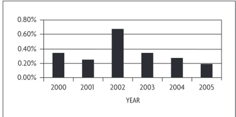
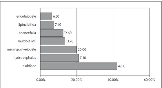
of the 26,588 births from January 2000 to decem-ber 2005, malformations were observed in 975 newborns (3.67%–95%CI 3.44–3.9) and 95 presented with CNS mal-formations (3.67% (IC=95%, 3.44–3.9)). The annual distribu-tion of these malformadistribu-tions is illustrated in Figure 1 and the prevalence of speciic malformations may be evalu-ated in Figure 2.
Clinical characteristics of both cases and controls are presented in Table. Maternal age ranged from 15 to 44 years. Forty-six per cent of mothers were 15–20 years-old, and only 1.7% were over 40 years-old. Most families lived in the state capital (60.6%) and its metropolitan area (33.7%), while 5.7% came from other cities. Method of de-livery was vaginal in 56.8% of the births, cesarean in 42%
Fig 1. Annual prevalence of neurological malformations.
0.00% 0.20% 0.40% 0.60% 0.80%
2000 2001 2002 2003 2004 2005
and with the use of forceps in 1.1%. Most mothers were white (75.2%) and had only basic education (46.1%). Re-ported frequency of malformations in families was 11.7%, and of consanguinity 1.7%.
Most mothers used at least one medication during pregnancy (74.1%). Medications most frequently used are illustrated in Figure 3.
Malformation diagnosis was stablished during the irst day of life in 78.7% of the cases, and prenatally in 21.3%. The most frequent neurological malformations were: con-genital clubfoot, hydrocephalus and meningomyelocele
(n=95, Fig 2). other malformations included microceph-aly (3.2%), craniosynostosis with neurologic alterations (2.1%), holoprosencephaly (2.1%), macrocephaly (2.1%), Ar-nold Chiari syndrome (2.1%), ocular hypertelorism (1.1%), Moebius syndrome (1.1%), agenesis of the corpus callosum (1.1%) and squizencephaly (1.1%).
Comparing cases and controls, it is possible to observe that CNS malformations cases presented with younger maternal age, smaller birth weight, greater cephalic perim-eter, a greater proportion of premature births and smaller Apgar índices in the 1st and 5th minutes (Table).
42.10 21.10
20.00 13.70 12.60 7.40 6.30
0.00% 20.00% 40.00% 60.00%
encefalocele
Spina bifida
anencefalia
multiple MF
meningomyelocele
hydrocephalus
clubfoot
Fig 2. Prevalence of speciic neurological malformations.
0 20 40
Misoprostol Insulin Ferrous sulfate Nasal decongestant Glucocorticoids Antiparasitic Beta-agonists Antifungal Antispasmodic Antiemetic Antidepressant Antibiotics Antacid Antihypertensive Anxiolytics Analgesic Nosteroidal Antiinflamatory
Cases Controls
Fetal distress was more commonly observed in cases than in controls (28.73%, p=0.006).
There was no differences between cases and con-trols regarding premature birth, urinary infection or fetal growth restriction, alcohol use, drug use, maternal and pa-ternal schooling.
We observed a greater frequency of the other malfor-mations in the family of cases (16.1%) than controls (6.9%) (p=0.065). Consanguinity was very uncommon in the pres-ent sample and was not associated with neurological mal-formations.
Meningomyelocele was associated to female sex (73.7%, p=0.015), β-agonists (16.7%, p=0.016) and corti-coesteroids use (11.1%, p=0.029).
Insulin use (10.5%, p=0.029) was more frequent in cas-es with hidrocephalus than in controls .
discussioN
The global incidence of malformations was 3.67%, ac-cording to other reports in the literature that describe an incidence of 2-5% of significant structural anoma-lies13. In this study about malformations in southern Bra-zil, we observed that neurological malformations, includ-ing clubfoot, comprised almost ten percent of cases. In some studies, CNS malformations are described as the most common type of malformation, comprising up to 13% of cases14, second only to congenital heart disease4. These malformations contribute signiicantly to mortal-ity and morbidmortal-ity due to congenital disease during the irst year of life3.
NTds represent the most important group of CNS malformations, and occur due to failure of spontaneous closure of the neural tube beween the third and fourth week of gestational age1. Although the exact cause of NTds is still unknown, there is evidence suggesting that
many factors may adversally inluence fetal development, including radiation, drug use, chemical substances and ge-netic factors15.
The most frequent malformations in our study were clubfoot, hidrocephalus e meningomyelocele. In other studies, this order may vary slightly1. In the United States, anencephaly and spina bíida are the most commonly de-scribed CNS malformations, affecting 1:1000 gestations, and more than 300,000 patients worldwide10.
Both spina bífida and anencephaly are significant causes of infant and fetal deaths16. Children with anen-cephaly usually die at birth or with a few hours of life. Children with spina bíida that do survive are at increased risk for permanent incapacities and psychosocial diicul-ties17. Clinical problems such as paralysis, hydrocephalus Arnold-Chiari tipo II, siringobulbia, siringomielia and en-docrine dysfunction may result from the defect itself or from its repair17.
In this study, we included congenital clubfoot as a neurological malformation because it is known that neu-rological factors producing medullar or nerve alterations are paramount in its etiology. The normal joint develop-ment intrauterus depends on proper functioning of the fetal CNS, on its capacity to move inside the womb and on the amniotic luid quantity.
Along the last 25 years, many studies have shown that genetic and environmental factors play an important role in the etiology of NTds5. In the present work, we observed that maternal age was negatively associated with the pres-ence of these malformations. other authors show an U-shaped relationship, with higher incidences in ages below 20 years or above 35 years18.
Prenatal care is improving in Brazil and in many coun-tries worldwide. We observed that cases performed a higher number of ultrasonographic examinations, dem-Table. Clinical characteristics of cases and controls.
variable Case Control p
Maternal age (years) 24.82±7.042 27.81± 6.753 .005
Number of pregnancies 2.56±1.897 3.00±2.011 .131
Lenghth (cm) 49.37±3.08 49.96±1.72 .241
Toracic perimeter (cm) 32.88±2.52 33.32±1.76 .330
Weight (kg) 2801.64 3095.68 .015
Cefalic perimeter (cm) 36.86±5.69 34.53±1.35 .003
Gestational age 35.23±4.058 38.54±2. 074 .000
Number of prenatal follow up visits 7.27±4.506 7.06±3.237 .724
Number of ultrasonographic examinations during pregnancy
3.05±2.335 1.96±1.518 .001
Apgar 1 6.0247±3.35773 8.1667±1.06193 .000
onstrating the importance of careful screening and fol-low-up for these patients8.
our study failed to show a relationship between ma-ternal smoking and CNS malformations, although this rela-tionship is described in the literature. Some authors found a moderate increase in CNS malformations with daily ma-ternal exposition to passive smoking19.
There is a consistent body of evidence showing that the incidence of CNS malformations could be cut to half if folic acid supplementation would be implemented from the periconceptional period to the initial months of ges-tation20-22. Aiming to reduce the incidence of CNS malfor-mations, many countries are recommending to add folic acid to commonly consumed foods. In the United States, this lead to a 19% reduction of CNS malformations1.
In summary, it s very important to examine speciic causes of CNS malformations in our setting, since there is still many gaps in the understanding of their complex etiology. This kind of study may provide insights to a bet-ter planning of preventive policies that have shown to be effective in other contexts.
refereNces
1. Aguiar MJB, Campos AS, Aguiar RALP, Lana AMA, Magalhães RL, Ba-beto LT. Neural tube defects and associated factors among liveborn and stillborn infants. J Pediatr (Rio J) 2003;79:129-134.
2. Marks JD, Khoshnood B. Epidemiology of common neurosurgical dis-eases in the neonate. Neurosurg Clin N Am 1998;9:63-72.
3. Miura E, Failace LH, Fiori H. Perinatal and neonatal mortality at the Hospital de Clínicas de Porto Alegre, Brazil Rev Assoc Med Bras 1997; 43:35-39.
4. Pitkin RM. Folate and neural tube defects. Am J Clin Nutr 2007;85:285-288. 5. Kaufman BA. Neural tube defects. Pediatr Clin N Am 2004;51:389-419. 6. Sebold CD, Melvin EC, Siegel D, et al;NTD Collaborative Group. Re-currence risks for neural tube defects in siblings of patients with lipo-myelomeningocele. Genet Med. 2005;7:64-67.
7. Medical Research Council. Prevention of neural tube defects: results of
the Medical Research Council Vitamin Study. MRC Vitamin Study Re-search Group. Lancet 1991;338:131-137.
8. Czeizel AE, Dudas I. Prevention of the irst occurrence of neural-tube defects by periconceptional vitamin supplementation. N Engl J Med 1992;327:1832-1835.
9. Berry RJ, Li Z, Erickson JD, et al. Prevention of neural-tube defects with folic acid in China. China-U.S. Collaborative Project for Neural Tube Defect Prevention. N Engl J Med 1999;341:1485-1490.
10. MMWR. Use of dietary supplements containing folic acid among wom-en of childbearing age--United States, 2005. MMWR Morb Mortal Wkly Rep 2005;54:955-958.
11. Padmanabhan R. Etiology, pathogenesis and prevention of neural tube defects. Congenit Anom (Kyoto) 2006;46:55-67.
12. Castilla EE, Orioli IM. ECLAMC: The Latin-American Collaborative Study of Congenital Malformations. Community Genet 2004;7:76-94. 13. Amorim MMR de, Vilela PC, Santos ARVD, et al. Impact of
congeni-tal malformations on perinacongeni-tal and neonacongeni-tal morcongeni-tality in an university maternity hospital in Recife. Rev Bras Saude Mater Infant 2008;6:19-25. 14. Noronha L de, Medeiros F, Martins VDM, et al. Malformations of the
central nervous system: analysis of 157 pediatric autopsies. Arq Neu-ropsiquiatr 2000;58:890-896.
15. Behrman RE, Kliegman RM, Jenson HB. [tradução Vilma Ribeiro de Souza Varga, Nelson Gomes, Eleonora Silva Lins, et al]. Nelson, Trata-do de Pediatria. Tradução da 17º edição. Rio de Janeiro: Elsevier, 2005: 2103-2104.
16. Zurmohle UM, Homann T, Schroeter C, Rothgerber H, Hommel G, Ermert JA. Psychosocial adjustment of children with spina biida. J Child Neurol 1998;13:64-70.
17. Botto LD, Moore, CA, Khoury MJ, Erickson, JD. Neural-tube defects. N Engl J Med 1999;341:1509-1519.
18. Vieira AR, Taucher SC. Edad materna y defectos del tubo neural: evi-dencia para un efecto mayor en espina bíida que anencefalia. Rev Méd Chile 2005;133:62-70.
19. Li Z, Ren A, Zhang L, Guo Z. A population-based case-control study of risk factors for neural tube defects in four high-prevalence areas of Shanxi province, China. Paediatr Perinat Epidemiol 2006;20:43-53. 20. Liu S, West R, Randell E, et al. A comprehensive evaluation of food for-tiication with folic acid for the primary prevention of neural tube de -fects. BMC Pregnancy Childbirth 2004;4:20.
21. Shurtleff DB. Epidemiology of neural tube defects and folic acid. Cere-brospinal Fluid Research 2004;1:5.