Arq Neuropsiquiatr 2008;66(2-B):425-427
425
Duchenne gene carrier as cause of
asymptomatic hypercKemia
Diogo Fraxino de Almeida
2,3, Aluisio Cláudio Mentor Couto Melo Jr
2,
Paulo Rogério M. de Bittencourt
1gene Da Distrofia De Duchenne em portaDora assintomática como causa De cpK elevaDa
1UNINEURO, Unidade de Neurologia Clínica, Curitiba PR, Brazil; 2EMG LAB, Nossa Senhora das Graças Hospital (HNSG), Curitiba PR, Brazil; 3University
Hospital - Universidade Estadual de Maringá, Maringá PR, Brazil.
Received 18 December 2007, received in inal form 25 March 2008. Accepted 31 March 2008.
Dr. Diogo Fraxino de Almeida – Avenida Independência 258/405 - 87015-020 Maringá PR - Brasil. E-mail: [email protected]
Asymptomatic hyperCKemia can be a clinical chal-lenge for physicians, although its long term prognosis seems benign1. The causes usually include heavy exercise
practice, muscle injury, drug-induced myopathy, chron-ic alcoholism, hypothyroidism, clinchron-ically silent myop-athies, and susceptibility to malignant hyperthermia. A frequently missed cause in women is the carrier status of the Duchenne muscular dystrophy2.
We report a case and discuss its investigational approach.
case
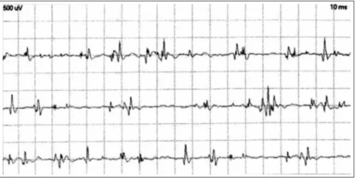
A 15 year-old girl was referred to us with asymptomatic hy-perCKemia after an extensive prior investigation. The hyperCK-emia was found after liver enzymes had been found mildly ele-veted during a fever work-up. There was no history of exercise intolerance, muscle injury, neuromuscular disease in the family, myotoxic drug use or personal and family history of anesthetic complications. She has an asymptomatic brother as well as no other family member has any history of neurological disorders. Resting CPK levels varied from 500 to 1400 U/L in the last three years. An electrophysiological study showed mild chronic myop-athy represented by normal nerve conduction studies, no abnor-mal spontaneous activity, and moderate amount of polyphasic
small amplitude and short duration motor unit potentials (MUP) with early recruitment (Fig 1). An extensive laboratory work-up was normal including CBC, sedimentation rate, ANA, TSH, free T4, PTH and electrolytes. A muscle biopsy performed in other medical center showed non-speciic myopathic changes and a diagnosis of myotonic myopathy had been proposed, despite the absence of clinical or electrophysiological myotonia and neg-ative genetic testing. A deletion in the exons 46-47 of the dys-trophin gene was detected by Southern-blotting and PCR tech-niques. The CPK levels in other family members were normal. A cardiologic evaluation including routine as well as 24-hours electrocardiogram and echocardiogram were normal. Further genetic study in family members was denied. Genetic counsel-ing and clinical follow-up are becounsel-ing provided on a regular basis.
The patient parents have agreed with this publication.
Discussion
Idiopathic hyperCKemia (IH) is deined as elevated se-rum CK and no clinical, electrophysiological or pathologi-cal evidence of neuromuscular diseases3. Further exclusion
criteria accepted nowadays include thyroid and parathy-roid disease, pregnancy, muscle injury, heavy physical ac-tivity, family history of neuromuscular disease and use
Arq Neuropsiquiatr 2008;66(2-B)
426
Duchenne gene carrier: hyperCKemia Almeida et al.
of myotoxic medications4. Some authors believe that the
diagnosis of IH only can be made after a normal muscle biopsy5. Our patient did not fulill the criteria for IH, since
she had non-speciic myopathic changes in the needle EMG and muscle biopsy. A better term to be used in this context could be “asymptomatic pathologic hyperCKemia”. In fact, most patients with persistent asymptomatic ele-vated CPK levels have an underlying muscle disease7,
usu-ally with a benign course1, although in a minority of cases
the abnormality can precede an ominous neuromuscular disease6. Our patient had received a diagnosis of myotonic
dystrophy in other medical center based on the presence of excessive central nuclei in the muscle biopsy, despite the absence of clinical and electrophysiological myoto-nia and negative genetic testing for myotonic dystrophy.
Two usually underdiagnosed conditions associated with idiopathic or “asymptomatic pathologic” hyperCKemia are malignant hyperthermia susceptibility and dystrophin gene carrier status. Less frequently, caveolin-3 gene muta-tion and absence of dysferlin and sarcoglycans are found in patients with asymptomatic hyperCKemia6, as well as
patients with neuroacanthocytosis syndromes, including autossomal-recessive chorea-acanthocytosis and X-linked McLeod syndrome8. In all these diseases, the CK elevation
can predate by many years the neurological symptoms. Malignant hyperthermia (MH) is a rare autosomal dominant disease associated with halogenated anesthet-ics and depolarizing neuromuscular blocking agents use. A study found MH susceptibility in half of patients with possible idiopathic hyperCKemia and recommended the caffeine-halothane contracture testing in all asymptom-atic patients who undergo muscle biopsy due to persis-tently elevated CPK9. Just a few centers in Brazil are able
to perform this procedure.
Duchenne carrier status occurs in women with or with-out family history of Duchenne muscular dystrophy. Only a minority of them have “important weakness” and, when it is present, they are coined “manifesting genetic carriers”. Elevated CPK levels occur in up to 70% of carrier women of dystrophin gene10. Dystrophin immunohystochemical
staining should be included in the routine muscle biopsy
Arq Neuropsiquiatr 2008;66(2-B)
427 Duchenne gene carrier: hyperCKemia Almeida et al.
study in patients with hyperCKemia to rule out Becker muscular dystrophy in men or dystrophin gene carrier status in women, regardless of family history5,11. Typical
indings include partially dystrophin-deicient ibers and mosaic pattern (dystrophin positive and dystrophin nega-tive ibers) of staining12. If such staining had been done in
the muscle analysis of our patient, the correct diagnosis could have not been missed.
Genetic analysis of common deletions in the dystro-phin gene is a feasible alternative to avoid muscle biopsy in patients with hyperCKemia. Molecular techniques de-tect carriers and permit prenatal diagnosis in 95% of the patients13. The dystrophin gene is localized at the
chromo-some Xp21 and has 79 exons. About 70% of the patients have intragenic deletions of one or more exons, usually at the proximal or central regions of the gene. The deletions can be detected by PCR or Southern blotting using the patient’s blood. The diagnostic conirmation of the carrier status in females is hard by the presence of a normal copy of the gene in the other X chromosome and the relatively high incidence of point mutations. Another alternative is performing a DNA based linkage analysis using several polymorphic intragenic short tandem repeat markers. This method is a powerful helper in the diagnosis of the carrier status detection of deletional as well as non-deletional dystrophinopathy families. However, it requires a blood sample of the proband and other family members. In sus-pected patients with negative genetic tests, a muscle bi-opsy with dystrophin immunohistochemistry is indicated14.
In dystrophin gene carrier women, periodid cardiolog-ic evaluation is indcardiolog-icated, since some patients can show abnormalities in heart conduction and function15. In the
majority of the carriers cardiac abnormalities are asymp-tomatic, but close monitoring is recommended.
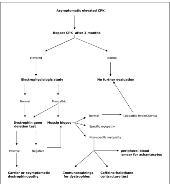
A suggested step by step approach for asymptomatic hy-perCKemia can be seen in the Figure 2. When all such work up is negative, asymptomatic hyperCKemia is reasonable and the patient can be assured that the prognosis is benign.
references
1. Reijneveld JC, Notermans NC, Linssen WHJP, Wokke JHJ. Benign prog-nosis in idiopathic hyperCKemia. Muscle & Nerve 2000;23:575-579. 2. Hoffman EP, Arahata K, Minetti C, Bonilla E, Rowland LP.
Dystrophi-nopathy in isolated cases of myopathy in females. Neurology 1992; 42:967-975.
3. Rowland LP, Willner J, DiMauro S, Miranda A. Approaches to the mem-brane theory of Duchenne muscular dystrophy. In Angelini C, Danieli GA, Fontanari D (Eds). Muscular dystrophy: advances and new trends. Amsterdam, Excerpta Medica, 1980:3-13.
4. Brewster LM, de Visser M. Persistent hyperCKemia: fourteen patients studied in retrospect. Acta Neurol Scand 1988;77:60-63.
5. Prelle A, Tancredi L, Sciacco M, et al. Retrospective study of a large population of patients with asymptomatic or minimally symptomatic raised serum creatine kinase levels. J Neurol 2002;249:305-311. 6. Dabby R, Sadeh M, Herman O, et al. Asymptomatic or minimally
symp-tomatic hyperCKemia: histopathologic correlation. IMAJ 2006;8:110-113. 7. Joy JL, Oh SJ. Asymptomatic hyperCKemia: an electrophysiologic and
histopathologic study. Muscle & Nerve 1989;12:206-209.
8. Walker RH, Peters JJ, Jung HH, Danek A. Diagnostic evaluation of clin-ically normal subjects with chronic hyperCKemia. Neurology 2007;68: 535-536.
9. Weglinski MR, Wedel DJ, Engel AG. Malignant hyperthermia testing in patients with persistently increased serum creatine kinase (CK) lev-els. Anesth Analg 1997;84:1038-1041.
10. Tachi N, Wakai S, Yutoh Y, Chiba S, Miura J. Asymptomatic hyperCKe-mia: detection of an isolated carrier of Duchenne muscular dystrophy. J Child Neurol 1990;5:351-353.
11. Oliveira ASB, Gabbai AA, Schmidt B, et al. Carrier detection of Duch-enne and Becker muscular dystrophy using muscular dystrophin im-munohistochemistry. Arq Neuropsiquiatr 1992;50:478-485.
12. Bonilla E, Schmidt B, Samitt CE, et al. Normal and dystrophin-deicient muscle ibers in carriers of the gene for Duchenne muscular dystrophy.
Am J Pathol 1988;133:440-445.
13. Panigrahi I, Mitall B. Carrier detection and prenatal diagnosis in Duch-enne/Becker muscular dystrophy. Indian Pediatr 2001;38:631-639. 14. Werneck LC, Scola RH, Maegawa GH, Werneck MC. Comparative
anal-ysis of PCR-deletion detection and immunohistochemistry in Brazilian Duchenne and Becker muscular dystrophy patients. Am J Med Genet 2001;103:115-120.