903
Arq Neuropsiquiatr 2008;66(4):903-905 Clinical / Scientiic note
HaSHimoto’S EnCEpHalopatHy witH
CliniCal FEaturES Similar to tHoSE
oF CrEutzFEldt-Jakob diSEaSE
Ana Cláudia Rodrigues de Cerqueira
1,2 A, José Marcelo Ferreira Bezerra
1B,
Gérson Canedo de Magalhães
1C, Márcia Rozenthal
3B, Antônio Egídio Nardi
2BEnCEFalopatia dE HaSHimoto Com CaraCtEríStiCaS ClíniCaS SEmElHantES à doEnça dE CrEutzFEldt-Jakob
Pedro Ernesto University Hospital of the University of the State of Rio de Janeiro, Rio de Janeiro RJ, Brazil (HUPE/UERJ): 1Neurology Service; 2Institute
of Psychiatry at the Federal University of Rio de Janeiro, Rio de Janeiro RJ, Brazil (IPUB/UFRJ); 3Federal University of the State of Rio de Janeiro, Rio de
Janeiro RJ, Brazil (Uni-Rio); AProbative training student for Master’s Degree in Psychiatry; BAssociate Professor; CFull Professor.
Received 18 June 2008, received in inal form 1 September 2008. Accepted 23 September 2008.
Dra. Ana Claudia Rodrigues de Cerqueira – Rua Trajano de Moraes 42 /701 - 24230-380 Niterói RJ - Brasil. E-mail: [email protected]
In 1966, Brain et al. described the irst case of Hashi-moto’s encephalopathy in a patient with stroke-like epi-sodes, seizure activities, and delirium. The symptoms were recurrent and reversible and were not associated with thy-roid dysfunction1. Since then, Hashimoto’s
encephalopa-thy has been recognized as a rare condition associated to Hashimoto’s thyroiditis and to the presence of high con-centrations of antithyroid antibodies, unlike those de-scribed in myxedema and in thyrotoxicosis2. Two
clini-cal forms have been described: the vasculitic form with stroke-like episodes, seizure activities, and slight cogni-tive decline; and the diffuse form with symptoms of de-pression, psychosis, myoclonus, tremors, delusion, luc-tuations in the level of consciousness, and dementia. The clinical manifestations can be luctuating or persistent, and reversible, if properly treated with immunosuppres-sive therapy3. In some cases, Hashimoto’s
encephalopa-thy can show rapidly progressive dementia, myoclonus, and electroencephalographic changes with periodic re-currence of the triphasic complexes similar to those of Creutzfeldt-Jakob disease, which makes a differential di-agnosis essential to both pathologies4.
We describe the case of a patient with Hashimoto’s encephalopathy who showed clinical manifestations simi-lar to those of Creutzfeldt-Jakob disease. The recognition of the condition was essential for the treatment and re-gression of the clinical presentation.
CaSE
A female patient, 68-years-old, Caucasian, widow, born in Rio de Janeiro, developed a clinical presentation of daily sad-ness, discouragement, decreased appetite, insomnia, and cogni-tive decline. The condition of the patient worsened with perse-cutory delusions, visual hallucinations, gait disturbance, tremor
of the extremities, muscle spasms, and luctuations in the level of consciousness. She had a history of hypothyroidism and did not use any medication. At admission, she was sleepy, disorient-ed, showing myoclonus, paratonia on the back of the head and arms, her deep relexes were symmetrical with plantar lexion response and suction and grasping relexes were present. The rest of the physical examination was normal.
The laboratory exams, including a complete blood count; biochemistry; arterial gasometry; hepatic function tests; auto-antibody tests, such as nuclear factor (ANF), Ro, anti-La, pANCA and cANCA, were either normal or negative. The
Arq Neuropsiquiatr 2008;66(4)
904
Hashimoto’s encephalopathy and Creutzfeldt-Jakob disease Cerqueira et al.
els of triiodothyronine (T3) and free thyroxine (T4 L) were within normal limits. The thyroid-stimulating hormone (TSH) was slight-ly elevated at 7.70 mU/L (reference value: 0.35–5.5 mU/L). The anti-thyroperoxidase (anti-TPO) concentration was very high at 1/6400 (reference value: up to 1/100). The presence of antithy-roglobulin antibody (Ac-anti-Tg) was not detected. The exami-nation of the cerebrospinal luid (CSF), including cytometry, bio-chemistry, immunoglobulins, detection of oligoclonal bands and testing for the presence of bacteria and fungi, immunological re-actions for syphilis, cysticercosis, schistosomiasis, toxoplasmo-sis, cytomegalovirus, HIV, HTLV1/2, herpes simplex and herpes zoster were either normal or negative.
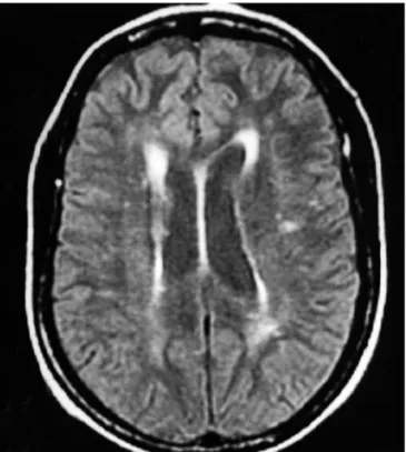
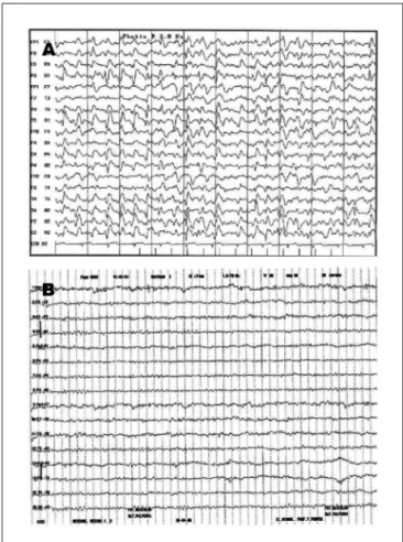
The magnetic resonance imaging (MRI) of the brain showed small changes that were not signiicant, such as the presence of hypersignal focus in the corona radiata and bilaterally semioval center (Fig 1). The electroencephalogram (EEG) at admission was abnormal, with slow, monomorphic, occasionally sharp, tripha-sic, with theta-delta frequency ranges from 2 to 3 Hz, intermit-tent, bilateral waves, predominantly in the temporoparietal re-gions (Fig 2A).
The diagnosis hypothesized was of Hashimoto’s encepha-lopathy and the treatment with methylprednisolone 1 g/day for three consecutive days, followed by prednisone with initial dose
of 60 mg/day, was established. In the irst two weeks, a consid-erable improvement in level of consciousness and myoclonus was observed. The prednisone dosage was gradually reduced in the following two months to 5 mg/day. In this period there was a regression of all signs and symptoms. This improvement in the clinical presentation can be correlated to the complete resolu-tion of the electroencephalographic changes (Fig 2B). The pa-tient authorized the publication of the case upon signature of an informed consent form.
diSCuSSion
Hashimoto’s encephalopathy is a rare condition asso-ciated with Hashimoto thyroiditis, the presence of high concentrations of anti-thyroid antibodies, without any evidence of thyroid dysfunction, and an excellent re-sponse to treatment with corticosteroids2. Recent
stud-ies have shown the presence of anti-thyroid antibodstud-ies in the CSF5 and alpha-enolase antibodies in the serum of
patients with Hashimoto’s encephalopathy, suggesting the involvement of an autoimmune mechanism in this con-dition6. However, the role of those antibodies and their
pathophysiology are unknown. A possible vasculitic pro-cess has been shown in biopsy studies 7.
The clinical presentation may suggest a diagnosis of Creutzfeldt-Jakob disease. In an epidemiological study made by Seipelt et al., six cases of Hashimoto’s encepha-lopathy were identiied and treated. The patients met the clinical criteria for Creutzfeldt-Jakob disease and were no-tiied8. The criteria established by World Health
Organiza-tion (WHO) for diagnosing to sporadic Creutzfeldt-Jakob disease (sCJD) include progressive dementia with a dura-tion of less than two years and two of the clinical indings – myoclonus, cerebellar or visual symptoms, pyramidal or extrapyramidal signs and akinetic mutism. The clinical manifestations associated with the presence of triphasic complexes in the EEG or the detection of the 14-3-3 pro-tein in the CSF make the diagnosis possible. However, the inal diagnosis is obtained through a neuropathological exam9. A recent study by Martins et al. described the irst
35 cases of Creutzfeldt-Jakob disease notiied in Brazil, 26 of these cases were initially classiied as possible sCJD, of which 51% fulilled the criteria for probable sCJD. The most frequent clinical manifestations were myoclonus (80%), pyramidal signs (68%), ataxia (65%) and early psy-chiatric symptoms (55%)10.
We related the case of a patient with Hashimoto’s encephalopathy and clinical features similar to those of Creutzfeldt-Jakob disease. The presence rapidly pro-gressive dementia, ataxia, myoclonus, and especially the presence of triphasic complexes in the EEG, make the di-agnosis a probable one for Creutzfeldt-Jakob disease. In this case, the Hashimoto’s encephalopathy diagnosis was
Arq Neuropsiquiatr 2008;66(4)
905
Hashimoto’s encephalopathy and Creutzfeldt-Jakob disease Cerqueira et al.
made based on the exclusion of other toxic-metabolic encephalopathic and neurological diseases of infectious or vascular origin, and on the detection of high concen-trations of anti-TPO antibodies. The recognition of this pathology and the treatment with immunosuppressant therapy determined the regression of the clinical presen-tation and changes in the EEG.
This report emphasizes the importance of a differen-tial diagnosis between the two pathologies. Creutzfeldt-Jakob disease has a progressive and inexorably fatal course, as opposed to Hashimoto’s encephalopathy, which has a luctuating course and excellent response to treat-ment with corticosteroids. We believe that this pathology is misdiagnosed, therefore, tests detecting the presence of anti-thyroid antibodies are recommended in a clini-cal presentation of encephalopathy of subacute onset or rapidly progressive dementia associated with myoclonus and triphasic complexes in the EEG, when the ethiology is not identiied.
rEFErEnCES
1. Brain L, Jellinek EH, Ball K. Hashimoto’s disease and encephalopathy. Lancet 1966;2:512-514.
2. Chong JY, Rowland LP, Utiger RD. Hashimoto encephalopathy: syn-drome or myth? Arch Neurol 2003;60:164-171.
3. Kothbauer-Margreiter I, Sturzenegger M, Komor J, Baumgartner R, Hess C W. Encephalopathy associated with Hashimoto thyroiditis: di-agnosis and treatment. J Neurol 1996;243:585-593.
4. Doherty CP, Schlossmacher M, Torres N, Bromield E, Samuels MA. Hashimoto’s encephalopathy mimicking Creutzfeldt-Jakob disease: brain biopsy indings. J Neurol Neurosurg Psychiatry 2002;73:597-603. 5. Ferraci F,Moretto G, Candeago RM et al. Antithyroid antibodies in the
CSF: their role in the pathogenesis of Hashimoto’s encefalopathy. Neu-rology 2003;60:712-714.
6. Ochi H, Horiuchi I, Araki N, et al. Proteomic analysis of human brain identiies alpha-enolase as a novel autoantigen in Hashimoto’s enceph -alopathy. FEBS Lett 2002;528:197-202.
7. Shibata N, Yamamoto Y, Sunami N, Suga M, Yamashita Y. Isolat-ed angiitis of the CNS associatIsolat-ed with Hashimoto’s disease. Rinsho Shinkeigaku 1992;32:191-198.
8. Seipelt M, Zerr I, Nau R, et al. Hashimoto’s encephalitis as a differen-tial diagnosis of Creutzfeldt-Jakob disease. J Neurol Neurosurg Psychi-atry 1999;53:162-163.
9. WHO (see http://www.who.int/entity/zoonoses/diseases/ Creutzfeldt.pdf)