Effects of iodinated contrast media in a novel
model for cerebral vasospasm
Efeitos do meio de contraste iodado em um novo modelo de vasoespasmo cerebral
Tatiana Nikitina1, Olga Zavaritskaya2, Vladimir Semenyutin3, Pontus B. Persson1, Andreas Patzak1, Mauricio Sendeski1
ABSTRACT
Objective:We developed anin vitromodel for vasospasm post subarachnoid hemorrhage that was suitable for investigating brain vessel autoregulation. We further investigated the effects of iodinated contrast medium on the vascular tone and the myogenic response of spastic cerebral vessels.Method:We isolated and perfused the superior cerebellar arteries of rats. The vessels were pressurized and studied under isobaric conditions. Coagulated blood was used to simulate subarachnoid hemorrhage. The contrast medium iodixanol was applied intraluminally.Results:Vessels exposed to blood developed significantly stronger myogenic tone (65.7 ± 2.0% vs 77.1 ± 1.2% of the maximum diameter, for the blood and the control group, respectively) and significantly decreased myogenic response, compared with the control groups. The contrast medium did not worsen the myogenic tone or the myogenic response in any group.Conclusion:Our results show that deranged myogenic response may contribute to cerebral blood flow disturbances subsequent to subarachnoid hemorrhage. The contrast medium did not have any negative influence on vessel tone or myogenic response in this experimental setting.
Keywords:brain ischemia, cerebral angiography, contrast media, hemodynamics, intracranial aneurysm, intracranial vasospasm, subarachnoid hemorrhage.
RESUMO
Objetivo:Desenvolvemos um modeloin vitropara vasoespasmo subsequente à hemorragia subaracnóide que foi adequado para investigar a autorregularão dos vasos cerebrais. Em seguida investigamos os efeitos o meio de contraste iodado no tônus vascular e na resposta miogênica dos vasos cerebrais espásticos.Método:Isolamos e perfundimos as artérias cerebelares superiores de ratos. Os vasos foram pressurizados e estudados em condições isobáricas. Sangue coagulado foi utilizado para simular hemorragia subaracnóide. O meio de contraste iodixanol foi aplicado intraluminarmente.Resultados:Os vasos expostos ao sangue desenvolveram aumento significativo do tônus miogênico (65.7 ± 2.0% vs 77.1 ± 1.2% do maior diâmetro, para o grupo de sangue e o grupo controle, respectivamente) com resposta miogênica significativamente menor do que aquela dos controles. O meio de contraste iodado não piorou o tônus miogênico ou a resposta miogênica em nenhum dos grupos. Conclusão: Nossos resultados mostram que uma resposta miogênica pode contribuir para as alterações de fluxo sanguíneo cerebral subsequentes à hemorragia subaracnóide. O meio de contraste iodado não teve nenhuma influência negativa no tônus vascular ou na resposta miogênica neste modelo experimental.
Palavras-chave:isquemia cerebral, angiografia cerebral, meio de contraste, hemodinâmica, aneurisma intracraniano, vasoespasmo intracraniano, hemorragia subaracnóide.
Cerebral delayed vasospasm is a severe complication following spontaneous subarachnoid hemorrhage (SAH). Vasospasm is an important cause of death and contributes 10-12% to the overall mortality after SAH, which reaches approximately 50% within the first month1,2,3.
There is nowadays no specific therapy or prophylaxis for vasospasm2,4,5. The low success of current treatment
strategies for vasospasm may be due to insufficient know-ledge about the pathophysiology of vasospasm, despite a large number ofin vitroandin vivostudies done to discover spastic mechanisms and to find an adequate treatment for cerebral vasospasm6.
The majority of studies on the mechanism of vasospasm were performed usingin vivoanimal models3,6,7. Whilein vivo
1Institut fuer Vegetative Physiologie, Charité-Universitaetsmedizin Berlin, Berlin, Germany; 2Research Division Cardiovascular Physiology, Medical Faculty Mannheim, Mannheim, Germany;
3Laboratory of Brain Circulation Pathology, Russian Polenov Neurosurgical Institute, Saint-Petersburg, Russia.
Correspondence: Andreas Patzak; Institut fuer Vegetative Physiologie, Charité-Universitaetsmedizin Berlin, Germany, Charitéplatz 1, 10117 Berlin; E-mail: [email protected]
Conflict of interest:There is no conflict of interest to declare.
Support:This work was supported by grants from the Deutsche Forschungsgemeinschaft (FG1368, PA479/10-1). Received 20 May 2014; Received in final form 22 October 2014; Accepted 10 November 2014.
DOI:10.1590/0004-282X20140222
studies successfully reproduce the clinical picture of vasos-pasm, there is high individual variability in their results6, what limits their application for studying effects of vasos-pasm on mechanisms of cerebral blood flow regulation. The use of in vitromodels, where conditions like
intralum-inal pressure, oxygen tension, and milieu surrounding the vessels are constant, might reduce these disadvantages. Unfortunately, current in vitro models of vasospasm that
use whole blood to simulate SAH8,9 are technically unsuit-able for investigation of the autoregulation of cerebral vessels, mostly because presence of blood prevents visualiza-tion of a pressurized vessel and diameter measurements. We developed a novel, highly reproducible and technically uncomplicated model of vasospasm in vitro using a
per-fusion myograph and videomicroscopy to investigate the myogenic response of cerebral vessels – one of the main
mechanisms of cerebral blood flow autoregulation10,11. Most diagnostic and therapeutic procedures needed for the treatment of SAH patients require the use of iodinated contrast media (CM)12,13,14. It has been shown that CM have adverse effects on vessels of the kidney and other vascular beds15,16. Further, CM influence some components of cereb-ral blood flow regulation in healthy subjects17. We thus tested the hypothesis that CM negatively influence the auto-regulation of cerebral blood flow after SAH.
METHOD
All animal handling and experiments were performed in accordance to the guidelines of the Office for Health and Social Matters of Berlin (Berlin, Germany).
Isolation and preparation of cerebral vessels
Adult male Sprague Dawley rats (Charles River, Germany), 150-200 grams (7-8 weeks old), were anaesthe-tized using isoflurane (Abbott, Baar, Switzerland) and decapitated. Brains were excised and placed in ice-cold pre-paration physiological salt solution. Segments of superior cerebellar arteries without branches in a diameter of about 250mm were isolated under magnification using sharpened forceps and microscopic scissors, and mounted on glass can-nulas within the myograph’s experimental chamber. As usual in experiments investigating myogenic response, there was no flow inside the vessel18.
Altogether 73 rats were included in the study. 50 success-ful experiments were performed. Excluding criteria were: intraluminal flow due to lacks or vessel branches and insuf-ficient development of spontaneous myogenic tone (,20%). Experimental groups and number of experiments are shown in Table.
Experimental conditions Intraluminal
The intraluminal solution consisted of experimental physiological salt solution (PSS) (146 mmol/l NaCl, 4.5 mmol/l KCl, 1.2 mmol/l NaH2PO4, 1.0 mmol/l MgSO4, 1.6
mmol/l CaCl2, 5.5 mmol/l glucose, 0.025 mmol/l EDTA, 5.0
mmol/l HEPES, pH 7.4 by temperature 37.0°C). The groups which received CM had an end concentration of 23 mg iodine/ml (1.8*10−4 mol/l) (iodixanol, GE Healthcare, Munich, Germany) in the intraluminal solution. The CM concentration is the same shown to cause constriction of renal vasa recta and afferent arterioles, and is within the range possibly reached during intravascular procedures in humans15,19,20.
Extraluminal inside the chamber
The experimental chamber was filled with PSS and warmed up to 37.0°C. The vessels were exposed to an initial intraluminal pressure of 80 mmHg.
Model of SAH: Fresh blood (2 ml) was collected through laparotomy and sectioning of the renal artery, and deposited into the experimental chamber (0.5 ml in each corner) of a perfusion myograph (model 110P, DMT, Aarhus Denmark). The blood was left to coagulate for 40 minutes by room tem-perature, during which suitable brain vessels were isolated and prepared for perfusion. The experimental chamber was then filled with the PSS, and the arteries were mounted onto the glass cannulas. There was no contact between the blood clot and the arteries.
Measurement of the vessel diameter and quantification of the myogenic response
The procedure for mounting of the artery, intraluminal pressure manipulation as well as measurement of vessel diameters was in accordance to the principles of investi-gating of myogenic responses in pressurized arteries21. Arterial diameter was recorded in a continuous manner
Table.The experimental groups according to experimental chamber and intraluminal content of the vessel.
Experimental groups Experimental chamber content Intraluminal content
1 Control(n = 22) PSS PSS
2 Blood (n = 13) PSS + blood clot PSS
3 CM (n = 9) PSS PSS + iodixanol
4 CM + blood (n = 6) PSS + blood clot PSS + iodixanol
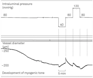
over time using an acquisition system consisting of a video camera assembled on an inverted microscope and con-nected to a software for automatic vessel diameter meas-urement (IonWizard 6.1, IonOptics, Milton, MA, USA) (Figure 1). Right after mounting of superior cerebellar arter-ies on glass pipets, continuous diameter measurement was started, and all vessels underwent a period of stabilization of 1 hour during which the development of myogenic tone was monitored. Only arteries which developed typical spon-taneous myogenic tone – for cerebral vessels more than
20% of constriction – were included into the study. The
value of the myogenic tone was expressed as the percent-age of the vessel diameter at the end of stabilization time in relation to the diameter in the maximal dilated state. After recording of baseline diameter measurements, the intraluminal pressure was changed in a controlled manner in 4 steps (each of 5 minutes): 1 step – from 80 to 40
mmHg; 2 step – from 40 to 80 mmHg; 3 step – from 80
to 120 mmHg; and 4 step – from 120 to 80 mmHg. The
myogenic response was quantified in micrometers (mm) as the difference between the diameter immediately pre-ceding the pressure step and the diameter measured 5 min-utes following the pressure step, when the vessel diameter is stable. All diameter values were measured at the external border of the vessel wall. Figure 2 shows a representative
tracing of a typical experiment, where the protocol steps are graphically depicted.
STATISTICAL ANALYSIS
Only one artery was used from each rat for each experi-ment. Calculation of power was previously performed to determine the optimal sample size. The IBM SPSS Statistics 22 software was used for statistical comparisons. Statistical significance was considered for p-value smaller than 0.05.
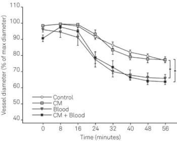
Data from spontaneous myogenic tone were reported in the text and in Figure 3 as average and standard error of the means (SEM). Two-way/repeated measurements ANOVA was used to compare groups for spontaneous myogenic tone. Bonferroni method was used as correction for multiple comparisons. Where ANOVA pointed to differences in myo-genic tone between groups, Tukey HSD test was used as a post hoc test to compare groups pairwise.
Data from the myogenic response were reported in the text as medians (with 25thand 75thpercentiles), and in the Figure 4 as box-plots. The myogenic response steps among all four groups were first compared with the Kruskal-Wallis test. Where differences among groups were detected, we used the Mann-Whitney-test to identify inter-group differ-ences pairwise.
Figure 1.Representative pictures of pressurized superior cerebellar arteries during experiments using an acquisition system consisting of a video system and digital imaging. The rectangle delimits the range to be analysed by the software performing automatic diameter measurement over time. Vertical lines show the vessel borders detected in real time. The tip of the glass cannulas inside of the vessel is indicated with an arrow at the bottom of the picture. (A) Development of normal spontaneous myogenic tone; (B) Development of spastic spontaneous myogenic tone in presence of the coagulated blood (volume 2 ml).
RESULTS
Spontaneous myogenic tone
Two-way/repeated measurements ANOVA followed by Tukey HSD test showed that exposure to blood significantly influenced the development of myogenic tone over time (p = 0.000002), while CM did not (p = 0.423). Vessels treated with blood developed a more pronounced myogenic tone compared to control groups:
N
Vessels treated with blood clot alone had a final diameterof 65.7 ± 2.0% of maximal diameter after stabilization time, compared to 77.1 ± 1.2% of the control group (average ± SEM, p = 0.00001);
N
Vessels exposed to CM and blood clot together had a finaldiameter of 62.1 ± 2.1% of maximal diameter, compared to 77.2 ± 1.7% of the CM group (average ± SEM, p = 0.00024).
Myogenic response
There were statistically significant differences between the experimental groups in all four steps of the myogenic response measurement (Kruskal-Wallis test, p , 0.05). Figure 4 shows the magnitude of variation in vessel diameter in response to controlled changes in intraluminal pressure, as well as which experimental groups differed from another in each step of the protocol (Mann-Whitney-test). Exposition to blood alone impaired the myogenic response in all steps of pressure. CM alone did not influence the myogenic response in comparison to the control group. In the groups exposed to blood, CM influenced the myogenic response only in the third step of the protocol (80 to 120 mmHg) when compared to blood alone.
In the control group, the change of intraluminal pressure from 80 to 40 mmHg dilated vessels by 11.9 mm (25th and 75thpercentiles, 8.2
mm and 16.5mm, respectively), while the change from 40 to 80 mmHg constricted vessels by -13mm (-19 mm, -7.7 mm). Increasing the intraluminal pressure from 80 to 120 mmHg changed the diameter by 1.4mm (-1.8mm, 4mm). Decreasing the intraluminal pressure from 120 to 80 mmHg dilated vessels by 0.6mm (-2.9mm, 4.2mm) (Figure 4). CM alone did not significantly change the myogenic response (80-40 mmHg: 9.1mm, 6.2mm, 10.8 mm, p = 0.28; 40-80 mmHg: -9.3 mm, -13.1 mm, -7.1 mm, p = 0.24; 80-120 mmHg: -0.1 mm, -4.1 mm, 4 mm, p = 0.84; 120-80 mmHg: 1.7mm, -2.8mm, 4.3mm, p = 0.89) compared to the control group, respectively (Figure 4).
Vessels treated with blood showed a significant decrease of both the dilatory and the constrictor myogenic response (80-40 mmHg: -3.3 mm, -7.9 mm, -1.9 mm, p = 0.00004; 40-80 mmHg: 1.7mm, 0.5mm, 3mm, p = 0.0002; 80-120 mmHg: 10.9 mm, 7 mm, 18 mm, p = 0.0006; 120-80 mmHg: -6.4mm, -23.1mm, -2.5mm, p = 0.008) in comparison to the con-trol group, respectively (Figure 4).
Vessels treated with blood and CM together showed a significant decrease of the first and second steps of the myo-genic response (80-40 mmHg: -3.3 mm, -4.3 mm, -0.6 mm, p = 0.0014; 40-80 mmHg: -1.1mm, -1.4mm, 0.4mm, p = 0.0014; 80-120 mmHg: 1.1 mm, -1.3 mm, 2.5 mm, p = 0.93; 120-80 mmHg: -1.6 mm, -3.1 mm, 0 mm, p = 0.33) in comparison to CM alone, respectively (Figure 4).
The myogenic response differed between vessels treated with blood and CM together compared to blood alone only at the third step of pressure (80-40 mmHg: p = 0.55; 40-80 mmHg: p = 0.072; 80-120 mmHg: p = 0.0032; 120-80 mmHg: p = 0.099; Figure 4).
DISCUSSION
The focus of our study was to assess the effect of a con-temporary, widely used CM on the myogenic response of spastic cerebral vessels. A novelin vitromodel was developed
to simulate vasospasm post SAH. It consisted of deploying a controlled amount of blood clot in the experimental chamber without mechanical interaction with the vessel.
The mechanisms of vasospasm post SAH have been inves-tigated using different models. There arein vivoandin vitro
approaches. The most commonin vivotechniques are:
injec-tion of blood into the brain cisternae and vessel avulsion22,23. Some investigators placed blood clots into the brain3,6.In vitro models include application of whole blood8,9or of vasoactive substances which are hypothetically involved in the devel-opment of vasospasm18into the organ bath solution.
In our experience models of SAH using whole blood8,9 were not suitable for the investigation of spontaneous Figure 3.Development of spontaneous myogenic tone during
myogenic tone and myogenic response, primarily because whole blood hinders the visualization of pressurized arteries. The use of coagulated blood does not muddle the solution and allow the measurement of vessel diameter. It is possible to use coagulated blood of different ages, over longer periods of time. Importantly, we can control the exact proportion between the volume of the blood clot and PSS surrounding the vessel, and avoid the effects of irregular distribution of blood around the brain cisterns. By careful and controlled isola-tion of each vessel we can also exclude that vessel dysfuncisola-tion happens due to mechanical damage by bleeding. Although mechanical irritation of vessels or brain damage are considered as possible contributing factors4,8,24,25to clinical vasospasm, in our study we intended to separate the mechanical effects from the effects of blood clot presence and ageing.
We found that exposition to coagulated blood significantly increases the spontaneous myogenic tone of cerebral arteries.
This indicates that ourin vitromodel successfully reproduces changes in the vessel tone which correspond to the initial events of vasospasm post SAHin vivo. The data support the
assumption that the causal agents of vascular spasm originate largely from the clotted blood23. We thus believe that the use of coagulated blood immersed in PSS provides a nearer approximation of the environmental conditions in the brain cisterns following SAH in comparison to the models using either whole blood or individual vasoconstrictors.
intraluminal pressure was increased to what the authors con-sidered as supraphysiological levels (i.e., above 140 mmHg)26. In contrast, in our experiments only the vessels exposed to blood clot showed an impaired myogenic response, and in all levels of pressure. These apparently contradictory findings may result from the use of different models of vasospasm, dif-ferent time points for investigating the myogenic response during the development of vasospasm, and different protocols for quantifying the myogenic response.
We found out that CM did not significantly influence ves-sel tone and did not negatively influence the myogenic response, in both healthy as well as in spastic vessels. This is important because patients with SAH have disturbances of cerebral blood regulation25, and many diagnostic and therapeutic procedures needed for their treatment may require the use of CM12,13,14. It has been shown that several types of CM may have potentially deleterious effects on the tone and reactivity of vessels from several vascular beds15,16. Correspondingly, there are evidences that CM affect regional cerebral blood flow in healthy subjects17. Interestingly, Rosengarten et al.17showed that the dynamic cerebral blood flow regulation – the increase of regional
cerebral blood flow caused by brain activity–was negatively
affected by CM, while stable regional brain blood flow was not affected. We think that these results are compatible with our findings, given that the stable regional blood flow is directly dependent on the myogenic response. Although our results do not show statistical differences for vessels treated with blood clot and CM, we have to consider a small number of samples and multiple comparisons. Moreover,
when translating the current findings intoin vivocondition,
we can meet various limitations.
Interestingly, we found that vessels exposed to blood clot and CM showed a lower degree of functional impairment in comparison to vessels exposed to blood clot alone. A pos-sible explanation for this finding could be endothelial damage induced by the CM, with consequent decreased nitric oxide release and increased superoxide production15,27. Although the influence of the endothelium on the myogenic response is usually not marked, some experimental models show that the myogenic response of cerebral arteries might be modulated by the endothelium10.
In summary, we developed a novel, reproducible in vitro
model of vasospasm post SAH which is adequate to invest-igate cerebral autoregulation. Our finding that the myogenic response was deranged in our model is compatible with clin-ical studies showing that the autoregulation of cerebral blood flow is impaired after SAH. There was no negative influence of CM on myogenic tone and myogenic response in cerebral vessels with acute vasospasm. The influence of CM on other mechanisms of regulation of cerebral blood flow in patients with vasospasm and SAH is still unknown, and warrants further investigation using other types of models.
Acknowledgements
We thank A. Gerhardt for the technical assistance during our experiments.
References
1. Odom MJ, Zuckerman SL, Mocco J. The role of magnesium in the management of cerebral vasospasm. Neurol Res Int. 2013;2013:ID943914.
2. Ciurea AV, Palade C, Voinescu D, Nica DA. Subarachnoid hemorrhage and cerebral vasospasm - Literature review. J Med Life. 2013;6:120-5.
3. Titova E, Ostrowski RP, Zhang JH, Tang J. Experimental models of subarachnoid hemorrhage for studies of cerebral vasospasm. Neurol Res. 2009;31(6):568-81. http://dx.doi.org/10.1179/174313209X382412
4. Athar MK, Levine JM. Treatment options for cerebral vasospasm in aneurysmal subarachnoid hemorrhage. Neurotherapeutics. 2012;9(1):37-43. http://dx.doi.org/10.1007/s13311-011-0098-1
5. Adamczyk P, He S, Amar AP, Mack WJ. Medical management of cerebral vasospasm following aneurysmal subarachnoid hemor-rhage: a review of current and emerging therapeutic interventions. Neurol Res Int. 2013;2013:ID462491. http://dx.doi.org/10.1155/2013/ 462491
6. Marbacher S, Fandino J, Kitchen ND. Standard intracranial in vivo animal models of delayed cerebral vasospasm. Br J Neurosurg. 2010;24(4):415-34. http://dx.doi.org/10.3109/02688691003746274
7. Chen S, Feng H, Sherchan P, Klebe D, Zhao G, Sun X et al. Controversies and evolving new mechanisms in subarachnoid hemorrhage. Prog Neurobiol. 2014;115:64-91. http://dx.doi.org/ 10.1016/j.pneurobio.2013.09.002
8. Simeone FA, Vinall P. Mechanisms of contractile response of cerebral artery to externally-applied fresh blood. J Neurosurg. 1975;43(1):37-47. http://dx.doi.org/10.3171/jns.1975.43.1.0037
9. Linder M, Alksne JF. Prevention of persistent cerebral smooth muscle contraction in response to whole blood. Stroke. 1978;9(5):472-7. http://dx.doi.org/10.1161/01.STR.9.5.472
10. Schubert R, Mulvany M. The myogenic response: established facts and attractive hypotheses. Clin Sci. 1999;96(4):313-26. http://dx.doi. org/10.1042/CS19980403
11. Duchemin S, Boily M, Sadekova N, Girouard H. The complex contribution of NOS interneurons in the physiology of cerebrovas-cular regulation. Front Neural Circuits. 2012;6:51. http://dx.doi.org/ 10.3389/fncir.2012.00051
12. Kellner P, Stoevesandt D, Soukup J, Bucher M, Raspé C. Aneurysmatisch bedingte Subarachnoidalblutung. Anaesthesist. 2012;61(9):792-814. http://dx.doi.org/10.1007/s00101-012-2077-2
13. Seibert B, Tummala RP, Chow R, Faridar A, Mousavi SA, Divani AA. Intracranial aneurysms: review of current treatment options and outcomes. Front Neurol. 2011;2:45. http://dx.doi.org/10.3389/ fneur.2011.00045
15. Sendeski M, Patzak A, Persson PB. Constriction of the vasa recta, the vessels supplying the area at risk for acute kidney injury, by four different iodinated contrast media, evaluating ionic, nonionic, monomeric and dimeric agents. Invest Radiol. 2010;45(8):453-7. http://dx.doi.org/10.1097/RLI.0b013e3181d77eed
16. Sendeski MM. Pathophysiology of renal tissue damage by iodinated contrast media. Clin Exp Pharmacol Physiol. 2011;38(5):292-9. http://dx.doi.org/10.1111/j.1440-1681.2011.05503.x
17. Rosengarten B, Steen Müeller MK, Müeller A, Traupe H, Voss RK, Kaps M. Contrast media effect on cerebral blood flow regulation after performance of cerebral or coronary angiography. Cerebrovasc Dis. 2003;16(1):42-6. http://dx.doi.org/10.1159/000070114
18. Seker F, Hesser J, Neumaier-Probst E, Groden C, Brockmann MA, Schubert R et al. Dose-response relationship of locally applied nimodipine in an ex vivo model of cerebral vasospasm. Neuroradiology. 2013;55(1):71-6. http://dx.doi.org/10.1007/s00234-012-1079-8
19. Liu ZZ, Viegas VU, Perlewitz A, Lai EY, Persson PB, Patzak A et al. Iodinated contrast media differentially affect afferent and efferent arteriolar tone and reactivity in mice: a possible explanation for reduced glomerular filtration rate. Radiology. 2012;265(3):762-71. http://dx.doi.org/10.1148/radiol.12120044
20. Seeliger E, Flemming B, Wronski T, Ladwig M, Arakelyan K, Godes M et al. Viscosity of contrast media perturbs renal hemodynamics. J Am Soc Nephrol. 2007;18(11):2912-20. http://dx.doi.org/10.1681/ ASN.2006111216
21. Schubert R. Isolated vessels. In: Dhein S, Mohr FW, Delmar M. Practical methods in cardiovascular research. Berlin: Heidelberg; 2005. p. 198-211.
22. Brunner E, Puri ML. Nonparametric methods in factorial designs. Stat Papers. 2001;42(1):1-52. http://dx.doi.org/10.1007/s003620000039
23. Crowley RW, Medel R, Kassell NF, Dumont AS. New insights into the causes and therapy of cerebral vasospasm following subarachnoid hemorrhage. Drug Discov Today. 2008;13:254-60. http://dx.doi.org/ 10.1016/j.drudis.2007.11.010
24. Zheng M, Zhu H, Gong Y, Wang D, Xie Q, Tang H et al. Involvement of GMRP1, a novel mediator of Akt pathway, in brain damage after intracerebral hemorrhage. Int J Clin Exp Pathol. 2013;6(2):224-9.
25. Koide M, Sukhotinsky I, Ayata C, Wellman GC. Subarachnoid hemorrhage, spreading depolarizations and impaired neurovascular coupling. Stroke Res Treat. 2013;2013:ID819340. http://dx.doi.org/ 10.1155/2013/819340
26. Ishiguro M, Puryear CB, Bisson E, Saundry CM, Nathan DJ, Russell SR et al. Enhanced myogenic tone in cerebral arteries from a rabbit model of subarachnoid hemorrhage. Am J Physiol Heart Circ Physiol. 2002;283(6):H2217-25. http://dx.doi.org/10.1152/ajpheart.00629.2002